• N&PD Moderators: Skorpio

Pharmacology Psychedelic Pharmacology

This thread contains discussion about a Pharmacology-related topic

mr peabody

Moderator: PM
Staff member
Joined
Aug 31, 2016
Messages
6,038
Location
Frostbite Falls, MN
shutterstock_186044744-1536x1028.jpg


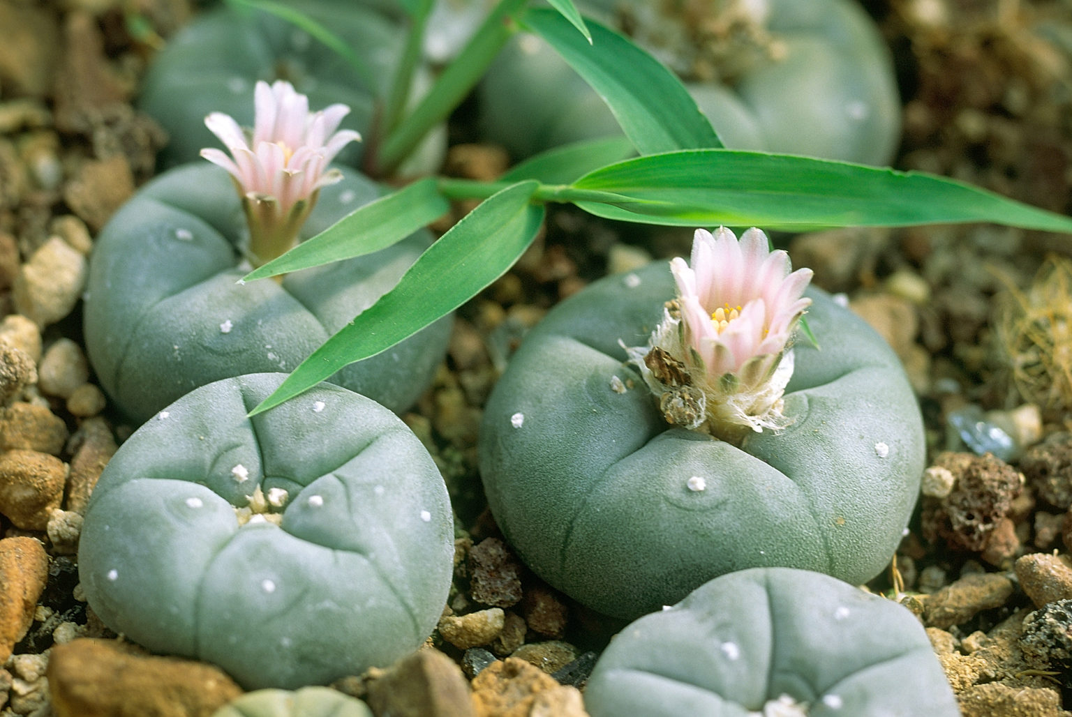
The Compounds in Psychedelic Cacti

Mescaline is the best-known phenethylamine in psychedelic cacti; but what about the other “minor” compounds in those cacti?

by Barbara E. Bauer, MS | Psychedelic Science Review

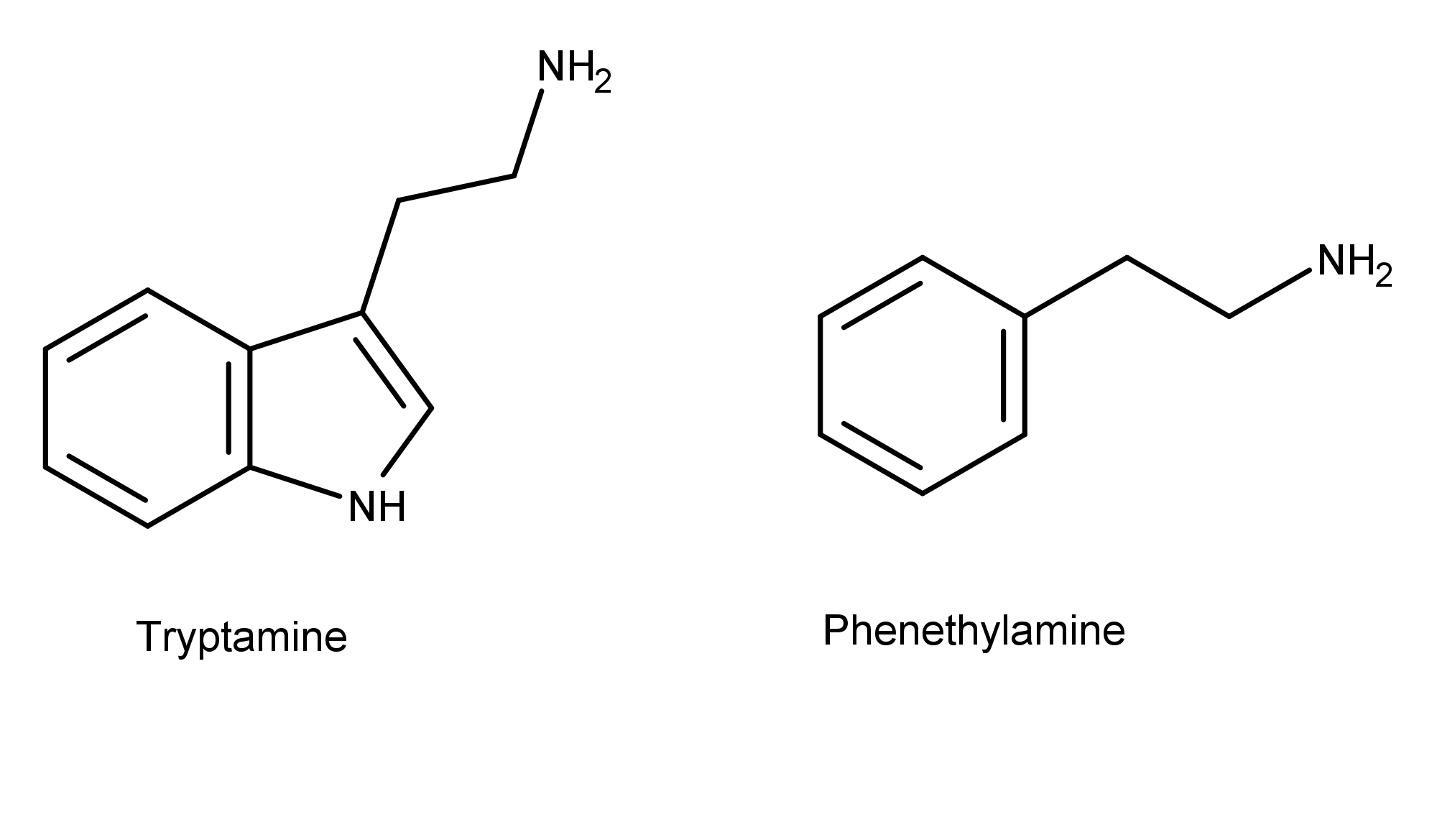
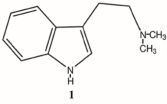
Scientists are learning that psychedelic compounds abound in nature. In addition to tryptamine compounds found in magic mushrooms and toad venom, psychedelic cacti provide a source of naturally occurring psychoactive phenethylamine compounds. The image below illustrates the difference in the chemical structures of tryptamine and phenethylamine.


Mescaline is probably the most well-known compound in psychedelic cacti. Species that contain it include the San Pedro cactus (Echinopsis pachanoi), Peyote cactus (Lophophora williamsii), and the Peruvian Torch cactus (Echinopsis peruviana). Peyote, in particular, is unique because archaeologists have found evidence of its use by Native Americans as far back as 5,700 years ago, making it the oldest known plant containing a bioactive drug compound. Mescaline was first synthesized by Ernst Späth in 1918.

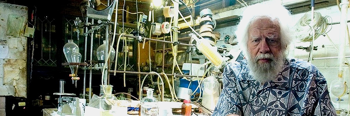
In their classic book “PiHKAL: A Chemical Love Story,” Alexander and Ann Shulgin called mescaline one of the “Magical Half Dozen” compounds that they considered the most important out of all those they synthesized and studied.

Other compounds in Psychedelic Cacti

In addition to mescaline, psychedelic cacti contain other psychoactive compounds. Many of those compounds remain untested (and probably many still undiscovered) so researchers don’t have a clear picture of which ones may have psychedelic/therapeutic effects and may be involved in entourage effects and/or allosteric modulation.

In 1969, Richard Evans Schultes stated in a paper in Science that, “Peyote contains at least 15 ß-phenethylamine and isoquinoline alkaloids.” He went on to say,
The intoxications induced by mescaline and by Peyote itself are very different, but they have unfortunately been confused in the literature.​

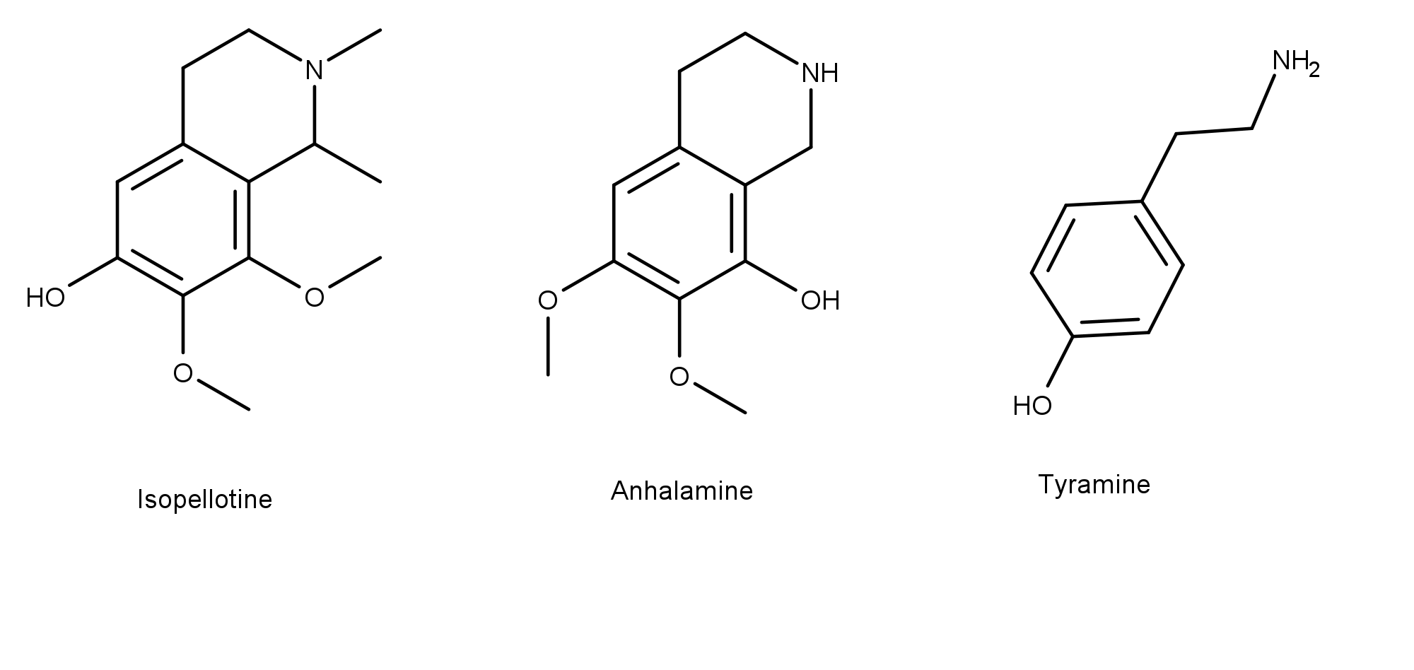
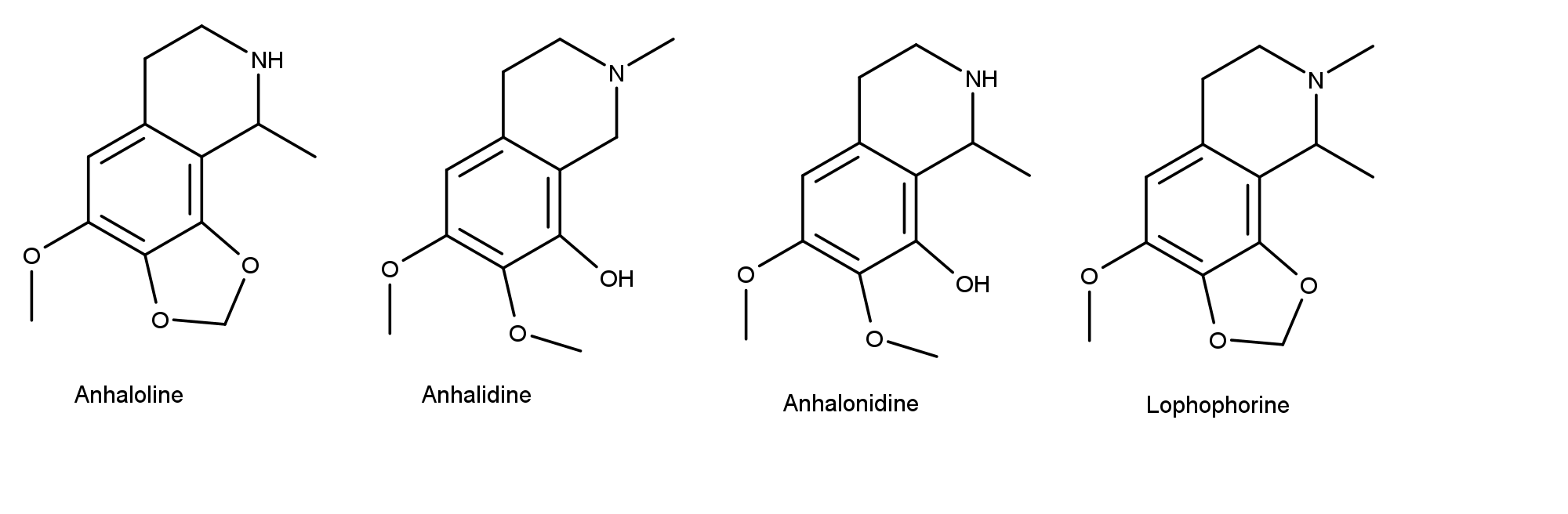
Research done in the 1970s identified compounds in the Peyote cactus including isopellotine, anhalamine, and tyramine.


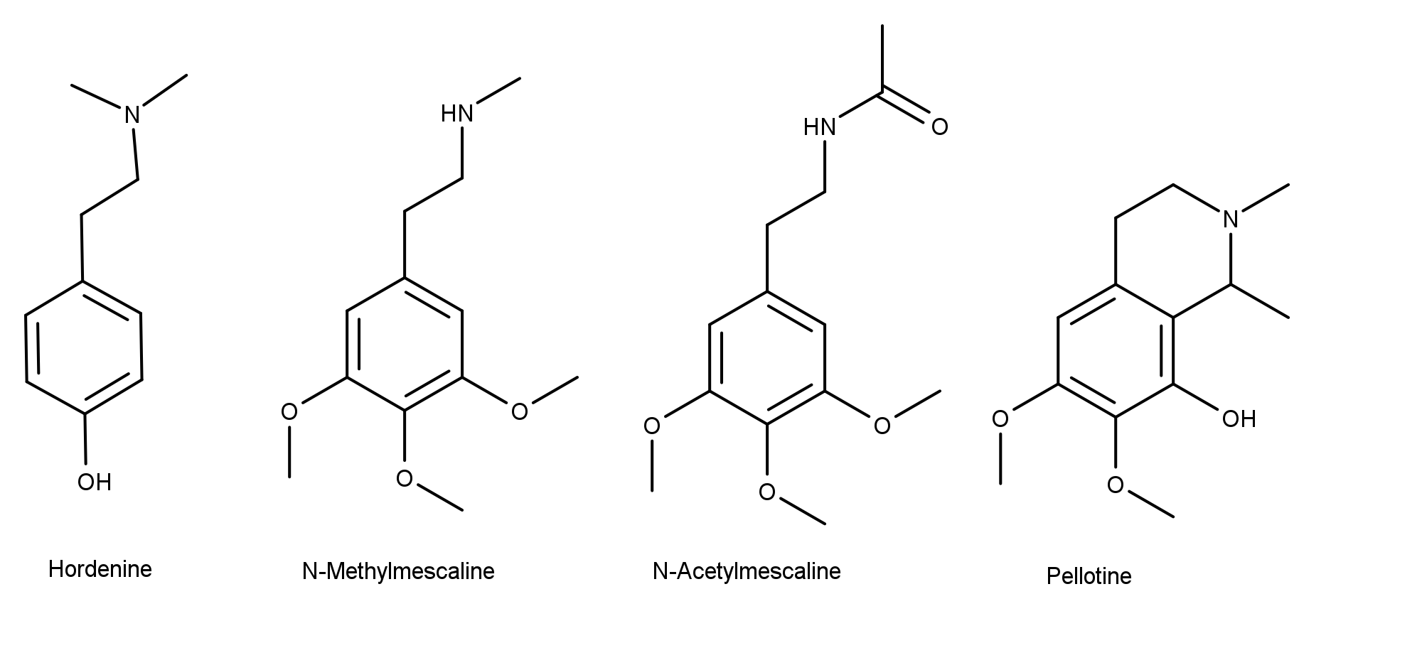
In 2015, Ibarra-Laclette et al. detected the following compounds in Peyote (and others identified previously) using GC-MS (gas chromatography-mass spectrometry):​
  • Mescaline​
  • Hordenine​
  • N-Methylmescaline​
  • N-Acetylmescaline​
  • Pellotine​
  • Anhalonine​
  • Anhalidine​
  • Anhalonidine​
  • Lophophorine​


Interestingly, the authors found that, while abundant in the “buttons,” mescaline was “barely present” in extracts obtained from Peyote roots, while hordenine (which has antibacterial properties) was found only in the roots.

Further, based on their literature review, Ibarra-Laclette et al. stated, “Not all of these substances exhibit psychopharmacological activity when administered singly, but in combination, they apparently potentiate the effects of the mescaline and definitely alter some characteristics for the experience.”

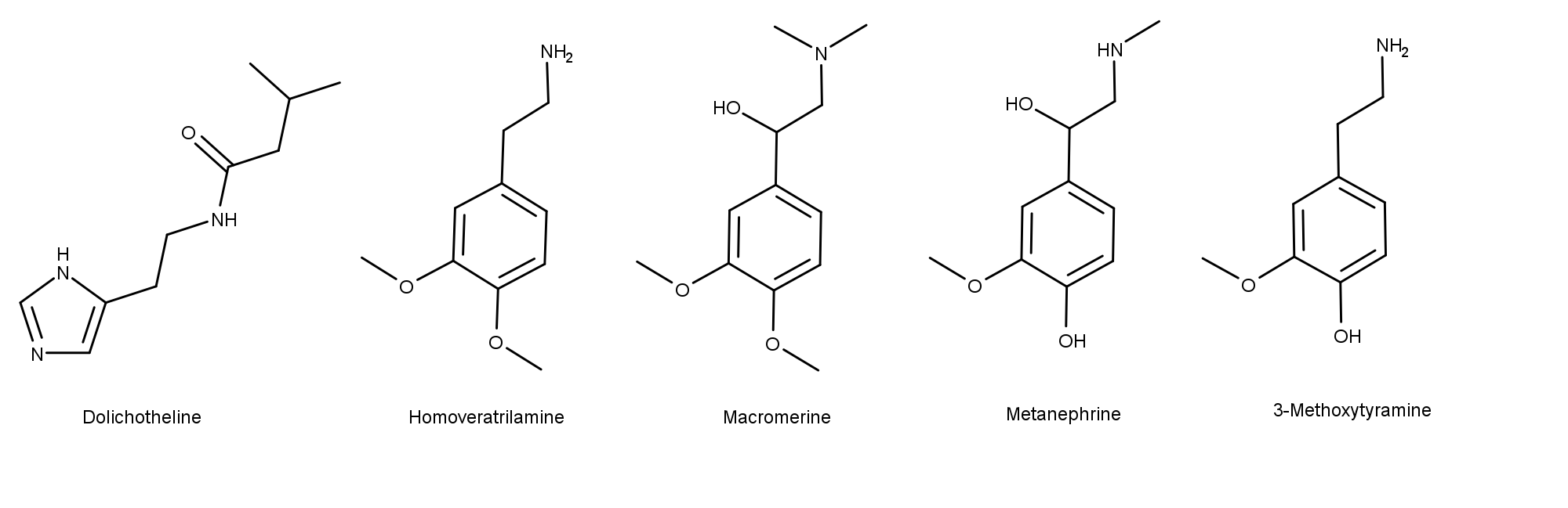
In addition to those listed so far, in the 1977 book “Peyote and Other Psychoactive Cacti,” Adam Gottlieb listed the following compounds in his ‘Dictionary of Cactus Alkaloids.’​
  • Dolichotheline​
  • Homoveratrilamine​
  • Macromerine​
  • Metanephrine​
  • 3-Methoxytyramine​
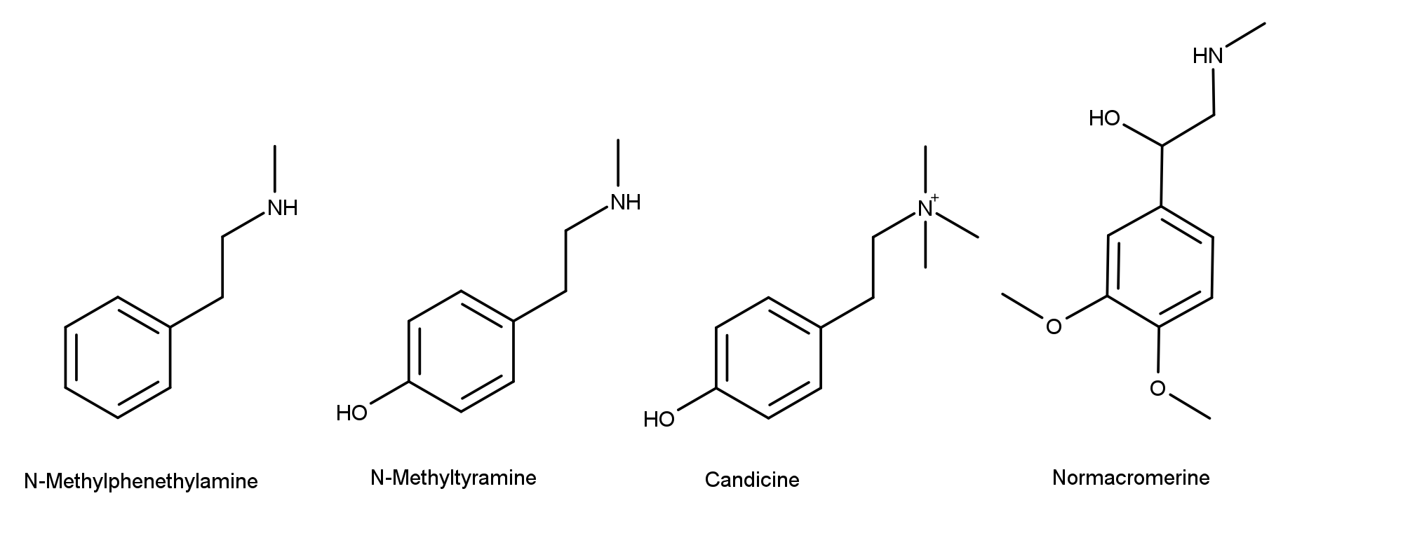
  • N-Methylphenethylamine​
  • N-Methyltyramine​
  • Candicine​
  • Normacromerine​


Here, the compound candicine is particularly interesting because it is a quaternary ammonium compound. The magic mushroom compounds aeruginascin and 4-HO-TMT also have this functional group.

Like magic mushrooms, the chemical composition of naturally occurring cacti appears to be highly variable. In 2010, Ogunbodede et al. analyzed the mescaline levels in the cortical stem from several San Pedro cactus samples. They observed that “The range of mescaline concentrations across the 14 taxa/cultivars spanned two orders of magnitude, from 0.053% to 4.7% by dry weight.” These data may bring to mind how much the levels of a single compound can vary in magic mushrooms and toad venom, and likely in other organisms.

Continuing research on Psychedelic Cacti

Clearly, there is much mystery still surrounding the chemical composition of psychoactive cacti– including the identity of all the active compounds and the concentrations of those compounds. Just like magic mushrooms and toad venom, it is feasible that these compounds have synergistic effects on the body. But it’s all speculation until scientists identify all the compounds in these cacti, elucidate their pharmacology, and understand how they work together to produce certain effects.
 
Last edited:
Cybin_Manhattan_Psychedelic_Clinic_Underserved_Minority_Communities_Psychedelic_Spotlight.jpg


New biosynthesis of psilocybin and related tryptamines

by Barb Bauer | Psychedelic Science Review

Researchers took the psilocybin-making genes from P. cubensis and put them into S. cerevisiae allowing them to produce psilocybin, several of its analogs, and a “new to nature” compound.

Psychedelic research is experiencing many scientific advances for producing psychedelic compounds. This is because trying to get meaningful and usable amounts of psilocybin and psilocin from magic mushroom flesh, for example, is not practical. The levels of these compounds in dried mushrooms are only 0.2% – 1.0% of their dry weight. Having ways to make meaningful amounts of these compounds at a reasonable cost is essential for conducting scientific studies from receptor assays to clinical trials in humans.

The Novo Nordisk Foundation Center for Biosustainability in Denmark recently published a study in the journal Metabolic Engineering describing how they bioengineered Saccharomyces cerevisiae to produce psilocybin and other related tryptamine derivatives.

PSR previously reported on several papers describing both synthetic and bioengineering methods for producing psychedelic compounds and derivatives. Both the new techniques (like the one described herein) and improvements on existing methods are critical for the advancement of psychedelic science. Two of these articles are Scientists Engineer E. Coli to Produce Psilocybin and Scientists Synthesize and Test the Magic Mushroom Compounds Baeocystin, Norbaeocystin, Norpsilocin, and Aeruginascin.

What is S. cerevisiae and why use it?

S. cerevisiae is a type of yeast. Milne et al. chose to work with it “due to its long use in industrial production as well as the fact that it naturally produces very few secondary metabolites or other tryptophan derivates, thereby facilitating simple downstream processing and purification.”

They also chose this yeast species because it naturally expresses cytochrome P450 enzymes that convert tryptamine to 4-hydroxytryptamine (read more about this in the section below, Optimizing the Synthesis of Psilocybin). The presence of the enzyme is a significant cost-savings, according to the authors. It eliminates an extra synthesis step used by other yeasts, which require an expensive substrate during production.

Modifying yeast to produce psilocybin analogs

The research team inserted genetic material from magic mushrooms into S. cerevisiae. Specifically, they cut out certain psilocybin-making genes from the magic mushroom Psilocybe cubensis and put them into S. cerevisiae. They did this by using plasmids as vectors for transporting the genes into the target yeast cells. Once inside, the plasmid incorporates the new genes into the yeast’s DNA (the plasmids are “programmed” to know where to insert them). The modified yeast then starts making psilocybin according to the instructions encoded in the magic mushroom’s DNA.

The yeast also makes psilocybin derivatives

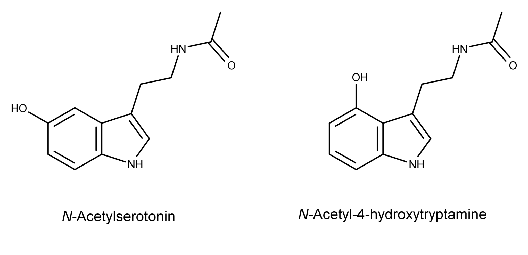
Notably, the researchers also detected several psilocybin derivatives being produced by their bioengineered yeast. The observed psilocybin analogs included psilocin, norpsilocin, baeocystin, norbaeocystin, 4-hydroxytrimethyltryptammonium (i.e., dephosphorylated aeruginascin), and the “new to nature” N-acetyl-4-hydroxytryptamine.

Why was only the dephosphorylated version found and not aeruginascin itself? The researchers noticed that the amount of psilocybin that S. cerevisiae made “was accompanied by a concomitant production of psilocin.” They theorized that the same phosphatase enzymes (of which there are several in S. cerevisiae) acting on psilocybin to produce psilocin were doing the same thing to aeruginascin. The action of these enzymes may also explain the presence of norpsilocin, which is dephosphorylated baeocystin.

Another result of this study was the creation of a biosynthetic method for producing a “new-to-nature” tryptamine derivative, N-acetyl-4-hydroxytryptamine.

The researchers made the new compound by adding the gene for the enzyme serotonin N-acetyltransferase into the DNA of a different strain of S. cerevisiae that produces 4-hydroxytryptamine. The compound is structurally similar to the neurotransmitter N-acetylserotonin (normelatonin), differing only in the position of the hydroxyl group in the 4-position instead of the 5-position.

According to the authors, this was,

"…the successful production of a novel molecule structurally similar to both psilocin and normelatonin with potentially novel pharmacological activity."

3000-N-acetylserotonin-and-N-acetyl-4-hydroxytryptamine2-2048x989.png


Optimizing the synthesis of psilocybin

Initially, the amount of psilocybin produced by the genetically engineered S. cerevisiae was low. The researchers noticed that as the yeast was making psilocybin, there was an increase in extracellular tryptamine. The accumulation of tryptamine suggested to them that its conversion to 4-hydroxytryptamine inside the yeast was not very efficient.

The researchers improved the conversion of tryptamine to 4-hydroxytryptamine by giving the yeast a more efficient gene from P. cubensis. The gene, known as cytochrome P450 reductase, catalyzes the conversion of tryptamine to 4-hydroxytryptamine. After this gene was added to the DNA of S. cerevisiae, the data showed a 29-fold increase in the production of psilocybin and psilocin and significantly reduced levels of extracellular tryptamine.

Continuing research on magic mushroom compounds is needed

This research provides another method for producing meaningful amounts of psilocybin analogs for scientific study and downstream applications. Also, synthesis of the novel compounds N-acetyl-4-hydroxytryptamine and unphosphorylated aeruginascin by strains of S. cerevisiae provides more opportunities for research into their chemistry and pharmacology.

Taking genes from one organism and splicing them into another are well-known techniques. Nevertheless, the application of this technology to create psychedelic compounds represents an exciting new area of research. Other scientists have recently noted that supply problems have limited research into the “minor” tryptamines in magic mushrooms. They also hypothesize that these compounds could have substantial therapeutic value alone or when combined with psilocybin. This recent work discussed above could offer new possibilities for supplying minor tryptamines for further research and development.

psychedelicreview.com​
 
Last edited:
Enhanced-MRI-scan-of-the-brain-by-Wellcome-Images-Creative-Commons.jpg


The pharmacology of psilocybin and psilocin

by Barb Bauer | Psychedelic Science Review

Psilocybin and psilocin are two psychedelic compounds found in magic mushrooms. Calling these and other magic mushrooms compounds hallucinogens is an incorrect generalization. A hallucinogenic effect is not always present in users. Therefore, the terms psychedelics or psychotomimetics are the preferred designations put forth by experts in the field.

Several genera of magic mushrooms contain psilocybin and psilocin including Inocybe, Conocybe, Panaeolus, Gymnopilus, and Pluteus. However, psilocin is not the only psilocybin derivative in magic mushrooms. Others include norpsilocin, baeocystin, norbaeocystin, and aeruginascin. The derivatives and amounts present vary in different parts of the mushroom. They also vary in between species and even within batches of the same species.

This article examines the chemical structure of these psychedelics, discusses how the body transforms them, and how they interact with receptors in the brain.

Chemical structures of psilocybin and psilocin

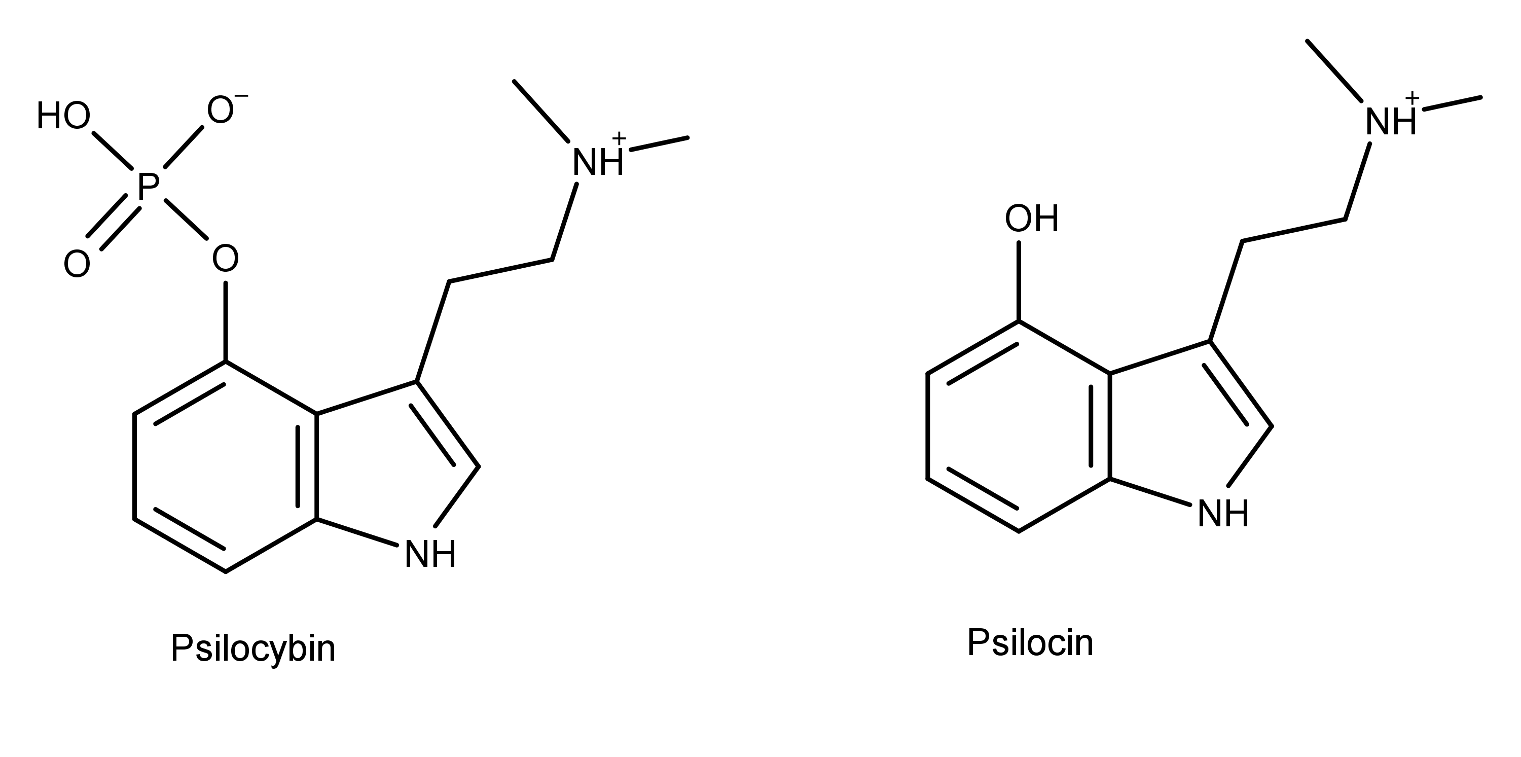
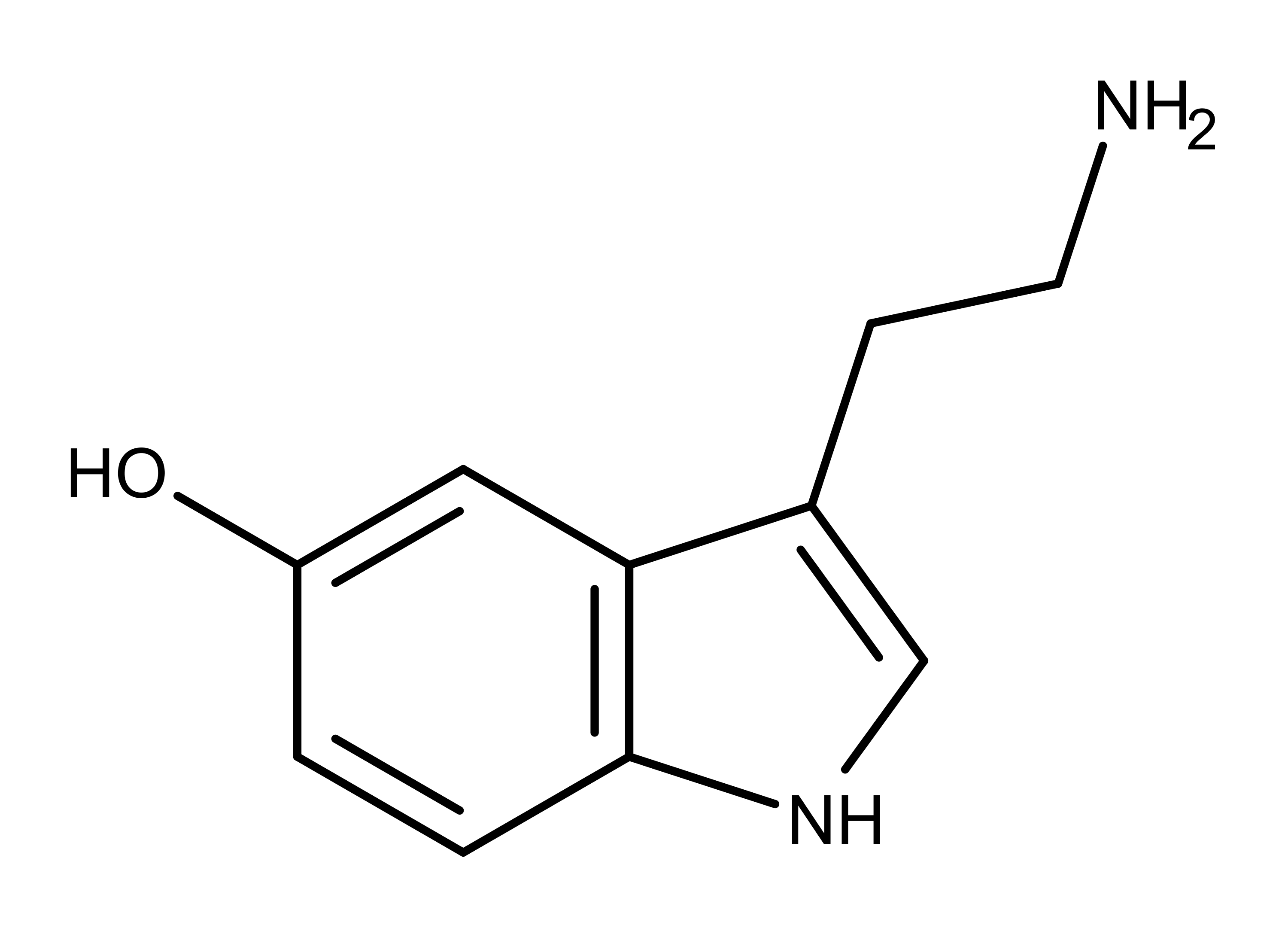
Psilocybin and psilocin (Figure 1) are tryptamine alkaloids and structural analogs of the neurotransmitter serotonin (Figure 2). Psilocybin and psilocin differ from each other at position 4, having a phosphate and hydroxyl group, respectively. As structural analogs of serotonin, both compounds differ from serotonin by having two methyl groups on the amine group on carbon 3. Also, the substituent groups on the benzyl ring of psilocybin and psilocin are in position 4, while the hydroxyl group on serotonin is in position 5.

3000-psilocybin-and-psilocin.png

Figure 1: The chemical structure of psilocybin and psilocin, two compounds found in magic mushrooms.


3000-serotonin.png

Figure 2: The chemical structure of the neurotransmitter serotonin.

The similarities and differences between the chemical structures of these compounds is key to understanding the effects of psilocybin and psilocin.

Absorption, metabolism, and excretion in mammals

Psilocybin is a prodrug of psilocin. This means the prodrug psilocybin undergoes changes in the body which convert it into the active form, psilocin. Specifically, a chemical process called dephosphorylation removes the phosphate group on psilocybin, creating psilocin.

The dephosphorylation of psilocybin occurs in two ways in different areas of the body.

- The acidic environment in the stomach is a favorable environment for the rapid dephosphorylation of psilocybin.

- Enzymes such as alkaline phosphatase and other non-specific esterases dephosphorylate psilocybin in the intestines, kidneys, and the blood.

The phosphate group on psilocybin in Figure 1 is highly polar. This polarity along with the positively charged amine group makes the molecule zwitterionic and more soluble in water than psilocin. Without the phosphate group, psilocin becomes more lipid soluble than psilocybin, making it metabolically available in the body and more easily absorbed in the intestines.

At this point, psilocin is distributed all over the body via the bloodstream. Being lipid soluble allows psilocin to cross the blood-brain barrier and elicit its effects (see the Biological Interactions section below). Researchers can detect psilocybin and psilocin in human blood plasma 20-40 minutes after oral administration of psilocybin. They find maximum levels in the blood 80-105 minutes after administration.

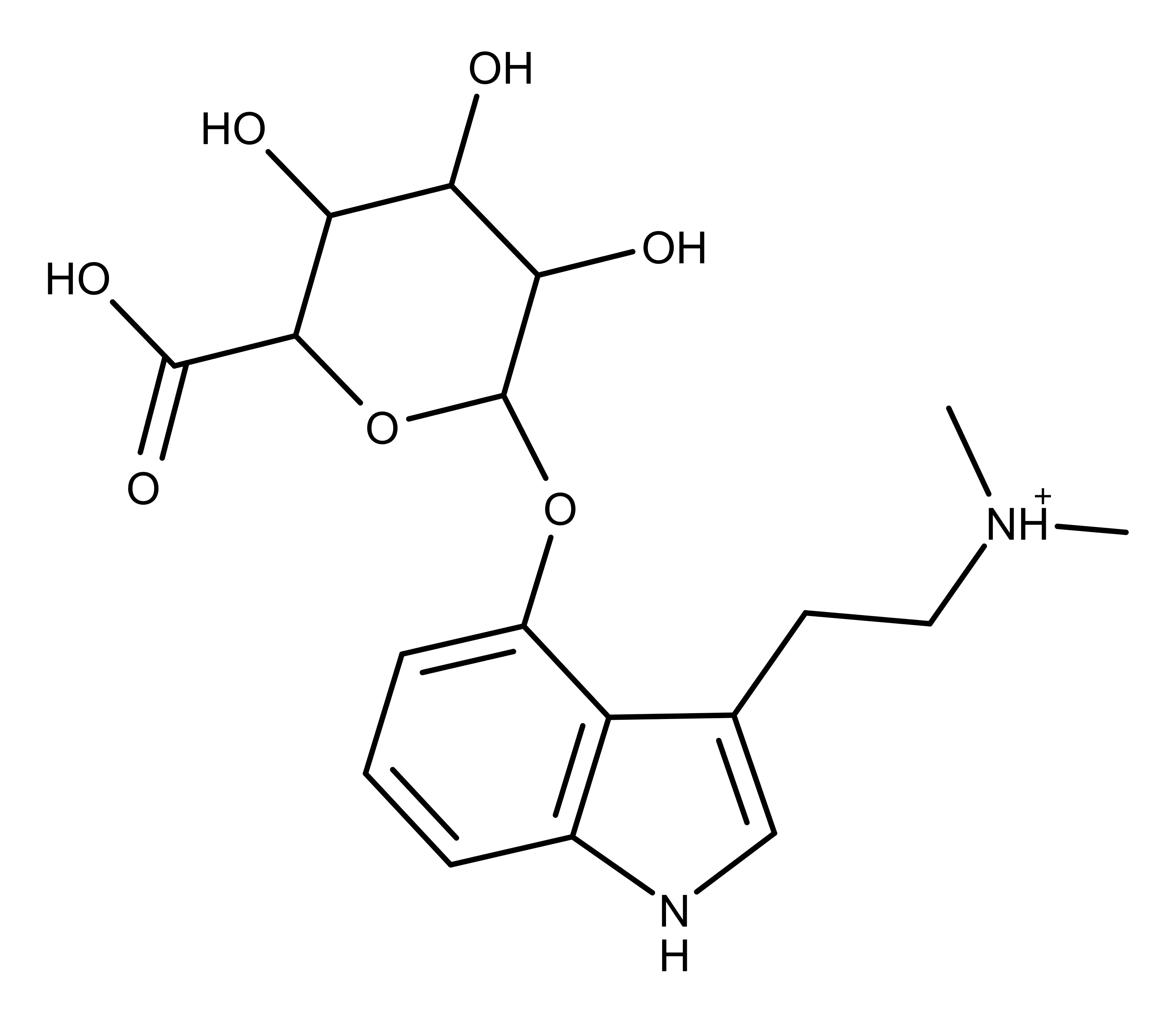
About 80% of metabolized psilocybin gets excreted in the urine as a compound called psilocin-O-glucuronide (Figure 3). Some psilocin and psilocybin (only 3-10 percent are excreted in the urine, mostly in a conjugated form with glucoronic acid.


psilocybin-O-glucuronide.png

The chemical structure of psilocin-O-glucuronide, a urinary metabolite of psilocybin.

Biological receptor interactions

At last count, there are seven main classes of serotonin receptors containing 14 subtypes. Of these, the 5-HT2A is the serotonin receptor (5-HT is an abbreviation for serotonin, 5-hydroxytryptamine) is studied the most in relation to psychedelic effects. It is important to keep in mind that scientists are not currently studying how the interactions of psilocybin and psilocin with other receptors impacts the psychedelic effect of these compounds.

Psilocybin has a low binding affinity as an agonist for the 5-HT2A receptor compared to the active form of the molecule, psilocin. The difference in the binding affinities of psilocybin and psilocin on the human 5-HT2A receptor is striking. The Ki (disassociation constant) for psilocybin is >10,000 nM, while for psilocin, Ki = 102.7 nM. The lower the Ki value, the less likely the molecule is to dissociate from the receptor. Therefore, these data clearly show that psilocin has a greater agonistic affinity for the 5-HT2A than psilocybin. This clarifies that psilocin is primarily responsible for the psychedelic effect of magic mushrooms, not psilocybin.

Other studies confirm the role of the 5-HT2A receptor in the psychedelic experience. For example, in 1998, Vollenweider, et al., found the 5-HT2A receptor antagonist ketanserin blocks the effects of psilocybin and psilocin. More recently, a study used PET (positron emission tomography) scans and monitored the levels of psilocin in the blood plasma of volunteers after they took psilocybin. The researchers found the subjective experiences of the volunteers were directly related to the levels of psilocin in their blood plasma and the number of occupied (with psilocin) 5-HT2A receptors in their brains.

Signal transduction pathways

Signal transduction is a scientific term for the biochemical reactions that occur when a molecule binds to a receptor on a cell. Different molecules have different signal pathways inside cells that use different types of receptors. Scientists who study psychedelics such as psilocybin and psilocin are interested in serotonin receptors. Understanding these receptors is crucial because psilocybin and especially psilocin, use these same serotonin receptors to bring about their effects in users.

When it comes to the exact cell signal transduction pathway(s) used by psilocin to elicit its psychedelic effects, researchers have an understanding of several aspects of 5-HT2A. Several serotonin receptors including 5-HT2A belong to a family called G-protein coupled receptors (GPCRs). GPCRs are one of the largest and most diverse families of proteins in mammals. These receptors have a unique structure consisting of seven protein helices that span the cell membrane. When a compound binds to the 5-HT2A receptor on a cell, it stimulates an enzyme called phospholipase C. This, in turn, activates another enzyme called protein kinase and also stimulates the release of calcium ions from storage areas in the cell. This enzymatic and biochemical activity culminates in cellular changes that result in the overall perceived effect.

This pathway is only a small piece of the big picture showing how psychedelic drugs work. At a higher level, Baumeister et al. reviewed the pharmacodynamics of classical hallucinogens and summarized three mechanisms by which they are known to work, or may work, in the brain:

- Hallucinogens may disrupt normal serotonin pathways in the brain in several ways. One way may be by binding to presynaptic 5-HT1A receptors in the Raphe nuclei that project into the prefrontal cortex and subcortical structures. This disruption would alter the serotonergic output to the rest of the brain.

- When hallucinogens bind with postsynaptic 5-HT2A receptors in cortical neurons, this changes cellular signaling and functioning via the excitatory amino acid glutamine. The effect is what the authors describe as “complex manifestations in consciousness.”

- Normally, neurons in the reticular nucleus of the prefrontal cortex regulate sensory input into the brain. Hallucinogens disrupt this process by binding with reticular postsynaptic 5-HT2A receptors. The binding causes another cascade of events, altering the sensory information received by the cortex. The result is the disruption of the normal activity of the sensory cortices.

The need for more research

It is interesting to note from this receptor binding data that psilocin binds well to several other serotonin receptors besides 5-HT2A. For example, notice the Ki values for psilocin compared to psilocybin for 5-HT1A and 5-HT1B. How is this impacting the subjective effects the user is experiencing? Also, what is the effect of the low Ki values both compounds have for 5-HT2B? It is clear that more research is needed in this area to understand these psychedelics and their receptor interactions. Compounding this whole mystery is psilocybin and psilocin being just two of many potentially active compounds in magic mushrooms.

Conclusion

The pharmacology of psilocybin and psilocin is fascinating and complicated. The chemistry of these compounds dictates how the body metabolizes them and how they interact with receptors. Research has made great strides in understanding how psychedelics work with the 5-HT2A receptor, but endless avenues of exploration still exist for the curious researcher.

psychedelicreview.com​
 
SS.jpg


Psychedelic Pharmacology

by James Kent

Most psychedelic molecules are structurally similar to neurotransmitters that modulate signal flow in the brain. If we take a close look at the structure of common neurotransmitters, the transmitters most closely related to the classic psychedelics are serotonin (5-HT), adrenaline (epinephrine), norepinephrine, and dopamine (DA), and all of these chemicals are classified as amines, meaning that they have a nitrogen (N+) containing amino group hanging off a root carbon ring. This nitrogen structure is the key element in any amino acid, carrying the energy needed for metabolic processes which do work. Since these transmitter chemicals have only one nitrogen group they are called monoamines, and they are the essential messengers of the aminergic neuromodulatory system.

Monoamines entering the bloodstream are normally kept out of the brain by the blood-brain-barrier, but psychedelic molecules have a neutral charge so they are able to pass. When these amine crystals pass through the blood-brain barrier, they brush against neural receptor sites; if the receptors are a good fit, the crystals get stuck for a short period of time. The bonding of amine ligands to serotonin and dopamine receptors is where psychedelic action begins.

Serotonin and the Tryptamines

Because of depressive mood disorders and pharmaceuticals like Prozac, the most well known neuromodulator is serotonin, or 5-HT (5-hydroxytryptamine). 5-HT is essential to many basic brain functions, linked to mood, depression, contentment, anxiety, sleep, appetite, and the regulation of involuntary smooth muscles that control blood pressure and digestive functions. Serotonin is an indoleamine and a variant of tryptamine, which is the most basic of all the indoleamines and the structural starting point for DMT (N,N-dimethyltryptamine), 5-MeO-DMT, psilocin, psilocybin, DPT, AMT, and most psychedelic drugs with acronyms ending in T (which stands for Tryptamine). LSD is also a tryptamine, but it is larger and more complex than the other tryptamines, and is in many ways structurally unique.

Dopamine and the Phenethylamines

Working in concert with serotonin is the neuromodulator dopamine (3-hydroxytyramine). Dopamine is synthesized from L-DOPA and is instrumental in modulating salient attention, motivational response, and fine motor control. Dopamine is central to the reward system, and dopamine release is stimulated by recreational drugs, food, gambling, sex, and physical risk taking. Dopamine imbalances are linked Parkinsons disease, ADD, compulsive risk behavior, and psychosis. The role of dopamine interruption is relevant to psychedelic activity in many aspects; psychedelics may affect sensuality and motor control, and may facilitate psychosis, mania, and compulsive behaviors.

Amphetamines and the phenethylamine group of psychedelics (mescaline, 2-CB, MDA, MDMA, and so on) are more structurally similar to dopamine, epinephrine, and norepinephrine, which are also monoamines but sometimes referred to as catecholamines since they are based on the single catechol ring structure. Epinephrine and norepinephrine are referred to as stress hormones because they prime the bodys energy production in response to stress and danger. The phenethylamines and catecholamines all have the six-carbon benzene ring backbone, simpler than the dual-ring tryptamine structure, with at least one amine group. The simplest form of this molecule is called phenethylamine, and is structurally similar to amphetamine.

In very general terms, the phenethylamine psychedelics are said to be more energetic, sensual, empathogenic, or entactogenic, while tryptamine psychedelics are thought to be more hallucinogenic, disorienting, and somatically heavy. These descriptions are very broad, but this is the popular distinction made between the two major classes of psychedelics.

Neuromodulators and Global Brain States

Serotonin, dopamine, and the other monoamines dont cause neurons to fire, they instead tune the spiking rate of neurons, which means they adjust global network polarity over time to make neural assemblies more or less responsive to stimulus. Serotonin and dopamine are projected into higher areas of the brain from nuclei in the brainstem and middle brain, meaning they are primal signaling mechanisms for modulating many areas of the brain simultaneously. The axons from these aminergic clusters reach upward to many areas of the cortex, affecting the thalamus (sensory filter), amygdala (fear and survival), hypothalamus (homeostatic regulator), hippocampus (memory and learning), and neocortex (sensory and logic processing). Neuromodulators synchronize the neural response to incoming stimulus and keep local competing brain circuits functioning smoothly and in unison. These neuromodulators produce a one-way bottom-up effect, which means they are switched on and off reflexively and unconsciously by glands in the brainstem and basal forebrain in direct response to internal conditions or external stimulus. Using neuromodulators the brainstem can exert global homeostatic control over organism mood and behavior. The effect of the aminergic modulators projected upward by the brainstem are tonic, which means their signaling effects are sticky and persist over the duration of many incoming spike trains.

Generally serotonin is thought to have a polarizing effect on neurons, making them less likely to fire and thus having an overall relaxing effect on the brain. This is why many depression and anxiety remedies focus on increasing the supply of serotonin; to decrease anxiety and increase satisfaction. If we assume psychedelics are mimics for neurotransmitters and apply this analogy to DMT, we would expect DMT to have a calming effect on the brain because it looks similar to serotonin. But a flood of DMT does not calm the brain, it makes it hallucinate. Psychedelics act on the same receptors as serotonin and dopamine, but as partial agonists. Since DMT binds to the same receptor sites as serotonin but does not produce a relaxing effect, it would be logical to assume that DMT is a 5-HT antagonist, meaning it blocks serotonin and depolarizes neurons, making them more excitable. This is not the case. There are many different types of 5-HT receptors, some inhibit neural activity and some promote neural activity. Like most psychedelics, DMT is classified as a selective 5-HT2A partial or full agonist; also active at other 5-HT subtypes, at adrenal receptors, at Sigma-1 receptors, and at tertiary amine receptors. This means that DMT is active at many 5-HT sites and can mimic some of the agonistic functions of serotonin with varying frequency and efficacy.

5-HT partial agonism can be described as a subtle form of aminergic modulatory signal interference. In the most general case it can be assumed that psychedelic activity is due to interference at 5-HT receptor subtypes. In more specific cases we can assume that visual hallucinogenic effect is associated with 5-HT2A and 5-HT2C receptor interaction. Somatic heaviness and dreaminess is associated with broader aminergic interaction; and more sensual, entactogenic, or compulsive effects are associated with DA and adrenergic receptor interaction. Psychedelics can have a wide affinity and interact as partial or full agonists at multiple receptor subtypes to produce a wide range of effects. Because psychedelics are full or partial agonists acting on the same modulatory pathways as 5-HT, the synergistic interaction between these competing agonists can be described in terms of a modulatory wave interference pattern. Agonistic interference at 5-HT subtypes promotes disinhibition and extreme excitability between feedback-coupled autonomic neural assemblies in the cortex, midbrain, and brainstem. Excitation in the autonomic neural assemblies which process sensation and memory lead to spontaneous hallucination; excitation in autonomic assemblies which process thought and self-awareness lead to expanded states of psychedelic consciousness.

Molecular Shape and Receptor Affinity

The strength and duration of the bond a ligand forms with a receptor is referred to as receptor affinity or potency, and is described in terms of pharmacodynamics. The higher the affinity the stronger and longer the ligand bonds with a receptor and influences charge moving across the neural membrane. Research has shown that 5-HT2A receptor affinity is an accurate measure of the potency of any psychedelic compound; the higher the affinity the higher the potency and psychedelic effect. Another thing we know is that the conformational shape of the amine determines how long the molecule takes to metabolize and how sticky it will be at 5-HT receptor types. For instance, the amine tail of LSD is different from other tryptamines; it is long, complex, and connects back to the benzene ring, keeping it rigid instead of flexible like most amino groups or substitutions. Designer amines with a similarly rigid molecular structure have also shown a marked increase in psychedelic potency.

Using this information it can be assumed that the unique structure of LSD is what makes it so potent; giving it a high affinity across a wider range of receptor types; making it more difficult to metabolize; and giving it a broader range of effect over a longer duration. DMT also binds to a wide variety of 5-HT receptor types, but it is smaller and metabolizes very quickly. When DMT is taken with a monoamine-oxidase inhibitor (MAOi) in an ayahuasca mixture, the enzymes which metabolize DMT are blocked making the hallucinogenic effects of DMT orally active and longer lasting. Adding an MAOi to any tryptamine psychedelic will make it nearly twice as hallucinogenic. These few pieces of pharmacology tell us that the efficacy of modulatory interruption, or psychedelic potency, can be somewhat predicted by molecular shape, the rigidity of the molecular structure, and speed of metabolic pathways.

Breadth of psychedelic receptor binding
Mol/Target​
5ht1a​
5ht1b​
5ht1d​
5ht1e​
5ht2a​
5ht2b​
5ht2c​
5ht5a​
5ht6​
5ht7​
D1​
A-2A​
A-2B​
A-2C​
2C-E​
2.91​
3​
3.54​
2.6​
3.76​
4​
3.38​
0​
1.93​
2.77​
0​
2.71​
2.91​
3.44​
2C-B​
2.75​
3.11​
3.71​
3.05​
3.69​
4​
3.18​
0​
2.63​
2.81​
0​
2.64​
2.31​
3.12​
LSD​
3.73​
4​
3.7​
2.62​
3.54​
3.11​
3.11​
3.64​
3.75​
3.77​
2.34​
2.93​
0​
0​
DOI​
0​
2.31​
3​
2.66​
3.44​
3.13​
4​
0​
2.34​
1.9​
1.67​
3.79​
3.13​
2.88​
DMT​
0​
0​
3.91​
3.28​
2.58​
0​
3.42​
3.16​
3.35​
4​
3.51​
2.75​
3.53​
3.53​
Psilocin​
2.88​
2.19​
3.4​
3.03​
2.14​
4​
2.52​
2.83​
2.82​
2.82​
3.37​
1.36​
1.57​
1.03​
5-MeO-DMT​
4​
2.41​
3.48​
1.72​
0.98​
0.69​
1.55​
1.84​
2.73​
3.69​
2.38​
0​
0.86​
1.57​
DiPT​
4​
0​
2.51​
0​
0​
3.48​
0​
0​
0​
0​
0​
0​
2.62​
2.68​
Mescaline​
3.61​
0​
0​
3.16​
0​
3.97​
0​
0​
0​
0​
0​
2.92​
0​
4​
MDMA​
0​
0​
0​
0​
0​
3.64​
0​
0​
0​
0​
0​
2.94​
3.09​
3.21​
6-F-DMT​
2.81​
3.07​
3.66​
2.74​
2.47​
3.93​
2.58​
2.43​
4​
3.8​
2.67​
0​
2.99​
3.24​
Lisuride​
4​
2.27​
0​
0​
2.74​
3.01​
0​
2.99​
2.61​
2.64​
0​
3.22​
3.78​
3.88​
4C-T-2​
2.04​
0​
0​
1.77​
3.33​
4​
3.09​
2.56​
0​
2.18​
0​
0​
0​
0​
Table 1 lists the binding strength of popular psychedelic drugs at many 5-HT receptor sites listed in order of 5-HT2A affinity. This table should be an accurate representation of hallucinogenic potency in descending order. From subjective reports all substances at the top of this list are very hallucinogenic, but DMT, which is often considered to be the most hallucinogenic, actually falls somewhere in the middle. If we look at 5-HT2C affinity, which is also implicated in hallucination, we can see that all substances at the top of the list also have high 5-HT2C affinity, with DMT and DOI having slightly higher affinity than the rest. 5-HT7 receptor affinity, which stimulates cAMP activity and the reward system, also seems to be implicated in overall transcendent psychedelic action, with the mystically popular DMT, 5-MeO-DMT, and LSD topping the affinity list. In contrast, there are four non-visual psychedelics at the bottom of the list, 5-MeO-DMT, DiPT, Mescaline, and MDMA. These substances have very poor 5-HT2A,C affinity but oddly the bottom three all have a high 5-HT2B and adrenal affinity; this indicates they are effective at stimulating serotonin production, cardiovascular activity, and acute sensuality. It is interesting to note that DiPT, Mescaline, and 5-MeO-DMT all have a high 5-HT1A affinity, which is generally thought to work in contrast to 5-HT2A agonism. DiPT is unusual because is produces distinct audio hallucinations and little or no visual hallucinations, and predictably does not bond with targets associated with visual hallucination. By analyzing this affinity table it seems possible to predict the relative potency of any hallucinogen based solely on binding profiles, though the three control molecules at the bottom of the list (6-F-DMT, Lisuride, 4C-T-2) are reportedly non-hallucinogenic despite high 5-HT receptor promiscuity; this is likely because they are not active as agonists, they are antagonists, or their binding profiles somehow cancel each other out.

Dissociatives, anticholinergics, and other hallucinogens

Psychedelic tryptamines and phenethylamines are not the only hallucinogens, but all hallucinogens work by interrupting sensory binding pathways. Hallucinogenic dissociatives like ketamine (special K), phencyclidine (PCP), and dextromethorphan (DXM) interrupt NMDA glutamate sensory signaling pathways; these pathways mediate fast sensory signal projection through the brain. Anticholinergic deliriants like scopolamine and atropine interrupt cholinergic modulation of memory, recall, and dreaming; these pathways mediate the smooth input and output of memory from the hippocampus. Salvia divinorum interrupts Kappa-opioid tactile sensory pathways; these pathways mediate pain, gravity awareness, and feedback for determining physical orientation in space. Depressants like GHB and alcohol interrupt sensory binding via inhibitory GABA pathways, pathways which dampen and slow smooth sensory throughput. Nitrous Oxide (N20) is the simplest and perhaps the most promiscuous of hallucinogens, worming its way in between a number of rudimentary signaling channels to produce novel feelings of dissociation and out-of-body emergence. Although the pharmacological targets of hallucinogens differ, in all cases perceptual distortion is linked directly to interruption of seamless multisensory signaling and binding across the cortex. Any drug which interrupts pathways of multisensory signaling or binding will be considered psychedelic at high enough doses, this is why so many different types of plants and chemicals can be uniquely hallucinogenic across many different receptor targets.

http://psychedelic-information-theor...c-Pharmacology
 
Last edited:
LONDON.jpg


Rethinking serotonin could lead to a shift in psychiatric care

Imperial College London

A better understanding of how a key chemical messenger acts in the brain could lead to a radical shift in psychiatric care, according to a new research paper.

Serotonin is a neurotransmitter which helps brain cells communicate with one another, playing important roles in stabilising mood and regulating stress.

Despite its importance, current models to explain serotonin's function in the brain remain incomplete.

Now, in a review paper published this month in the Journal of Psychopharmacology, researchers from Imperial College London suggest that serotonin pathways are more nuanced than previously thought.

They argue that the existing view should be updated to incorporate a 'two-pronged' model of how serotonin acts.

The researchers believe their updated model could have implications for treating recalcitrant mental health conditions, including depression, obsessive compulsive disorder and addiction, and could exploit the therapeutic potential of psychedelic drugs.

In the brain, serotonin acts via a number of sites called 'receptors' and serotonin has at least 14 of these. Brain drugs such antidepressants, antipsychotics and psychedelics are known to interact with serotonin receptors and two of these are thought to be particularly important -- the so-called serotonin 1A and 2A receptors.

For patients with depression, commonly prescribed drugs called SSRIs (Selective Serotonin Reuptake Inhibitors) can help to relieve symptoms by boosting levels of serotonin in the brain. Evidence suggests an important part of how they work is to increase activity at the serotonin 1A receptor, which reduces brain activity in important stress circuitry, thereby helping a person cope better.

In contrast, psychedelic compounds such as LSD and psilocybin (the psychoactive component of magic mushrooms), are thought to act primarily on the serotonin 2A receptor. Accumulating evidence suggests that psychedelics with psychotherapy can be an effective treatment for certain mental illnesses and, with a focus on the 2A receptor, the authors' paper attempts to explain why.

Writing in the review paper, the researchers say that while the traditional view of developing psychiatric treatments has been focused on promoting 1A activity and often blocking the 2A, the therapeutic importance of activating the 2A pathway -- the mechanism by which psychedelics have their effect -- has been largely overlooked.

"We may have got it wrong in the past," said Dr Robin Carhart-Harris, Head of Psychedelic Research at Imperial and lead author on the paper. "Activating serotonin 2A receptors may be a good thing, as it makes individuals very sensitive to context and to their environment. Crucially, if that is made therapeutic, then the combination can be very effective. This is how psychedelics work -- they make people sensitive to context and 'open' to change via activating the 2A receptor."

According to the researchers, the 1A and 2A pathways form part of a two-pronged approach which may have evolved to help us adapt to adversity. By triggering the 1A pathway, serotonin can make situations less stressful, helping us to become more resilient. However, they argue that this approach may not always be enough, and that in extreme crises, the 2A pathway may kick in to rapidly open a window of plasticity in which fundamental changes in outlook and behaviour can occur.

Growing evidence shows that in conditions such as treatment-resistant depression, obsessive compulsive disorder and addiction, certain brain circuitry may become 'stamped in' and resistant to change. The researchers suggest that in such cases, activating the 2A pathway -- such as through psychedelics -- could potentially offer a way to break the cycle, helping patients to change negative behaviours and thought patterns which have become entrenched.

By enabling the brain to enter into a more adaptive or 'plastic' state and providing patients with a suitably enriched clinical environment when they receive a drug treatment, clinicians could create a window for therapy, effectively making patients more receptive to psychotherapy.

According to the authors, their updated model of how serotonin acts in the brain could lead to a shift in psychiatric care, with the potential to move patients from enduring a condition using current pharmacological treatments, to actively addressing their condition by fundamentally modifying behaviours and thinking.

Professor David Nutt, Director of Neuropsychopharmacology in Imperial's Division of Brain Sciences, explained: "This is an exciting and novel insight into the role of serotonin and its receptors in recovery from depression that I hope may inspire more research into develop 5-HT2A receptor drugs as new treatments."

Dr Carhart-Harris added: "I think our model suggests that you cannot just administer a drug in isolation, at least certainly not psychedelics, and the same may also true for SSRIs. We need to pay more attention to the context in which medications are given. We have to acknowledge the evidence which shows that environment is a critical component of how our biology is expressed."

He added: "In psychiatry, as in science, things are rarely black and white, and part of the approach we're promoting is to have a more sophisticated model of mental healthcare that isn't just a drug or psychotherapy, it's both. I believe this is the future."

'Serotonin and brain function: a tale of two receptors'
by Robin Carhart-Harris and David Nutt is published in the Journal of Psychopharmacology.

https://www.sciencedaily.com/releases/2017/09/170904093724.htm
 
SP.jpg


Dopamine release and duration of action: Predicting the addictive potential of phenethylamines

by Benjamin Malcolm | Spirit Pharmacist

Phenethylamine (PEA) is both a naturally occurring neurotransmitter and chemical backbone for several other types of medications and neurotransmitters. Legendary psychedelic chemist Alexander Shulgin synthesized dozens of novel psychedelic phenethylamines that he documents in his and his wife’s book PIHKAL (Phenethylamines I have Known and Loved). He is credited with re-synthesizing MDMA as well as inventing designer phenethylamines such as 2CB.

Phenethylamines are much broader than a class of psychedelics and are primarily known as ‘sympathomimetics’ because of their actions on the sympathetic (fight or flight) nervous system. Catecholamine neurotransmitters like norepinephrine and dopamine as well as the hormone epinephrine are the chemical mediators of sympathetic neurotransmission and drugs derived from phenethylamine traditionally used of therapeutic purposes mimic their actions.

Non-psychoactive phenethylamines include bronchodilator medications used for asthma such as albuterol while psychoactive phenethylamines include psychostimulants such as amphetamine, methamphetamine, and cathinones (‘bath salts’). Psychostimulants such as amphetamine act as ‘releasers’ of norepinephrine and dopamine, which stimulate the sympathetic nervous system. It is release of dopamine in the central nervous system that is thought to drive the habituating and addictive potentials of amphetamine. Thus, paying attention to dopamine releasing effect of phenethylamines is an important clue in understanding risk for habituation, re-enforcement, or addiction.

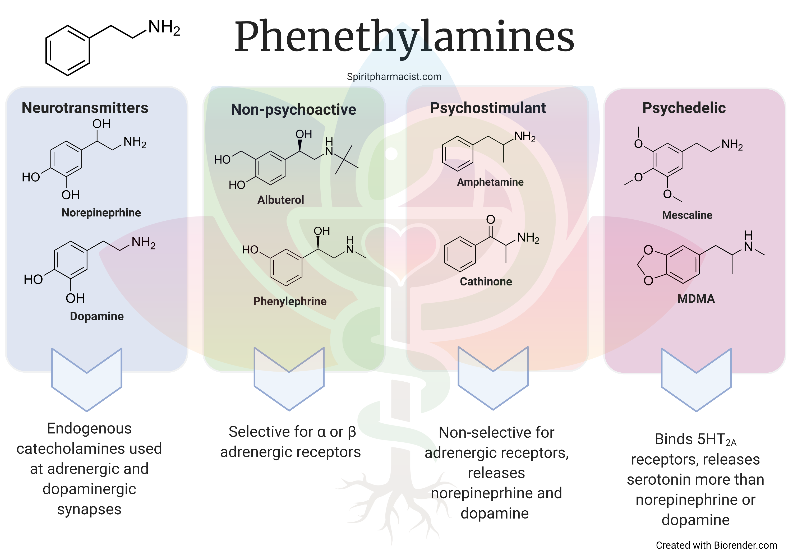
Phenethylamines3.png


The psychedelic phenethylamines tend to act as ‘releasers’ of serotonin and be able to bind 5HT2A receptors, although preserve some of the effects of traditional stimulants by acting on norepinephrine or dopamine. They are hybrids between traditional stimulants and psychedelics. The prototype psychedelic phenethylamine is 3,4-methylendioxymethamphetamine (MDMA). MDMA tends to be euphoric and preserve the ego structure. It carries some risks of habituation, dependence, or addiction linked to release of dopamine. MDMA tends to distort sensory perceptions or cause hallucinations much less than classical tryptamine psychedelics (e.g. psilocybin, LSD, DMT). For these reasons one may consider MDMA more of a serotonergic amphetamine than a true psychedelic.

The degree of effect on dopamine neurotransmission and duration of action appear to help guide understanding of habituation. When comparing phenethylamine psychedelics like MDMA with novel designer phenethylamines, substances with higher release of dopamine and shorter duration of action can tempt the user to frequently re-dose. For example, when comparing the actions of mephedrone (4-methylmethcathinone or 4-MMC) with MDMA it is noted that MDMA has considerably less dopamine release than mephedrone and that mephedrone has dopamine release similar to amphetamine. In addition, mephedrone also has a much shorter half-life than MDMA meaning that pleasurable effects wear off sooner. Frequent re-dosing creates a stacking of physical effects, the risks of behavioral reinforcement and addiction as well as adverse reactions such as cardiovascular events, seizures, or psychosis increases.

None of this is to raise alarm bells about the addictive potential of MDMA or demonize amphetamine and cathinones. On the contrary, if we know traditional stimulants can be therapeutic and know that serotonergic psychedelics can also be therapeutic, drugs that hybridize their effects deserve thorough exploration for therapeutic potentials. The point is that the mechanisms of phenethylamine psychedelics are rather broad and tends to differentially effect serotonin, norepinephrine, and dopamine neurotransmitter systems. Psychedelic actions and effects occur due to release of serotonin and modulation of ‘psychedelic’ 5HT2A receptors while propensity for re-dosing and habituation is linked to duration of action and effects on dopamine. Chemical fingerprinting of psychedelic phenethylamines across neurotransmitter systems and measurement of basic pharmacokinetic parameters may guide harm reduction efforts as drugs with high potential for habituation and re-dosing are identified among the sea of alphabet-amines (phenethylamine psychedelics) available in today’s clandestine marketplaces.
 
qlzbyRaYRXaU2zUGn0pP_PhenethylamineIntroBlog.png

An introduction to Psychedelic Phenethylamines​

by Ben Malcolm | Spirit Pharmacist | 7 May 2021

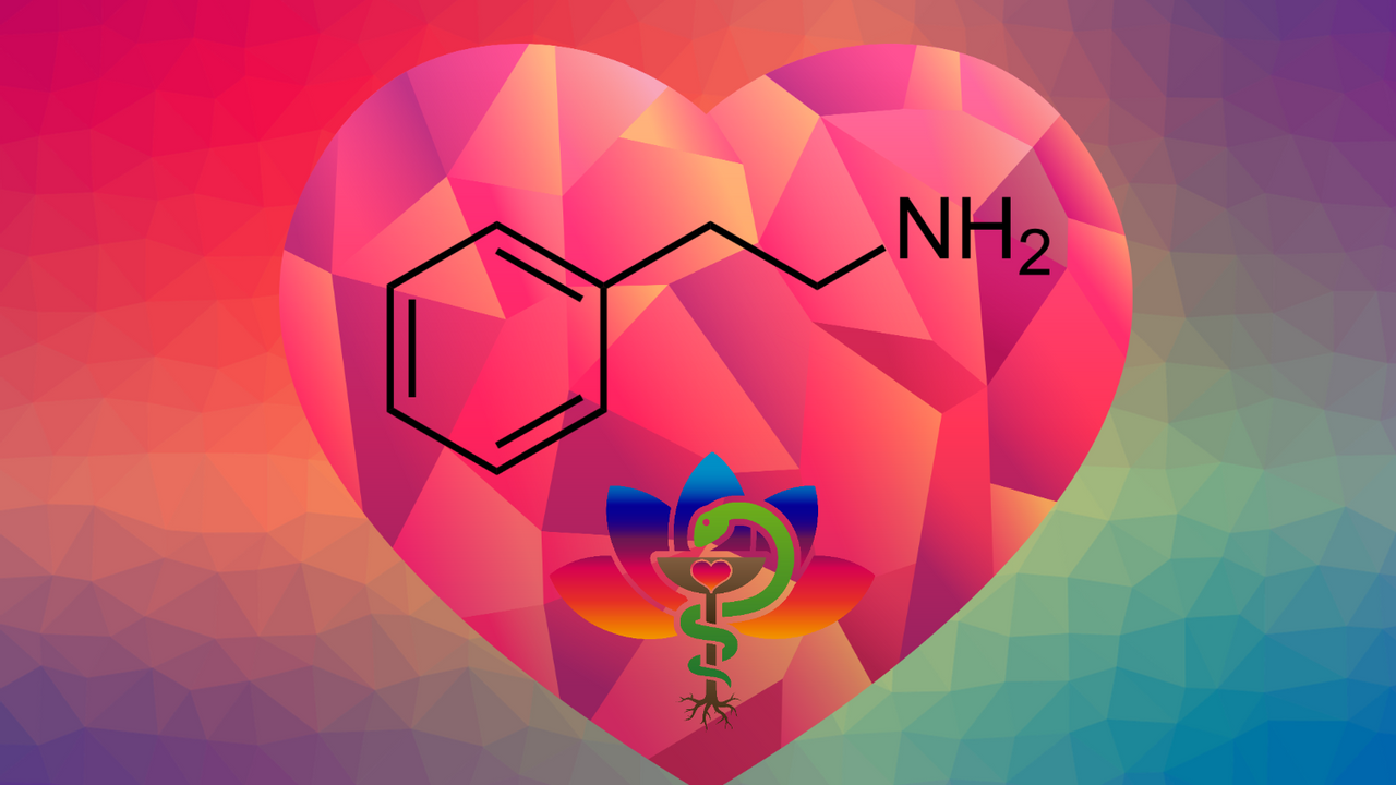
Phenethylamine (PEA) is a neurotransmitter that is naturally produced by our bodies. The chemical structure of phenethylamine also acts as a backbone for a variety of sympathomimetics with activity at sympathetic and central nervous systems.

How do phenethylamines act?

Phenethylamines can exert a variety of effects that varies from agent to agent based on the way it interacts with its neuronal target. Phenethylamine can cause neurotransmitter release through interactions with neurotransmitter packaging and reuptake pumps. It can also block those neurotransmitter reuptake pumps to increase the amount of neurotransmitter in the synapse. Phenethylamine can also directly stimulate the postsynaptic receptors to exert its effects.

wVQ8wkIRUeEouGV8UePa_Phenethylamines_5_.png


Classes of phenethylamines

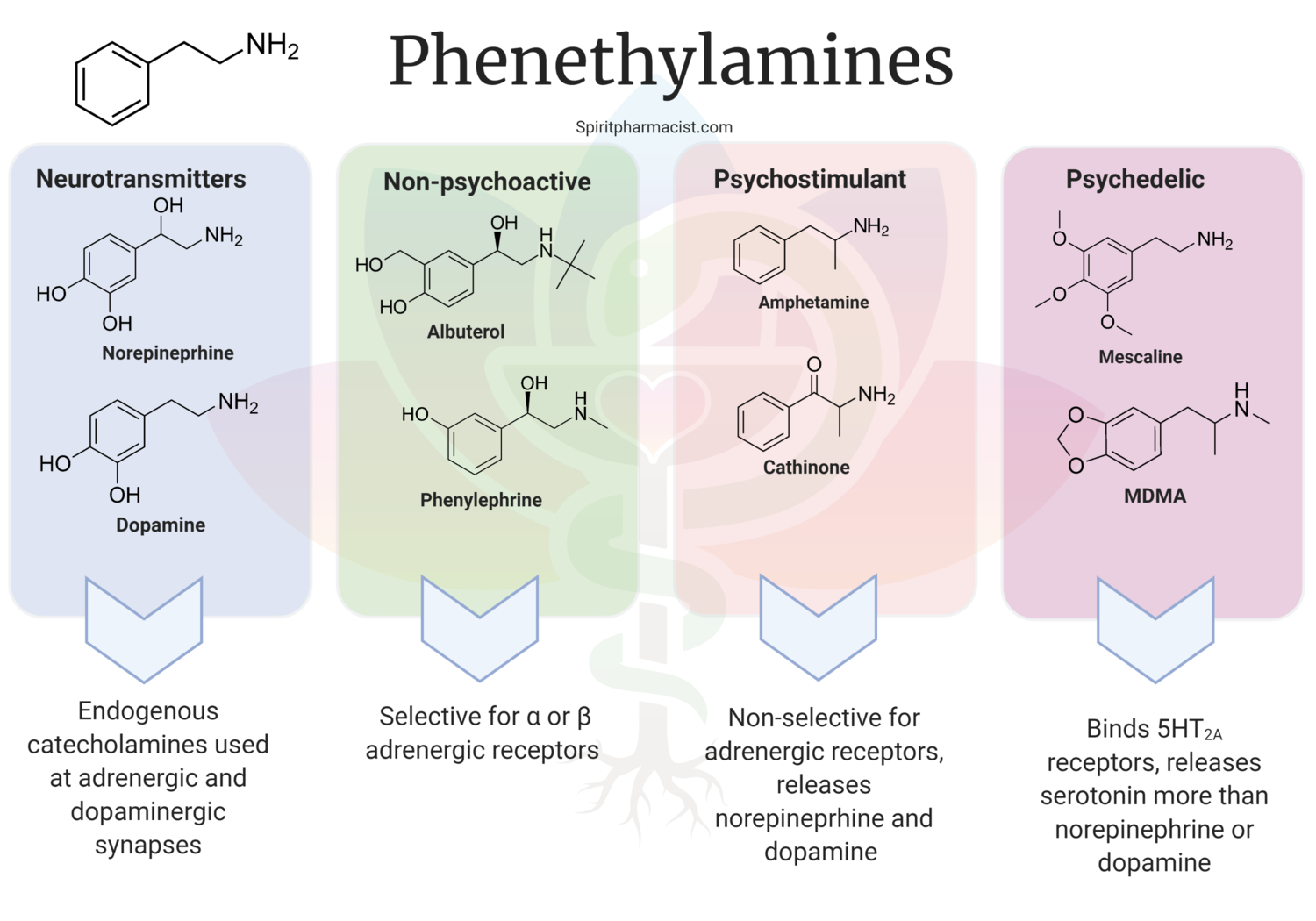
Phenethylamines could be broadly categorized into four classes: neurotransmitters, non-psychoactive sympathomimetics, psychostimulants, and ‘psychedelics’

The neurotransmitter class includes phenethylamine itself as well as norepinephrine, dopamine, and epinephrine which are endogenous catecholamines that act as chemical messengers at adrenergic and dopaminergic synapses.

Some phenethylamines have been selectively engineered for specificity at certain adrenergic receptors. For example, albuterol and phenylephrine are selective for postsynaptic adrenergic receptors. Phenylephrine specifically binds alpha-1 receptors which causes vascular smooth muscle contraction leading to vasoconstriction. Albuterol specifically binds the beta-2 receptors on smooth muscle to cause relaxation which leads to bronchodilation and airway opening. These agents are considered non-addictive and non-psychoactive.

Psychostimulants include amphetamine, methamphetamine, methcathinone and cathinone which are non-selective for adrenergic receptors in the sympathetic and central nervous system. Activation of receptors by these agents induces a release of norepinephrine and dopamine neurotransmitters from the synapse. The release of dopamine by the presynaptic cell is thought to give rise to the addictive potential of these agents.

Phenethylamine psychedelics

There is only one phenethylamine that is a classic psychedelic, which is mescaline. MDMA and other novel designer psychostimulants often include serotonergic pharmacology and many act to increase serotonin or bind ‘psychedelic’ 5HT2A receptors. These substances often have considerable pharmacology at other neurotransmitter systems such as dopamine and norepinephrine. These drugs typically release serotonin more readily than norepinephrine or dopamine and are thought to provide a hybrid of psychostimulant and psychedelic properties. MDMA and many others may be arguably better classified as enactogens ‘to touch within’ or empathogens ‘generating a state of empathy’ than psychedelics ‘mind-manifesting’. They are colloquially considered “heart openers” due to effects increasing emotional openness and expansiveness. Novel psychedelic phenethylamines and their ranges in effects are described in Alexander and Ann Shulgin’s work PIHKAL.

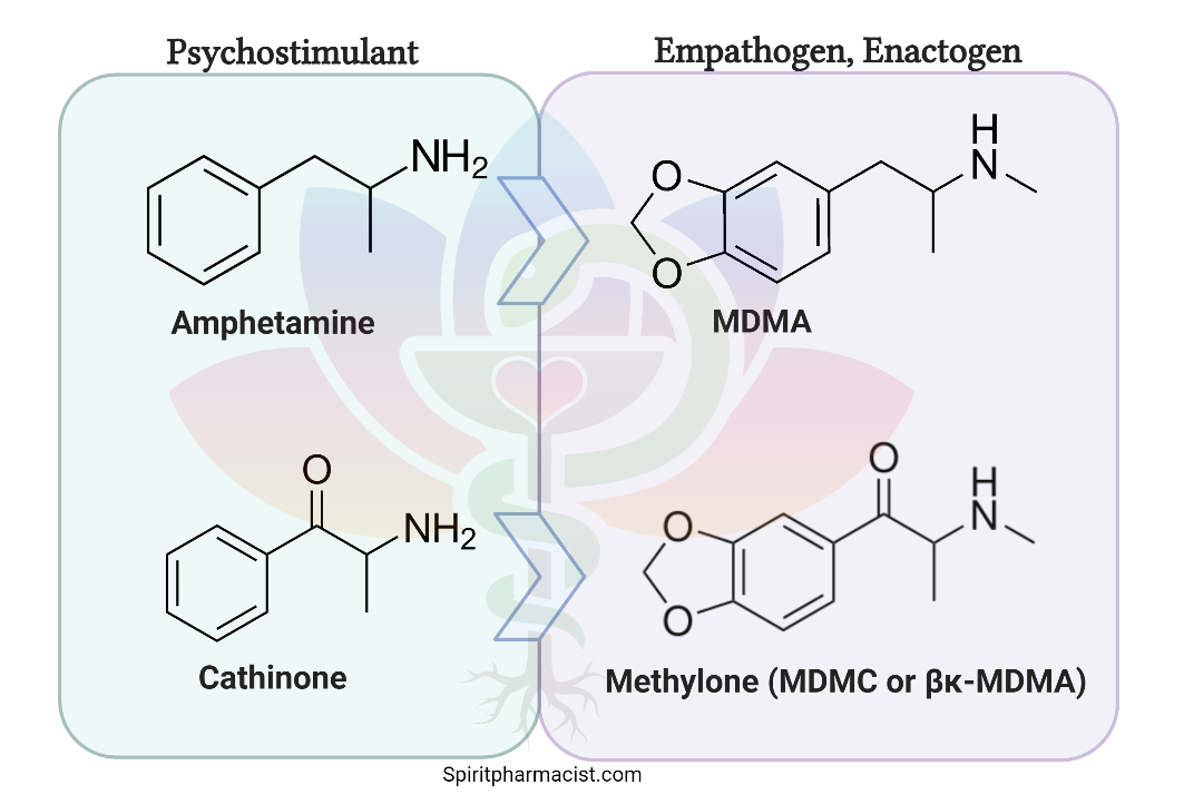
More recent trends in designer psychedelic stimulants have utilized cathinone rather than amphetamine as the chemical backbone to create a new genesis of analogous compounds to various amphetamine-based psychedelic stimulants. For example, methylone is the cathinone-version of MDMA. These analogues may share much overlapping pharmacology, although there are also significant differences in their effects.

dmUeGKI3SG2pK5UKWUDS_MDMA_cathinone_methylone.png


Psychedelic phenethylamines | Targets and effects

Psychedelics phenethylamines have a wide variety of target receptors, although commonly have serotonergic action and it could be considered a pharmacologic definition of their class that they have activity or bind with 5HT2A receptors. Activity at other post-synaptic receptors (e.g. 5HT1A, 5HT2C) may also influence effects. Their activity at norepinephrine and dopamine neurotransmitter systems is shared with traditional psychostimulants.

Psychedelic phenethylamines commonly act to increase neurotransmitter levels, oftentimes by interrupting storage of neurotransmitters in vesicles and reversing the flow the neurotransmitter reuptake pumps. Some may appreciably block neurotransmitter reuptake themselves or have inhibitory activity at monoamine oxidase.

The actions on the monoamine systems (serotonin, norepinephrine, dopamine) differ between substances, ultimately causing variations in subjective experience, safety, and addictive potential. Psychedelic phenethylamines often comes in a 'series' of compounds (e.g. DOx, 2Cx, 25x-NBOMe). Let’s take a quick look at MDMA as a ‘reference’ compound.

MDMA

MDMA could be considered a reference compound for psychedelic phenethylamine activity. It has a complex mechanism of action which is primarily serotonergic. MDMA causes a release of primarily serotonin from presynaptic vesicles through VMAT2 inhibition. The SERT receptor reverses the normal reuptake flow of serotonin and releases 5HT back into the synapse causing an intrasynaptic increase in 5HT. The process of MDMA’s release of serotonin has been termed ‘carrier-mediated’ release. This increase in serotonin neurotransmission increases activation to the 5HT2A receptors, which MDMA also weakly binds. Due to its ability to increase serotonin there is a potential for serotonin toxicity to occur with MDMA overdose or when mixed with monoamine oxidase inhibitors (MAOIs). Contraindication with MAOIs is common to psychedelic phenethylamines due to so many having significant effects in increasing neurotransmitters. There are additional neurohormonal effects of MDMA to consider due to the release of oxytocin and antidiuretic hormone. Neurohormonal effects could play a role in therapeutic effects (oxytocin - enhanced bonding and trust) or adverse effects (antidiuretic hormone - SIADH).

Addiction potential of psychedelic phenethylamines

The addiction potential of phenethylamines is likely driven by pleasurable and euphoric effects in addition to dopamine-mediated reinforcement. The variability in the ability of psychedelics to reinforcement properties creates a spectrum of addictive potential among agents:​
  • Mescaline is not considered to be reinforcing and has low addictive potential​
  • MDMA has some reinforcing properties and addictive potential, although release less dopamine than amphetamine​
  • Novel designer phenethylamines with short durations of action and heavier release of dopamine carry significant addictive potential​
Phenethylamine toxicology

In addition to the addiction potential of these agents there are also toxicities associated with these agents when high doses are used chronically. Neurotoxicity can result in damage to serotonergic and/or dopaminergic neurotransmitter systems resulting in depression, anxiety, fatigue, insomnia, irritability, and difficulty with emotional regulation. As with any agent, overdose is a possibility with psychedelic phenethylamines. Overdose toxidromes may have features of stimulant overdose or serotonin syndrome present including hyperthermia, seizures or myoclonus, muscle rigidity, and complications such as rhabdomyolysis.

High notes

Psychedelic phenethylamines are part central nervous system stimulant and part psychedelic. Some may be more appropriately termed entactogens or serotonergic amphetamines than psychedelics. They are pharmacologically diverse in actions, yet all bind 5HT2A receptors. MDMA is being advanced in clinical trials although there are hundreds of other designer psychedelic phenethylamines that could have therapeutic potential. With widespread use of novel designer substances, processes of discovery could be accelerated, however uncertainty in risk profiles creates potential for significant risks to public health.
 
21151628_10159330574940118_1160796148041186312_n.jpg

Faan Rossouw

An Introduction to Psychedelic Tryptamine Chemistry

by Faan Rossouw

This paper is intended for the general reader that has an appreciation for the beauty of chemistry, and/or desire to learn more about it. I am going to be pedantic throughout the paper, deconstructing technical terms and “dirty pictures” with the assumption that you do not know what they mean. That way we can learn them as we go along. If you are already fluent in Chemistrian, it goes without saying that you are free to skip over these and peruse selectively. This first section is an introductory exploration of the tryptamine class, and will be followed by further forays into other interesting aspects related specifically to this class before I move on.

The three main classes of psychedelics

There are three classes to which most psychedelic compounds belong – tryptamines, phenethylamines, and ergolines. The tryptamines include most of the well-known naturally-occurring psychedelics, including compounds derived from entheogenic fungi (psilocybin and psilocin), DMT, 5-MeO-DMT, bufotenin, and ibogaine. Mescaline is the only common naturally-occurring phenylethylamine, yet the class includes numerous well-known synthetic compounds such as MDMA and the 2-C’s. Ergolines most notable representatives include the naturally-occurring LSA and the semi-synthetic compound that turned on a generation, LSD.

Tryptamines

Psychedelics of this class are all derived from tryptamine, a ubiquitous endogenous ligand and agonist of the human trace amine-associated receptor 1 (TAAR1). The name tryptamine is derived from its structural similarity to l-tryptophan, an essential amino acid and the precursor to both serotonin and melatonin.

Substituted tryptamines

Although the “template” for psychedelics tryptamines is the molecule with all the various positions presented in Figure 2, in actuality there are limitations to how this manifests in psychedelic compounds. This is either because certain modifications are either difficult to impossible, or they lead to inactive compounds. An example of this is if something is attached to position 2 (Figure 2) the compound becomes a serotonin-2A receptor antagonist therefor losing its psychoactivity. Based on these restrictions we can simplify the template presented in Figure 2 to Figure 4, which is called the ‘substituted tryptamine’. The three main changes that synthetic chemists can make to derive psychedelic analogs is derived from this figure.


1572797929515.png


First, one can add side chains to either position 4 or 5, and those side chains have to contain an oxygen molecule. We can confirm this by looking at all the well known psychedelic compounds that have side chains attached to the ring – bufotenine has a hydroxyl (OH) group at position 5, 5-MeO-DMT has a methoxy (O-CH3) at position 5, psilocin has a hydroxyl (OH) group at position 4, and psilocybin has a phosphoryloxy (OPO3H2) at position 4. All at position 4 or 5, all with an oxygen included.

The second major change that can be made is a substitution at the α-position. Chemists can methylate (add a methyl group) the alpha-position to change a non-orally active species into one with orally active. We will explore this in full detail in the next article.

The final feasible change is adding sidechains to positions N1 or N2. All five of the major naturally-occurring species we have discussed thus far possess methyls at both positions (hence “dimethyl” from which the DM in DMT is derived – more below). These methyls may be substituted with more complex alkyls, another way in which chemists can turn non-orally active tryptamines into orally active species.

Tryptamine psychedelics

Now that we have an idea of the chemical “archetype” of tryptamine psychedelics and the possible changes chemists can make, let’s have a look at the five most well-known naturally-occurring examples: DMT, 5-MeO-DMT, bufotenin, psilocybin, and psilocin.

DMT

The substitutive name for DMT is N,N-dimethyltryptamine. One of the most magical parts of learning chemical language is that from it one can deduce what they actual molecule looks like, and vice-versa. Let’s explore that using DMT as an example. Starting from the back we have tryptamine, so we know that is the foundation of our molecule – the indole ring with an ethyl in position 3 attaching to an amine. Then we have “dimethyl,” meaning two methyls. Okay so now we know it’s the tryptamine molecule that has two methyls added to it. And where are these two methyls? They’re both positioned on the nitrogen of the amine, hence ‘N,N’.

What’s interesting about N,N-dimethyltryptamine is that it forms the foundation for all four other compounds we are going to discuss. In other words, all four of them are N,N-DMT with a little something extra. We can see that because the term is contained within the substitutive name of all four other molecules. Let’s have a look.

5-MeO-DMT

The substitutive name for 5-MeO-DMT is 5-methoxy-N,N-dimethyltryptamine (Figure 6). We can see that it has the whole name of DMT in it, so when we draw it we know we can start with that molecule – a tryptamine with two methyls on the amine (red and blue). What’s left is ‘5-methoxy’, which means that at position 5 we have a methoxy (green). A methoxy is a combination of a methyl and an oxygen – hence the name.


1572798088575.png


Bufotenin

The substitutive name for bufotenin is 5-hydroxy-N,N-dimethyltryptamine. As with 5-MeO-DMT, the molecule has DMT. But this time, instead of a methoxy at position five, we have the hydroxy, -OH.

Psilocin

The substitutive name for psilocin is 4-hydroxy-N,N-dimethyltryptamine. Same story, it starts with the structure of DMT (red and blue). If we compare them, we can see the psilocin is extremely similar to bufotenin, the only difference being where bufotenin had the hydroxy at position 5, here it is at position 4 (green). Later in this paper we learn why this small change is crucial to ensure that psilocin, unlike bufotenin, is an orally active species.


1572798143934.png


Psilocybin

The substitutive name for psilocybin is 4-phosphoryloxy-N,N-dimethyltryptamine (Figure 9). By now I’m sure you’ve grokked it – it’s a DMT molecule (red and blue) with a little something extra. As with it’s cousin psilocin, that something extra is at position 4, but here instead of a hydroxy, it’s a phosphoryloxy with the composition OPO3H2 (green).


1572798197751.png


The art of appetizing aromatics

In the previous section, I introduced the tryptamine class of psychedelics, and we discussed five well-known examples: DMT, 5-MeO-DMT, bufotenine, psilocybin, and psilocin. While the latter two, primary psychedelic constituents of Psilocybe mushrooms, are orally active, neither DMT, 5-MeO-DMT, nor bufotenine are. In this section we will explore two types of alterations that synthetic chemists can make to those molecules to bestow oral activity upon them. These alterations lead to the psychedelic tryptamine analogs (“research chemicals”): AMT (Indopan), MiPT, DiPT, 5-MeO-aMT (Alpha-O), 5-MeO-MiPT (Moxy), and 5-MeO-DiPT (Foxy Methoxy).

L-monoamine oxidase (MAO) are a family of enzymes that catalyze the oxidation of monoamines. Monoamines contain a single amine connected to an aromatic ring via a 2-carbon chain, and include neurotransmitters such as serotonin and norepinephrine, as well tryptamines such as DMT, 5-MeO-DMT, and bufotenin. The reason therefor that these compounds are not active after being consuming orally is because once they enter one’s gut they are inactivated by MAO.

If you want to experience the psychedelic effects of these compounds there are two basic strategies. The first is to use a route of administration that bypasses the gut. Smoking and vaporizing are by far the most common ways to achieve this, but are also the most intense (rapid onset) and shortest-lasting methods. Accordingly, some people favour other non-oral routes such as sublingual (under the tongue), insufflation (in the nasal passage), and rectal administration. Each of these administration routes has its own set of unique pharmacokinetic properties that may be favoured by certain people depending on the context and/or intention. Different strokes for different folks.

But that applies equally to oral delivery, which is unsurpassed in terms of its simplicity (swallow and then you’re done), ease and duration. Except for transdermal delivery, which is technologically complex and has severe restrictions on what can be administered, oral delivery is the longest lasting. Hence its popularity for journeyers that wish to go in deep. So even with a number of non-oral administration routes available, there is still good reason to utilize the oral route.

How to do so if we all walk around with an enzyme in our belly that will deactivate the psychedelic? Simple – consume another compound, called a monoamine oxidase inhibitor (MAOI), that will deactivate that enzyme. Ayahuasca is a prime example of this, though there are a number idiosyncratic formulas of the brew, in essence it is based on two core ingredients. One contains DMT, the most common being chacruna (Psychotria viridis), and the other contains the MAOI, which is always the ayahuasca vine (Banisteriopsis caapi).

Synthetic chemists love to ask “what if” questions. Like “what if” I make this simple change to the molecular nature of the compound, how does that then affect its properties? These type of questions are explored not only in the name of scientific curiosity, but also because studying how simple changes affect the properties of compounds informs us about its structure-activity relationship, as well provide intimations of what the target receptor looks and behaves like. To the specific question of whether or not a simple alteration to DMT/5-MeO-DMT can actuate oral activity chemists have thus far provided two answers – α-methylation and N-alkylation.

As we covered previously, DMT is a tryptamine molecule with two methyls at the N-position. So what would happen if, instead of adding two methyls to the N-position of the tryptamine, we added a single methyl to the alpha-position? This yields AMT (alpha-methyltryptamine), a molecule originally developed in the ‘60s by a Michigan-based pharmaceutical company called Upjohn and which was prescribed in the USSR as an antidepressant. It is at once psychedelic, entactogenic (like MDA/MDMA), and a stimulant with an oral dose typically lasting upwards of 12 hours.

The same goes for 5-MeO-tryptamine (mexamine) – if instead of adding two methyls to the N-position to form 5-MeO-DMT we add a single methyl to the alpha-position, we get 5-MeO-AMT – 5-methoxy-alpha-methyltryptamine (Figure 5). This orally-active and potent psychedelic, commonly known as ‘Alpha-O’, is sometimes peddled as faux-LSD. This is problematic as, unlike LSD with no known lethal toxicity, 5-MeO-AMT has lead to deaths at fairly low doses. It’s not a War on Drugs, it’s a War on People.

With both AMT and 5-MeO-AMT there is a chiral centre at the alpha-position. Attaching a single methyl to the alpha position potentially yields either an S- or R-configuration. Both are psychoactive, both orally active, but work by Dr. David Nichols lab has found that the S-enantiomer is more potent.

N-Alkylation

With N-alkylation we manipulate DMT and 5-MeO-DMT as the departure point to realize oral activity. Both these molecules possess two methyls on the amine nitrogen. Work again by Dr. Nichols’ lab has found that if you replace one, or both, these methyls with isopropyl, the molecule becomes orally active.

In the case of DMT, if a single methyl is replaced by an isopropyl it results in MiPT (N-methyl-N-isopropyltryptamine), an obscure psychedelic with indistinct effects first introduced to the world in TiHKAL. In the case of 5-MeO-DMT, the same single substitution results in 5-MeO-MiPT (5-methoxy-N-methyl-N-isopropyltryptamine). Commonly known as “Moxy”, it is an extremely potent (4 to 6 mg p.o.) psychedelic with stimulating properties.

As my articles on chemistry are intended for the general reader, I just want to take a brief moment here to remind you that the reason I always write out the substitutive name of each compound is because it describes the actual molecule. If we know the substitutive name, we can draw the molecule, and vice-versa. Let’s briefly review this by using Moxy as an example, but please feel free to skip over to the next paragraph if this is old news for you by now. Starting from back we have tryptamine, so our “foundational” structure is an indole ring with an ethyl chain at 3 which connects to an amine group. Then we start from the front – at position 5 we have a methoxy group, at N1 we have a methyl (fuschia), and then at N2 we have an isopropyl.

If both methyls are substituted by isopropyl, in the case of DMT the result is DiPT (N,N-diisopropyltryptamine), another bizarre creation of Sasha that primarily produces audial distortions. With 5-MeO-DMT the double substitution leads to 5-MeO-DiPT (5-methoxy-N,N-diisopropyltryptamine) which likely has the most endearing street name of any psychedelic – “foxy methoxy”. Note that in both cases, though making the additional isopropyl substitution retains oral activity, it decreases potency.

What’s Going On Here?

So why is it that in both the case of DMT and 5-MeO-DMT replacing a methyl with a slightly larger and more complex compound makes it impervious to deamination by MAO thereby giving it oral activity? To give us a clue we need to look at the nitrogen in the amine group – Figure 9. In order for MAO to deaminate a molecule it needs to access the lone electron pair of electrons (blue) on the nitrogen. A change in the molecule, such as substituting functional groups, changes its 3D-conformation. In the case of substituting a methyl with an isopropyl group on the amine, it changes the molecule’s 3D shape in such a way that shields the lone pair of electrons from MAO, thus giving it oral activity.

How do we know this is the case that it’s the molecule’s 3D shape that protects the lone pair from attack by the MAO and thus allows it to retain oral activity? Earlier in this article I said that MAO break down tryptamines. We then spoke about DMT and 5-MeO-DMT, but what about psilocybin and psilocin? They are naturally-occurring tryptamines, yet they are also orally active – how so? Pioneering work by Dr. David Nichols in the ‘80s using NMR spectroscopy showed that the fact that psilocin has a substitution at position 4 and not 5 (as with DMT/5-MeO-DMT) causes a critical change in the molecule’s 3D structure which ensures the compound is orally active. This study and all the profound implications for psychedelic chemistry gleamed from it will be the topic of the next section.

Afterword:

If it is your intention to consume DMT, and especially 5-MeO-DMT, orally by combining it with an MAOI please do your homework. And once you’ve done your calculations, double-check them. Terence McKenna used to quip that the only real danger with DMT is “death by astonishment.” Though that is the case for smoking it, overdoing orally-administered DMT/5-MeO-DMT can lead to serotonin shock, convulsions, and in some cases, death. The Psychedelic Ship is leaving the harbour, please don’t drop any cannonballs on the deck.

Why is psilocin orally active?

In the previous section we learned that though DMT and 5-MeO-DMT lack oral activity, chemistry wizards are able to change that. By making one of a variety of simple alterations to their structure they may be changed into analogs (“research chemicals”, or RCs), each possessing their own unique subset of characteristics including oral activity. That’s because the chemists changed the three-dimensional configuration of the molecules in such a way that the lone pair of electrons situated on the amine’s nitrogen became shielded, thereby preventing their degradation by MAO. To recap, if one consumes monoamines (such as certain tryptamines) orally, MAO transforms them in the gut and by the time they enter the bloodstream they are no longer psychoactive.

This section is going to unpack a study that showed, by comparing the structures of the naturally-occuring molecules psilocin and bufotenin why the former is orally active while the latter is not. This is another pioneering study from the lab of Dr. David Nichols, who is, along with Albert Hoffman and Sasha Shulgin, in my estimation one of the three true giants of psychedelic chemistry. Its his work and excellent lectures from ESPD50, Psychedelic Science (2013 and 2017), and Breaking Convention that restoked my appreciation for chemistry and inspired me to not only deepened my knowledge, but also to start this series of articles. The outpourings from his majestic mind has fundamentally shaped the topics and content of these articles… Shout out Big D, whut-whut!

The structure and atomic composition of a chemical is obviously critical to our understanding, and the progression of, chemistry and pharmacology. The problem with that is that molecules are small – really small. Even with today’s stupefying repertoire of advanced scientific analytical instruments there is still no practical way for us to observe their structure directly. So instead we have devised sophisticated methods in which to do so indirectly. One of these methods is called Nuclear Magnetic Resonance (NMR) Spectroscopy, which uses information about the spin of atomic nuclei to determine what a compound’s structure looks like.

In 1980 the team at Purdue University used NMR spectroscopy to investigate how the three-dimensional structures of bufotenin and psilocybin differ from one another. Even though these two compounds are constitutional isomers, there is a critical difference in their activity – psilocin is orally active, whereas bufotenin is not. This tiny change, moving the hydroxyl group from position 5 to 4 made this critical difference in the way they are absorbed by a human body. Though 2D-representations of the respective molecules are too low resolution to allude to the reason for the disparity, the researchers (correctly) suspected that by looking at their 3D-structures they would be able to understand why one molecule could resist deamination by MAO, while the other could not.

NMR spectroscopy revealed that the ethyl sidechain of bufotenin is able to rotate freely, meaning it can spin around on its own axis (Figure 5). That is however not the case for psilocin, something locks it in place, preventing it from rotating freely. The ethyl sidechains of the molecules are identical, which means that whatever is preventing the free rotation of psilocin’s ethyl sidechain is related to the hydroxyl group being situated at position 4, and not 5. To find out exactly what that was, the researchers used specialized software called LAOCN3. Before we explore what they found it would be useful to our interpretation of the results if we brushed up on a couple of elementary concepts in chemistry.


1572799179987.png


There are two basic types of bonds that atoms can form with one another. The first, called an ionic bond, forms when atoms exchange electrons with one another. This happens if the encountering atoms possess large differences in their respective affinities for electrons (called electronegativity), one atom really wants to lose an electron, while the other really wants to gain it. So an electron (or electrons) are exchanged, and because it is negatively charged the transfer changes the charge of the each atom. The atom that gains the electron gains a negative charge and thus becomes negative, while the atom that loses the electron loses a negative charge and thus becomes positive. And as the old adage goes, opposites attract – the oppositely-charged atoms come together and form a stable bond with one another.

The other type of bond that can unite atoms is a covalent bond. This happens when atoms with similar affinity for electrons encounter one another, neither really wants to lose/gain an electron so they reach a compromise – they share their electrons among each other. Both atoms pretend that the electron that it shares, as well as the electron shared by the other atom, belongs to it. It’s this overlap of shared electrons that connects the atoms together into a single molecule.

Because there are no electrons that are transferred in the covalent bond the atoms don’t assume a charge as was the case with ionic bonds. However, that’s only partially true… In certain cases the atoms that take part in a covalent bond do have some difference in their affinity – not enough for them to exchange electrons and form an ionic bond, but enough so that when they form a covalent bond and share electrons those shared electrons are closer to one atom than the other. This is known as a polar covalent bond. The atom to which the shared electrons are in closer proximity has a higher electronegativity and thus becomes partially negative (δ-). Conversely, the atoms with lower electronegativity is further from the shared electrons and is partially positive (δ+). Because of this asymmetrical charge, polar molecules are able to form weak bonds with other polar molecules, or with compounds that have a net charge. Now that we’ve covered some basic concepts let’s get back to the results of the study and apply what we’ve learned by taking a closer look at psilocin.

Taken together: psilocin has hydroxyl group at position 4 with a partially negative oxygen and a partially positive hydrogen, and an amine with a nitrogen that is partially negative and carbons that are partially positive. Because of these partial charges something interesting happens – the partially positive hydrogen from the hydroxyl group and the partially negative nitrogen from the amine attract one another.

But what has this to do with the difference in oral activity between the two molecules? Turns out, everything. It’s this hydrogen bond and closed loop formation in psilocin which shields the lone pair of electrons situated on the nitrogen. Because MAO cannot access the electrons it cannot deaminate the molecule – this is why it can pass through the gastrointestinal system unchanged.

The hydrogen bond and resulting closed loop formation also lead to several other important changes in the property of the molecule which further accentuates its efficacy and potency as an orally-active psychedelic tryptamine. After generating 3D-models of the respective molecules, the researchers went on to compare their pKa, and Log P values.

When they measured the pKa and the Log P for both psilocin and bufotenin they found the following:

The pKa for Bufotenin is 9.67, meaning that at that specific pH-value equal amounts of the the molecule will be present in both the ionized (water soluble) and protonated forms (lipid soluble). When the molecule is in the blood, which has a pH of about 7.4, almost all of it (99.5 percent) is in the ionized form. In contrast, psilocin has a pKa of 8.47, closer to the pH of blood. So for psilocin, only about 52% is in the ionized form. That means that in the blood, 48% of psilocin will be in its unionized form versus only about 0.5% when it comes to bufotenin. As it is only the unionized form of the drug that can cross cell-membranes, this has profound implications for the potency of these two drugs – psilocin is not only able to better withstand degradation by MAO, but once it is in the blood there is also much more of it available in a form that can cross cellular membranes and thus can reach the target receptors and exert an effect.

The difference in pKa is also related to the shielding of the electron lone pair by the hydrogen bond. Amines possess a nitrogen with a lone pair of electrons. These free electrons, which carry a negative charge, are all too happy to snap up positively-charged protons (H+) from a solution they are in. This is, according to the Bronsted-Lowry acid-base theory, the very definition of a base – something that accepts protons. When it comes to psilocin the lone pair of electrons are shielded and are thus much less likely to accept protons. As a consequence, psilocin is less basic that is bufotenin.

The researchers also detected a difference in the Log P values – 1.19 for bufotenin, and 1.45 for psilocin. In the Log P scale a negative value indicates a compound which is hydrophilic, whereas a positive value indicates one that is lipophilic. Both these compounds are thus lipophilic, and psilocin, with the higher value, is more lipophilic. For drugs in general it is preferable for them to be lipophilic so as to be able to cross cell membranes, but not too lipophilic because then they immediately migrate to, and are stored in, the body fat. Research indicates that a Log P value of about 3.0 is the “sweet spot”, so psilocin is closer to this number, again indicating that its properties are more favourable once it enters the body.

The researchers started with a simple question: how is it that two isomeric compounds with such a small difference have such widely different properties when they are consumed orally? With NMR Spectroscopy we learned that it all has to do with the fact that because the hydroxyl group of psilocin is a little bit closer to the amine it was able to form a hydrogen bond between the two groups. This hydrogen bond shields the electron lone pair from deamination by MAO, which means that, unlike bufotenin, psilocin is orally active. The hydrogen bond also decreases the molecule’s proton-accepting capacity thereby decreasing its pKa value which means that at blood pH there is more of psilocin in the non-ionized (lipid soluble) form which is able to cross cell membranes and thus enter the central nervous system (CNS). Finally, we saw that it also affected the Log P value, and that psilocin is a more lipophilic compound, closer to an ideal value for drugs to effectively enter and bind to the appropriate receptors in the CNS.

http://altdotmind.com/an-introduction-to-psychedelic-tryptamine-chemistry/
 
sonoran-toad-psychedelic-spotlight.jpeg


Scientists create potential cell-based 5-MeO "bio-factory"*

by David E. Carpenter | LUCID

Medical doctor and researcher Leonard Lerer, along with scientists at the biotechnology company he founded, Back of the Yards Algae Sciences (BYAS), are on their way to making what they claim is the first important scientific contribution toward helping to conserve toad populations. The company says they have successfully reproduced toad parotoid glands and the world’s first known cell-based 5-MeO-DMT, essentially recreating the basic parts of the cell’s structure to carry out the physiological processes of the organism.

“This is breakthrough work,” says Lerer, “as not only is it now possible for researchers to get naturally derived 5-MeO-DMT without milking toads, but it also makes a contribution to the conservation of Incilius alvarius.”

According to Lerer, his company is forging a new trail in creating sustainable, cruelty-free 5-MeO-DMT with a near-term plan of providing researchers — completely free of cost — the cell-based toad material to study. His company plans to operate in the spirit of open science, similar to a statement recently signed by hundreds of scientists working with psilocybin, MDMA, and similar substances, to share in the common good rather than for private gain. “We will give the extract free of charge to researchers on the basis of a commitment to open science. That is, that they make their results publicly available,” says Lerer.

Providing cell-based toad venom to an admittedly limited number of researchers studying 5-MeO-DMT may seem a small contribution relative to the amount of people using toad venom in non-clinical settings. But BYAS’s longterm plan could make a significant difference, says Lerer. He says his company looks to deploy a combination of “cellular agriculture, computational biology, and pre-clinical technologies for the discovery and development of safe, effective, active psychedelic molecules.”

The drug development and clinical testing work for this effort will be done through BYAS’ partner company, Parow Entheobiosciences, which was established to advance their cell-based psychedelics into clinical development for major psychiatric and neurological conditions. According to Lerer, the BYAS-PEB partnership is intended to take the “FDA route” and anticipates an initial meeting with the FDA before the end of 2022 for its first indication of OCD.

Cell-based psychedelics

BYAS was established in 2018 to develop and sell natural food colorant (a booming market that’s projected to be worth $3.2 billion by 2027). It is now focused on sustainable, zero-waste innovations in algae and mushroom extracts for food and agriculture. While investigating the transformative potential of algae and mycelia in food, agriculture, and health, the company expanded their portfolio of sustainable solutions and began producing a line of plant-based products. This includes what the company says is a breakthrough algal heme analog to make plant-based meats taste better (a direct competitor to the Impossible burgers’s GMO soy-based heme).

Then COVID-19 hit, says Lerer, creating a pause and an opening for his current work with cell-based psychedelics. He says their cell-perpetuating mindset was a natural fit for the company’s mission of waste reduction, sustainability, zero-cruelty and re-use of limited resources.

“The pandemic forced us to slow down our production,” says Lerer. “But it has been a huge driver of innovative ideas and some incredible internal initiatives. We had time to grow our R&D program on cellular agriculture of psychoactive tryptamine derivatives for conditions such as PTSD, depression, OCD and addiction.”

Part of that shift towards tryptamines has included obtaining a coveted Drug Enforcement Administration analytical laboratory license to produce those robust cells from toad parotoid glands, which have proven to produce good yields of 5-MeO-DMT that keep on giving. “Our measurements of yield and concentration make us confident that we can upscale,” says Lerer. “We also have proof of concept that we can produce immortalized cell lines that are essentially a bio-factory.”

Lerer and his collaborators, including his brother Professor Bernard Lerer — a neuropsychiatrist and neuropharmacologist — were so confident of their innovation and its promise that in December, 2021, they announced the milestone at an annual meeting of the American College of Neuropsychopharmacology, the world’s leading forum for cutting-edge science on brain function and psychotropic drugs.

During this conference, the BYAS researchers announced the successful production of 5-MeO-DMT from parotoid gland cells taken from an unharmed Sonoran Desert toad. They used these cells to create immortalized cell lines, a technique used extensively in the cultured or lab grown meat industry.

According to Lerer, a paper is currently in preparation for submission to a peer-reviewed journal that will contain detailed methods to ensure that these experiments can be replicated and validated by other researchers. Lerer says that the procedure of harvesting these cells is as invasive as a human topical dermatological biopsy and the donor toad remains alive and thriving.

Is toad-derived 5-MeO equal to synthetic?

Candida Pino, who leads psychedelic ceremonies and is a proponent of using only synthetic 5-MeO-DMT, is a Sonoran Desert toad conservationist. She describes the intrusive practice of collecting toad-derived 5-MeO-DMT. “Incilius alvarius are captured when most active from late May to September when they come out of hibernation,” says Pino. “Held firmly in hand, toads are forced to expel self-protective toxins by squeezing the parotoid glands located behind their eyes. Collecting them to forcefully eject these toxins adds to the list of already significant environmental stressors they endure, including climate change, introduction of fungal pathogens, and habitat loss.”

She notes that a belief that toad-derived 5-MeO-DMT provides a purer experience than synthetic is not the same as scientific evidence. “Anecdotal evidence suggests that the effects of 5-MeO-DMT are the same whether it is delivered via toad toxins or synthetic,” says Pino. “A preference for ‘natural’ is based on a perception that is not scientifically based but is likely more of a placebo effect.”

Lerer’s team looks to get to the bottom of that. BYAS is examining the potential entourage effect of natural toad venom and checking if those anecdotal reports of its superiority are indeed factual. In collaboration with Hadassah BrainLabs in Israel — which studies non-human models of psychiatric and neurological diseases — BYAS and PEB are exploring whether natural, full-spectrum entheogens including psilocybin outperform synthetic psychedelics in conditions such as depression, OCD, and PTSD, including on measures of neuroplasticity. Lerer believes the data from those tests will inform their cell-based toad venom work moving forward. He says the first results will be available later this year and all results will be presented at conferences and published in peer-reviewed journals.

“In three to four months we’ll have the psilocybin comparison data,” says Lerer. “This platform will then be used for toad secretion to answer the similar question: is natural really better than synthetic or is this anecdotal?”

The bigger picture, says Lerer, includes BYAS and PEB collaborating to identify other natural entheogenic molecules or combinations of molecules that can treat specific conditions, including OCD (their lead indication), depression, PTSD, autism, schizophrenia, and TBI. With this goal in mind, he says BYAS is building up its library of entheogenic biomass including hundreds of different types of mushrooms, cacti, and plants.

To protect their Incilius alvarius research, BYAS has stated its intention to make its patented cell reproduction process available to other researchers in the future. The company is concerned that they and other researchers will have similar obstacles that psilocybin companies are currently experiencing. This includes trying to secure patents on natural molecules that are part of the planet’s entheogenic heritage. Lerer says he wants to prevent that, adding, “Our aim is to make our cell-based Incilius alvarius extract available to other researchers and focus on identifying novel molecules with therapeutic applications.”

 
Predicting the addictive potential of phenethylamines

by Benjamin Malcolm | Spirit Pharmacist

Phenethylamine (PEA) is both a naturally occurring neurotransmitter and chemical backbone for several other types of medications and neurotransmitters. Legendary psychedelic chemist Alexander Shulgin synthesized dozens of novel psychedelic phenethylamines that he documents in his and his wife’s book PIHKAL (Phenethylamines I have Known and Loved). He is credited with re-synthesizing MDMA as well as inventing designer phenethylamines such as 2CB.

Phenethylamines are much broader than a class of psychedelics and are primarily known as ‘sympathomimetics’ because of their actions on the sympathetic (fight or flight) nervous system. Catecholamine neurotransmitters like norepinephrine and dopamine as well as the hormone epinephrine are the chemical mediators of sympathetic neurotransmission and drugs derived from phenethylamine traditionally used of therapeutic purposes mimic their actions.

Non-psychoactive phenethylamines include bronchodilator medications used for asthma such as albuterol while psychoactive phenethylamines include psychostimulants such as amphetamine, methamphetamine, and cathinones (‘bath salts’). Psychostimulants such as amphetamine act as ‘releasers’ of norepinephrine and dopamine, which stimulate the sympathetic nervous system. It is release of dopamine in the central nervous system that is thought to drive the habituating and addictive potentials of amphetamine. Thus, paying attention to dopamine releasing effect of phenethylamines is an important clue in understanding risk for habituation, re-enforcement, or addiction.

Phenethylamines3.png


The psychedelic phenethylamines tend to act as ‘releasers’ of serotonin and be able to bind 5HT2A receptors, although preserve some of the effects of traditional stimulants by acting on norepinephrine or dopamine. They are hybrids between traditional stimulants and psychedelics. The prototype psychedelic phenethylamine is 3,4-methylendioxymethamphetamine (MDMA). MDMA tends to be euphoric and preserve the ego structure. It carries some risks of habituation, dependence, or addiction linked to release of dopamine. MDMA tends to distort sensory perceptions or cause hallucinations much less than classical tryptamine psychedelics (e.g. psilocybin, LSD, DMT). For these reasons one may consider MDMA more of a serotonergic amphetamine than a true psychedelic.

The degree of effect on dopamine neurotransmission and duration of action appear to help guide understanding of habituation. When comparing phenethylamine psychedelics like MDMA with novel designer phenethylamines, substances with higher release of dopamine and shorter duration of action can tempt the user to frequently re-dose. For example, when comparing the actions of mephedrone (4-methylmethcathinone or 4-MMC) with MDMA it is noted that MDMA has considerably less dopamine release than mephedrone and that mephedrone has dopamine release similar to amphetamine. In addition, mephedrone also has a much shorter half-life than MDMA meaning that pleasurable effects wear off sooner. Frequent re-dosing creates a stacking of physical effects, the risks of behavioral reinforcement and addiction as well as adverse reactions such as cardiovascular events, seizures, or psychosis increases.

None of this is to raise alarm bells about the addictive potential of MDMA or demonize amphetamine and cathinones. On the contrary, if we know traditional stimulants can be therapeutic and know that serotonergic psychedelics can also be therapeutic, drugs that hybridize their effects deserve thorough exploration for therapeutic potentials. The point is that the mechanisms of phenethylamine psychedelics are rather broad and tends to differentially effect serotonin, norepinephrine, and dopamine neurotransmitter systems. Psychedelic actions and effects occur due to release of serotonin and modulation of ‘psychedelic’ 5HT2A receptors while propensity for re-dosing and habituation is linked to duration of action and effects on dopamine. Chemical fingerprinting of psychedelic phenethylamines across neurotransmitter systems and measurement of basic pharmacokinetic parameters may guide harm reduction efforts as drugs with high potential for habituation and re-dosing are identified among the sea of alphabet-amines (phenethylamine psychedelics) available in today’s clandestine marketplaces.
 
Last edited:
20220127lnp1-structs.jpg


Psychedelic chemistry guides antidepressant drug discovery

X-ray crystal structures of psychoactive compounds bound to a key serotonin receptor suggest design strategies for non-hallucinogenic therapeutics

by Bethany Halford | C&EN

Scientists have long sought the secrets of the 5-HT2A serotonin receptor—a central nervous system receptor that binds hallucinogenic compounds, including LSD and psilocybin. Many hope to figure out why these molecules cause hallucinations when they bind to 5-HT2A while other compounds that bind, including serotonin, do not. LSD and psilocybin have been shown treat mood disorders, such as depression, and scientists wonder if they can design molecules that maintain that mood-altering ability without causing hallucinations.

Researchers now report a structural biology-guided strategy for making such molecules. A team led by Sheng Wang of the Chinese Academy of Sciences and Jianjun Cheng of ShanghaiTech University determined the crystal structures of the 5-HT2A receptor bound to LSD, psilocin (the active form of psilocybin), serotonin, and lisuride, a non-hallucinogenic treatment for Parkinson’s disease. By visualizing the differences in how those molecules bind, the researchers then designed several compounds that they hypothesized would interact with 5-HT2A without inducing hallucinations (Science 2022, DOI: 10.1126/science.abl8615).

The team’s strategy was to design rigid molecules that would reach into a pocket of the 5-HT2A receptor and mimic one of two poses that serotonin and psilocin strike in the receptor. The researchers also wanted their molecules to avoid binding within a hydrophobic pocket because that interaction might spur hallucinations. Tests in mice showed that two compounds, called IHCH-7079 and IHCH-7086, had antidepressant activity: mice given those compounds continued to struggle when suspended by their tails and swim when forced, unlike control mice that gave up and exhibited depression-like behavior. IHCH-7079 and IHCH-7086 also didn’t appear to make the mice twitch their heads—behavior that’s observed when the rodents take LSD or psilocybin.

David E. Olson, a professor at the University of California, Davis, who is also working on making non-hallucinogenic psychoactive compounds, says this work adds to the growing evidence that analogs of psychedelics may be effective antidepressants. “The 5-HT2A receptor is one of the most important targets in neuropsychiatry."

This additional structural data will aid efforts to design new antidepressants as well as antipsychotics,” he says in an email.

Wang says that their compounds still need to be optimized, noting that their required effective dose in mice is high. Nevertheless, he says, “we definitely want to prove our concept in a clinical trial.”

From the article here :

https://cen.acs.org/pharmaceuticals/drug-discovery/Hallucinogen-chemistry-guides-antidepressant-drug/100/web/2022/01
 
Still view SERT antagonism at 80 ~ 100%, Plays a role since DXM has no direct 5HT2 agonism/binding even at 1200mg. Before someone says It the NMDA antagonism the high amount of Indirect 5HT2A & 5HT2C would block any dopamine release. Also DMT unlike other Psych's Is a SRI + 5HT2A agonist, People report It being the strongest with effects reaching Deliriant levels that hard to mimic In Shrooms/LSD without adding a SRI/SNRI.

I wonder In DXM or DMT trips SERT forces the brain to overproduce bufotenine at levels where It binds to 5HT2 receptors. The same compound that thought to be very high In Autistic brains(SERT Is really low In those brains). bufotenine Infamous for being one of the more nausea inducing Psychdelic's.
 
Still view SERT antagonism at 80 ~ 100%, Plays a role since DXM has no direct 5HT2 agonism/binding even at 1200mg. Before someone says It the NMDA antagonism the high amount of Indirect 5HT2A & 5HT2C would block any dopamine release. Also DMT unlike other Psych's Is a SRI + 5HT2A agonist, People report It being the strongest with effects reaching Deliriant levels that hard to mimic In Shrooms/LSD without adding a SRI/SNRI.

I wonder In DXM or DMT trips SERT forces the brain to overproduce bufotenine at levels where It binds to 5HT2 receptors. The same compound that thought to be very high In Autistic brains(SERT Is really low In those brains). bufotenine Infamous for being one of the more nausea inducing Psychdelic's.​
^^

DMT, the endogenous psychedelic

Steven A. Barker

This report provides a historical overview of research concerning the endogenous psychedelic N, N-dimethyltryptamine (DMT), focusing on data regarding its biosynthesis and metabolism in the brain and peripheral tissues, methods and results for DMT detection in body fluids and brain, new sites of action for DMT, and new data regarding its possible physiological and therapeutic roles. Research that further elaborates its consideration as a putative neurotransmitter is also addressed. Taking these studies together, the report proposes several new directions and experiments to ascertain the role of DMT in the brain, including brain mapping of enzymes responsible for the biosynthesis of DMT, further studies to elaborate its presence and role in the pineal gland, a reconsideration of binding site data, and new administration and imaging studies. The need to resolve the “natural” role of an endogenous psychedelic from the effects observed from peripheral administration are also emphasized.

Introduction

Despite their presence in the human pharmacopeia for millennia, we have yet to resolve the biochemical mechanisms by which psychedelics so dramatically alter perception and consciousness. It is the only class of compounds that efficiently and specifically does so. For that matter, we do not fully understand the biochemistry of perception itself or how we live such a vivid and complex internal life in the absence of external stimulation. We do not understand the basic biochemical mechanisms of some of our most common experiences, such as the many human aspects of creativity, imagination or dream states. This is also true for extraordinary states of consciousness such as “visions” or spontaneous hallucinations or phenomena such as near-death experiences (NDE). And it is troubling that we have not sufficiently turned the scientific method on these latter subjects despite the profound role they have played in the evolution of our science, philosophy, psychology and culture.

The experiences derived from the administration of psychedelics are often compared to dream states. However, the experience of administered psychedelic substances is far more intense, robust and overwhelming than the subtlety of mere dreams. By comparison, the natural biochemical processes for our related psychedelic experiences are obviously far more highly regulated, occurring as an orchestrated and inherent function of the “normal” brain. Nonetheless, it is conceivable that attaining an explanation for these related natural human phenomena may lie in resolving the biochemical mechanisms involved in the more dramatic pharmacology of psychedelics, recognizing that the complexities and intensity of the “administered” experience are, essentially, an overdose relative to corresponding natural regulatory controls. Given their status, increased study of psychedelics, particularly with advanced brain imaging and molecular biology approaches, may provide a better understanding of the “common” biochemistry that creates mind.

Perhaps the science behind the discovery of endogenous opioids offers us a corollary. We came to better understand the common human experience of pain through examining the pharmacology of administered opiates and the subsequent discovery of endogenous opioid ligands, receptors and pathways that are predominantly responsible for and regulate the experience and perception of pain. Such may also be the case for understanding perception and consciousness. With the discovery of the endogenous DMT, perhaps, as with the endogenous opioids, we have a similar opportunity to understand perception and consciousness. Recent research has stimulated a renewed interest in further study of this compound as a neuro-regulatory substance and, thus, a potential neuro-pharmacological target. Taking results from these and more classical studies of DMT biochemistry and pharmacology together, this report examines some of the past and current data in the field and proposes several new directions and experiments to ascertain the role of endogenous DMT.

A brief history of DMT

In terms of Western culture, DMT was first synthesized by a Canadian chemist, Richard Manske, in 1931 but was, at the time, not assessed for human pharmacological effects. In 1946 the microbiologist Oswaldo Gonsalves de Lima discovered DMT's natural occurrence in plants. DMT's psychedelic properties were not discovered until 1956 when Stephen Szara, a pioneering Hungarian chemist and psychiatrist, extracted DMT from the Mimosa hostilis plant and administered the extract to himself intramuscularly. This sequence of events formed the link between modern science and the historical use of many DMT-containing plants as a cultural and religious ritual sacrament, their effect on the psyche and the chemical structure of DMT.

The discovery of a number of psychedelics in the 1950's and observations of their effects on perception, affect and behavior prompted hypotheses that the syndrome known as schizophrenia might be caused by an error in metabolism that produced such psychedelics in the human brain, forming a schizo- or psycho-toxin. The presence of endogenous psycedelic compounds, related mainly to those resembling dopamine (mescaline) or serotonin (DMT), were subsequently sought. Although several interesting new compounds were found, the only known psychedelics isolated were those derived from tryptophan (DMT, and 5-methoxy-DMT). Data were subsequently developed illustrating pathways for their endogenous synthesis in mammalian species, including humans. Over 60 studies were eventually undertaken in an attempt to correlate the presence or concentration of these compounds in blood and/or urine with a particular psychiatric diagnosis. However, there has yet to be any clear-cut or repeatable correlation of the presence or level of DMT in peripheral body fluids with any psychiatric diagnosis. Nonetheless, the discovery of endogenous psychedelics and the possibilities rendered in various hypotheses surrounding their role and function in mental illness, normal and “extraordinary” brain function spurred further research into the mechanisms for their biosynthesis, metabolism and mode of action as well as for their known and profound effects on consciousness.

DMT biosynthesis

After the discovery of an indole-N-methyl transferase in rat brain, researchers were soon examining whether the conversion of tryptophan to tryptamine could be converted to DMT in the brain and other tissues from several mammalian species. Numerous studies subsequently demonstrated the biosynthesis of DMT in mammalian tissue preparations in vitro and in vivo. In 1972, Juan Saavedra and Julius Axelrod reported that intracisternally administered TA was converted to N-methyltryptamine and DMT in the rat, the first demonstration of DMT's formation by brain tissue in vivo. Using dialyzed, centrifuged whole-brain homogenate supernatant from rats and humans, these same researchers determined that the rate of synthesis of DMT from TA was 350 and 450 pmol/g/hr, and 250 and 360 pmol/g/h, using NMT as substrate, in these tissues, respectively.

In 1973, Saavedra et al. characterized a nonspecific N-methyltransferase in rat and human brain, reporting a Km for the enzyme of 28 uM for TA as the substrate in rat brain. The highest enzyme activity in human brain was found in the subcortical layers of the fronto-parietal and temporal lobes and the cortical layers of the frontal parietal lobe. However, an INMT found in rabbit lung was shown to have a much higher Km than the brain enzyme in rats. This suggested that INMT may exist in several isoenzyme forms between species and possibly even within the same animal, each having different Km's and substrate affinities. INMT activity has subsequently been described in a variety of tissues and species. There have also been several reports of an endogenous inhibitor of INMT in vivo that may help regulate its activity and, thus, DMT biosynthesis.

Pathways for the biosynthesis and metabolism of DMT, 1. Biosynthesis: Tryptophan (2) is converted to tryptamine (TA, 3) by aromatic amino acid decarboxylase (AADC). TA is dimethylated to first yield N-methyltryptamine (NMT, 4) and then DMT (1) by indole-N-methyltransferase (INMT), using S-adenosyl-methionine (SAM) as the methyl source. Metabolism: TA, NMT and DMT are all substrates for monoamine oxidase, yielding indole-3-acetic acid (5, IAA) as both a common precursor metabolite and the most abundant metabolite of DMT itself. DMT is also converted to DMT-N-oxide (6) as the second-most abundant metabolite. Two 1,2,3,4-tetrahydro-beta-carbolines (THBCs) have also been identified as metabolites; 2-methyl-THBC (7, MTHBC) and THBC (8).

The combined data demonstrate that DMT is formed from tryptophan, a common dietary amino acid, via the enzyme aromatic L-amino acid decarboxylase (AADC) formation of TA and its subsequent N, N-dimethylation. The enzyme indolethylamine-N-methyltransferase (INMT) uses S-adenosyl-l-methionine as the methyl source to produce N-methyltryptamine and then DMT. Both AADC and INMT act on other substrates as well. As a historical and research note regarding DMT, there was initial confusion and misidentification of the products formed when using 5-methyltetrahydrofolate (5-MTHF) as the methyl source in INMT studies due to formation of indole-ethylamine condensation products with formaldehyde.

There has also been interest in the role of INMT and DMT biosynthesis in maturation and development. Relatively elevated levels of INMT activity have been found in the placenta from a variety of species, including humans. INMT activity in rabbit lung was reported to be elevated in the fetus and to increase rapidly after birth, peaking at 15 days of age. It then declined to mature levels and remained constant through life. In this regard, Beaton and Morris have examined the ontogeny of DMT biosynthesis in the brain of neonatal rats and rats of various ages. Using gas chromatography-mass spectrometry with isotope dilution for their analyses, DMT was detected in the brain of neonatal rats from birth. DMT levels remained low until days 12 and 17 at which time they increased significantly and then returned to the initial low levels for all subsequent ages. There has yet to be any follow-on research as to the significance of this change in DMT concentrations during rat brain neurodevelopment or correlation with possible changes of INMT activity in other developing tissues, specifically during days 12–17. Nonetheless, these findings correlate well with the Lin et al. data for INMT changes in rabbits and deserve further inquiry.

There is a significant literature concerning INMT, particularly in peripheral tissues. INMT and its gene have been sequenced, commercial antibodies for its detection have been developed and commercial probes exist for monitoring its mRNA and gene expression. A study using Northern blot detection of the INMT mRNA conducted by Thompson et al. in the rabbit suggested that INMT was present in significant quantities in the periphery, and particularly the lung, but that it was almost non-existent (low to absent) in the brain. These data became the foundation for several hypotheses that any neuropharmacological effects of endogenous DMT must lie in its formation in the periphery and its subsequent transport into the brain. This idea was strengthened by the fact that DMT has been shown to be readily, and perhaps actively, transported into the brain. However, the data concerning the apparent absence of INMT in brain would appear to be in conflict with the many earlier studies that demonstrated both in vivo and in vitro biosynthesis of DMT in the brain. Indeed, several studies had identified INMT activity or the enzyme itself in the central nervous system (CNS) including the medulla, the amygdala, uncus, and frontal cortex, the fronto-parietal and temporal lobes and, more recently, the anterior horn of the spinal cord as well as the pineal gland.

Thus, in 2011, Cozzi et al. sought to determine why earlier studies had not detected significant INMT in brain using Northern blots despite several reports that brain tissue had been shown to synthesize DMT from TA. One possibility was that INMT was “expressed in nervous tissue but that in some situations, INMT mRNA is not detectable by Northern analysis.” Examining primate nervous system tissues (Rhesus macaque spinal cord, pineal gland, and retina) probed with rabbit polyclonal antibodies to human INMT, all three tissues tested positive. INMT immunoreactivity in spinal cord was found to be localized in ventral horn motoneurons. The study also showed that INMT response was “robust and punctuate” in the pineal gland. Further, intense INMT immuno-reactivity was detected in retinal ganglion neurons and at synapses in the inner and outer plexiform layers. In 2012, Mavlyutov et al. reported that INMT is also localized in postsynaptic sites of C-terminals of rat motoneurons in close proximity to sigma-1 receptors, which have been linked to control of the activities of ion channels and G-protein-coupled receptors. It was proposed that the close association of INMT and sigma-1 receptors suggests that DMT is synthesized locally to effectively activate sigma-1 in motoneurons. It has been further proposed that DMT is an endogenous sigma-1 receptor regulator.

Taking these newer data together with historical in vitro and in vivo results regarding INMT enzyme activity in the brain and CNS, it is now clear that the work of Thompson and Weinshilboum is not the final word on DMT biosynthesis in the brain.

Future research on the biosynthesis of DMT

Considering that tryptamine formation, itself a trace biogenic amine, is essential for the formation of DMT and given its own rapid metabolism by monoamine oxidase (MAO) as well, demonstrating its availability for the biosynthesis of DMT is also relevant to a complete elucidation of the overall pathway. Indeed, demonstrating the co-localization of AADC and INMT should be a necessary endeavor in any future research regarding DMT biosynthesis in both the brain and periphery. The co-localization of AADC in discreet brain cells and areas with INMT permits TA and, subsequently, DMT formation locally. With demonstration of co-localization of the necessary biosynthetic machinery in the brain, both AADC and INMT, mechanisms for a rapid biochemical response to signaling and DMT formation may be shown to exist. Furthermore, the demonstration of mechanisms for the protection, storage, release and re-uptake of DMT would demonstrate that higher concentrations of DMT could be reached in the synaptic cleft and at neuronal receptors than would have to occur from, based on previous thought, formation and transport from the periphery. Pursuit of research of these mechanisms, as well as detailed mapping of INMT-AADC in the brain, is needed. We should not rule out the possibility that the biosynthesis and transport of DMT can and does occur from the periphery, however.

Peripheral DMT, especially if synthesized in tissues that bypass liver metabolism on first pass, may also serve as a signaling compound from the periphery to the brain. Such signaling may occur in maintaining homeostasis or in response to extreme changes in physiology. However, the immediate availability of TA for the biosynthesis of DMT in the periphery should also be demonstrated and studies examining the co-localization of AADC and INMT in the periphery should also be performed. This will require using highly sensitive and well validated antibodies and probes for detection of INMT and/or its mRNA in brain and/or peripheral tissues as well as those for aromatic-L-amino acid decarboxylase (AADC). Demonstration of co-localization with AADC has not been previously conducted in any other study seeking to identify INMT's presence or to demonstrate INMT activity. Such a determination may prove fruitful since a preliminary examination for the possible co-localization of INMT and AADC in the brain is supported by the data provided in the Allen Brain Atlas, mapping INMT and AADC gene expression.

A thorough re-examination of possible peripheral DMT biosynthesis is needed. Indeed, INMT actually methylates other substrates, such as histamine. Thus, much of the INMT in the periphery may be involved to a greater degree with methylation of other substances than TA alone. In this regard, in vitro studies of INMT as it relates to DMT biosynthesis necessarily added TA to their incubations, making TA “artificially” available in regions where natural levels may be absent or at significantly lower levels. Without a source for TA, the hypotheses regarding the formation of DMT in the periphery and its transport to the brain as a mechanism of action/function of endogenous DMT may be seen to be based on a less significant pathway than previously thought. Failure to demonstrate co-localization of INMT and AADC in the periphery would alter, to some degree, the focus of studies of peripheral synthesis and detection for understanding the role of endogenous DMT.

At least one study has now shown that the pineal gland has high concentrations of INMT. These data are underscored by the findings of Barker et al. demonstrating the presence of DMT in pineal perfusates from free-moving rats. Clearly, further research into the biosynthesis and role of DMT in the pineal is needed, as is a further assessment of our current knowledge of pineal function.

We will also need to examine protein and gene arrays to determine the factors that assist or work in concert with the up and down regulation of the INMT system in brain and how it responds to selected physiological changes. Such analyses will be essential in examining the possible role of DMT biosynthesis in changing biochemical and physiological events. We will also need to create brain-specific INMT KO animals, to further understand DMT biosynthesis and the “normal” role of DMT in vivo. It would also be of interest to better understand the possible role of DMT in neurodevelopment as suggested by the work of Beaton and Morris and Lin et al. in rats and rabbits, respectively. While DMT appears to clearly be biosynthesized in the pineal, mechanisms for its biosynthesis and release may exist in other brain areas as well and research into these other possibilities will also need to proceed.

DMT metabolism

The metabolism of DMT has been thoroughly studied, with a great deal of newer data being provided from studies of ayahuasca administration. All of the in vivo metabolism studies have shown that exogenously administered (IV, IM, smoking, etc). DMT is rapidly metabolized and cleared, with only a small fraction of IV or IM administered DMT subsequently being found in urine. For example, 0.16% of an intramuscular dose of DMT was recovered as the parent compound following a 24 h urine collection. DMT administered in this manner reached a peak concentration in blood within 10–15 min and was below the limits of detection within 1 h. It was estimated that only 1.8% of an injected dose was present in blood at any one time. Due to rapid metabolism in the periphery, DMT is not orally active, being converted to inactive metabolites before sufficient penetration to the brain can occur. DMT is only orally active if co-administered with a monoamine oxidase inhibitor (MAOI). DMT is pharmacologically active following administration by injection or smoking, pathways which can avoid first-pass metabolism by the liver to some degree. The time to onset of effects is rapid (seconds to minutes) by these routes and short lived.

The primary route of metabolism for DMT is via monoamine oxidase A (MAO-A), yielding indoleacetic acid. The other metabolites formed include DMT-N-oxide, the second most abundant metabolite, and lesser amounts of N-methyltryptamine, which, along with TA, is also a substrate for MAO-A, with both yielding IAA. Inhibition of MAO leads to a shift in favor of the amounts of DMT-NO and NMT formed. Other metabolites have been reported, such as 6-hydroxy-DMT (6-OH-DMT), as well as products from a peroxidase pathway, reported to yield N, N-dimethyl-N-formyl-kynuramine, and N, N-dimethyl-kynuramine. However, these latter metabolites have yet to be identified in vivo. Metabolites also result from the cyclization of an intermediate iminium ion that forms during demethylation of DMT, yielding 2-methyl- 1,2,3,4- tetrahydro-beta-carboline and THBC.

The primary role of MAO-A in the metabolism of DMT has been further confirmed by pre-treatment of experimental subjects with the MAO inhibitor (MAOI) iproniazid as well as other MAOIs, the ability of the MAO-inhibiting harmala alkaloids of ayahuasca to make DMT orally active and the increased half-life and extended effects of an α, α, B, B-tetradeutero-DMT, which is less susceptible to MAO-A metabolism due to the kinetic isotope effect.

Future research on the metabolism of DMT

While the metabolism of DMT has been thoroughly studied and a number of metabolites have been identified, one of the complications in understanding the role and function of endogenous DMT has been the fact that, to date, no study examining body fluids (blood, urine, saliva) has ever been conducted to correlate such data with human physiological events, such as circadian changes, sex differences, etc. Of greater impact is the fact that, despite DMT's rapid metabolism and multiple metabolites, no study has fully assessed all of these compounds simultaneously to better understand DMT's overall occurrence or rate of endogenous synthesis, release, clearance and/or the overall assessment of the relevance of endogenous levels in the brain or periphery. All of these factors need to be examined. Given that peripherally administered DMT, at what must be considered as much higher doses than would be expected to occur naturally in the entire organism, is rapidly metabolized and cleared, measuring endogenous DMT alone in an attempt to assess its role and function is probably doomed to failure. This is particularly true if endogenous DMT is mainly produced, stored and metabolized in discreet brain areas and that DMT and its metabolites so produced never attain measurable levels in peripheral fluids.

To the degree that DMT is produced peripherally, measurement of IAA, DMT-NO, N-methyltryptamine and the precursor for the synthesis of both DMT and NMT, tryptamine, would be advantageous. These compounds have been variously reported in tissue, blood and urine samples. However, this approach is complicated by the fact that the major MAO metabolite of all three of these latter compounds, IAA, is also derived from dietary sources and is produced from the action of bacteria in the gut. It is not unreasonable to question whether measurement of DMT and its metabolites, and thus the role and function of endogenous DMT, can be understood by simply trying to measure these compounds in the periphery. This is particularly true in understanding DMT production in the CNS. Peripheral measurements may not be the way to determine the central role of DMT and DMT produced in the brain may never be available for measurement in the periphery. Nonetheless, additional studies should determine if there is validity in such measurements and examine possible circadian, ultradian or diurnal variations in DMT synthesis as well as the changes that may occur due to alterations in other physiological parameters.

DMT detection in blood, urine, and cerebrospinal fluid

Barker et al. have published a thorough overview of the 69 published studies examining blood, urine and cerebrospinal fluid detection of endogenous N, N-dimethylated tryptamines [N, N-dimethyltryptamine (DMT), 5-hydroxy-DMT (bufotenine, HDMT), and 5-methoxy-DMT (MDMT)]. Nearly all of the studies were directed at establishing a relationship between the presence and/or level of these compounds and a psychiatric diagnosis. In total, the 69 studies examined DMT in thousands of subjects. A critical review of these data determined, however, that most early studies reporting rather high concentrations of these compounds in blood and/or urine were most likely in error and any correlations based on these data were likewise probably incorrect. The reasons for this conclusion were: (1) Based on current analytical requirements for unequivocal structure identification, it is highly probable that many of these studies misidentified the target analyte. (2) If properly identified, the studies showed that a psychiatric diagnosis was not a necessary or sufficient criterion for finding one or more of these psychedelics in various body fluids; “normal” controls were also positive (and sometimes higher) for these compounds. Nevertheless, it was also concluded that, particularly where mass spectral evidence was provided, DMT and HDMT are endogenous and can often be successfully measured in human body fluids. The evidence was less compelling for MDMT where the only two MS-based positive studies—in CSF—were performed by the same research group. There was no mass spectral data demonstrating detection of MDMT in blood or urine. There was also no study that attempted a determination of HDMT in CSF.

In conducting studies to determine the natural occurrence of a compound as being endogenous, it is also necessary to eliminate other possible dietary or environmental sources. Of the 69 studies reviewed, many addressed the possible source of DMT as being from diet or gut bacteria by using special diets. Of those conducted, it was determined that neither was a source but additional research in this area using more modern technology and a more standard diet across studies is a necessity. There have also been only a few efforts to examine the many variables that may influence the levels of these compounds, such as circadian or diurnal variations, sleep stages and gender-age-related differences. Indeed, most of the studies collected only a single time point or were from 24 h collections (urine). Such infrequent sampling makes it impossible to assess central DMT production from peripheral measurements and suggests, perhaps incorrectly, that DMT only appears intermittently or not at all. In trying to compare the results, interpretations and correlation of the data were hampered by variability in sampling methods, amount of sample assayed, type of sample (plasma, serum and/or whole blood), divergent techniques and analytical methodology that also had highly variable or unspecified limits of detection.

Future research measuring DMT in the blood, urine, and/or cerebrospinal fluid

In terms of pursuing future research on the presence of the endogenous indolealkylethylamines, further studies are necessary to determine whether MDMT actually exists in humans. Similarly, there are no data on the possible presence of HDMT in CSF although it has been routinely identified in urine. Future analyses to determine endogenous N, N-dimethyl-indolethylamines should also include a search for their major metabolites. The methodology applied in such analyses must include rigorous validated protocols for sample collection, storage, extraction and analyte stability and appropriate criteria for unequivocal detection and confirmation of the analytes using validated methods. Modern exact-mass liquid chromatography-mass spectrometry instrumentation should be the analytical method of choice. Such capabilities may then be applied to address the many variables that may influence the ability to measure DMT and/or its precursors and metabolites in the periphery.

Measurement of DMT in the brain

Many studies have been conducted to detect and/or quantitate DMT in blood and urine and only a few in the CSF of humans. However, the CSF studies made no effort to quantitate the DMT detected. In fact, there have been no efforts to quantify the actual levels of endogenous DMT and its metabolites in human brain and only a few have attempted to address the issue in rats. Barker et al. described the presence of DMT in pineal gland perfusates from free-moving rats but no quantitation was conducted since the perfusates were essentially dilutions of the surrounding tissue effluent and were collected at a single point-in-time. As noted, no circadian studies of DMT production or release from the pineal as a function of time have ever been conducted. In Karkkainen et al., using multiple extraction and clean-up steps and an LC/MS method for analysis, reported the level of DMT in whole rat brain (n = 2) taken from animals pre-treated with a MAOI as being 10 and 15 ng/kg. This study's information is unfortunately quite limited in terms of sample number and did not address extraction recoveries, method validation or brain distribution of DMT.

As noted earlier, one study, using rat pups of different ages and conducted using a validated extraction/gas chromatographic-mass spectrometric analysis of whole-brain extracts, examined the ontogeny of DMT in rat brain and found significant changes in the concentrations of DMT as a function of age. The highest levels were 17.5 +/- 4.18 ng/g of brain (wet weight) at day 17. Values for other days ranged from undetected to 1, 2 or as high as 11 ng/g. A n = 6 and a total of 4–6 brains were pooled for each day-post-birth analysis. Since pooled whole brain was used for the analysis, it is still not known how the DMT was distributed in the brain or if the DMT observed actually arose from a discrete brain area or areas alone. The data necessarily expressed the DMT concentration as if it was homogeneously distributed. Rats were also sacrificed at constant times during the study and no accounting was made for possible circadian or ultradian variations.

Given these facts, any speculation that attempts to dismiss the relevance of DMT in vivo because the concentrations in brain are too low necessarily ignores the fact that data concerning the actual levels of DMT in brain, particularly humans and levels that may be observed in different brain areas, simply does not exist.

Future research to determine the concentration of DMT in brain tissues

While more research into the brain concentrations and distribution of DMT is obviously warranted, it is possible, as with many other substances, that it may only be found in specific brain areas or cell types. For example, the pineal gland of an adult rat weighs between 0.9 and 1.56 mg and the total brain weight is approximately 2.0 g. If all of the DMT found, on average, at day 17 in the Beaton and Morris study were to be located solely in the pineal, the tissue concentration would range between 18.9 and 10.9 ug/g or, converting ug to moles and gram to liter, the concentrations would be about 0.1 umoles/g or 0.1 mmoles/L to 0.06 umoles/g or 0.06 mmoles/L. While converting g to ml regarding tissue is by no means exact, the point to be made is that DMT in brain could have significant concentrations in discrete brain areas and exist in sufficient concentrations in such areas to readily affect various receptors and neuronal functions. Lower concentrations could occur in other brain areas as well with their concentrations being enhanced by mechanisms for DMT uptake and vesicular storage. What is obvious from these speculative calculations is the fact that more research into DMT brain distribution and concentrations is needed, recognizing its rapid metabolism and possible sequestration. It is quite clear that we have no good estimates at present concerning brain/neuronal distribution or concentration of endogenous DMT, particularly in humans, that will permit informed decisions or conclusions to be drawn regarding its function or the relevance of in vitro binding studies and relative Km's to endogenous levels. As with measurements in other matrices, well validated and sensitive methods for such quantitative analyses will be required.

Receptor binding of DMT: 5-HT2A, TAARs, and sigma-1 receptors

There is a significant literature correlating the binding affinity of DMT and related psychedelics for the 5HT2A receptor and its subset of receptors with other psychedelics and their subsequent behavioral effects. However, DMT has been shown to interact with a variety of ionotropic and metabotropic receptors. While the subjective behavioral effects of exogenously administered DMT appear to be primarily acting via 5-HT2A receptors, the interaction of other receptors, such as other serotonergic and glutaminergic receptors, may also play a synergistic and confounding role. Indeed, the activation of frontocortical glutamate receptors, secondary to serotonin 5-HT2A receptor-mediated glutamate release, appears to be a controlling mechanism of serotonergic psychedelics. However, although this type of receptor research is quite mature, these findings have yet to define and accurately correlate what makes a compound psychedelic vs. compounds that have similar binding characteristics that are not psychedelic. Clearly, we are missing some pieces to the psychedelic receptor/mode-of-action puzzle.

For example, Keiser et al. have shown that DMT binds to a variety of 5-HT receptors and that such binding does have physiological relevance. In their study, the role of 5-HT2A agonism in DMT-induced cellular and behavioral effects was examined in both cell-based and 5-HT2A knock-out mouse models. It was reported that “DMT binds to 5-HT1A, 5-HT1B, 5-HT1D, 5-HT2A, 5-HT2B, 5-HT2C, 5-HT5A, 5-HT6, and 5-HT7 receptors with affinities from 39 nM to 2.1 uM.” Nonetheless, it was observed that DMT was not only a potent partial agonist at 5-HT2A but also that the DMT-induced head twitch response, a common measure of psychedelic activity, occurred only in wild-type mice but not in 5-HT2A knockout mice. However, it has been shown that the mixed 5-HT1A/1B antagonist pindolol markedly potentiates the subjective effects of DMT in humans. Furthermore, DMT-enhanced inositol trisphosphate production has been shown to persist even in the presence of the 5-HT2A antagonist ketanserin, suggesting other receptor sites for DMT's effects. Of interest is the finding of Urban et al. that receptors, such as the 5-HT family, can couple to multiple effectors, which allows receptor agonists to produce different pharmacological endpoints. Thus, certain compounds may selectively activate a specific subset of effectors producing a functional selectivity that complicates the interpretation of observed psychopharmacological or biochemical effects. In this regard, Carhart-Harris and Nutt have recently offered a novel bipartite model of serotonin neurotransmission involving co-modulation of the 5-HT1A and 5-HT2A receptors. This bipartite model purports to explain how different serotonergic drugs (including psychedelics) modulate the serotonergic system in different ways to achieve their observed pharmacology.

Clearly the 5-HT2A receptor is involved in the mode of action of DMT and other psychedelics, but is it also clear that this is not the sole receptor on which we should rely for an overall explanation.

Despite the failure of serotonin receptor binding theory to completely explain psychedelic activity, these observations support the 5-HT2A receptor as being a possible primary target for DMT's psychedelic effects. While DMT has been shown to bind to the 5-HT2A receptor with relative high affinity, many other compounds that lack DMT's visual effects have a higher affinity for the 5-HT2A receptor.

In examining the possible complex interaction of multiple systems that may be necessary to explain the effects of compounds such as DMT, attention has also turned toward additional possible binding sites. Another set of functionally relevant binding sites for DMT is the family of trace amine-associated receptors (TAARs). DMT has been shown to be an agonist in binding to TAAR-1 with high affinity, causing activation of adenylyl cyclase and cAMP accumulation in TAAR1 transfected HEK293 cells. However, as is the case with the 5-HT2A receptor, other psychedelics and non-psychedelics also stimulate cAMP production following binding at TAAR1. There has yet to be sufficient research of TAAR to determine what role, if any, this class of receptors plays in the pharmacology or endogenous function of DMT. Thus, the research to date regarding the role of TAAR receptors suffers from the same lack of explanation for the mode of action of the psychedelics as the 5-HT2A but may comprise a piece of what is obviously a complex set of interactions.

Another receptor family has also been implicated; the sigma-1 receptor. One of the possible roles of the sigma-1 receptor appears to be to act as an intracellular chaperone between the endoplasmic reticulum (ER) and mitochondria. In this role, it is involved in the transmission of ER stress to the nucleus. This process would be expected to result in the enhanced production of anti-stress and antioxidant proteins, with the activation of sigma-1 mitigating the possible damage done by hypoxia or oxidative stress. Using in vitro cultured human cortical neurons (derived from induced pluripotent stem cells), monocyte-derived macrophages, and dendritic cells, Szabo et al. have shown that DMT greatly increases the survival of these cell types in severe hypoxia, apparently via its interaction with sigma-1 receptors. A decreased expression and function of the alpha subunit of the hypoxia-inducible factor (HIF-1) was also observed, suggesting that DMT-mediated sigma-1 activation may alleviate hypoxia-induced cellular stress and increase survival via decreased expression and function of the stress factor HIF-1α in severe hypoxia. Such a mechanism has relevance to stroke, myocardial infarct or similar arterial occlusive disorders, cardiac arrest, and perinatal asphyxia, all conditions associated with hypoxic consequences. Szabo et al. and Szabo and Frecska have speculated that DMT may also contribute to neuroregenerative and neurorestorative processes by modulating the survival of microglia-like cells.

These sigma-1 associated effects may also be related to findings that DMT affects the rate of genetic transcription associated with synaptic plasticity, increased expression of brain-derived neurotrophic factor (BDNF) expression associated with synaptic plasticity, cognitive processes such as memory and attention, and modulation of efficacy and plasticity of synapses.

The sigma-1 receptor has been implicated in several neurobiological disorders and conditions and is found widely distributed though out the body, including in the CNS. However, both psychedelics and non-psychedelics bind to sigma-1 receptors, again complicating an attribution to this receptor as the primary site of DMT's action. Further, DMT binds to sigma-1 receptors at what should be considered as a high concentration but does, nonetheless, have agonist activity. INMT has been shown to be co-localized with sigma-1 receptors in C-terminals of motor neurons and such intracellular synthesis would allow for DMT accumulation and storage, producing the necessary μM concentrations for its action. It is also important to consider that the role of endogenous DMT is not necessarily to produce the same effects as observed from exogenous administration and such a “normal” role may be one of its biological assets.

It has also been observed that sigma-1 receptor agonists are potentially neuroprotective. DMT has been shown to reduce neuronal inflammation via the sigma-1 receptor and can also induce neuronal plasticity, a long-term recuperative process that goes beyond neuroprotection. Sigma-1 receptors can also influence cell survival and proliferation, and Frecska et al. have suggested that DMT is protective during cardiac arrest and perinatal development. With respect to the ontogeny of DMT, Lin et al. and Beaton and Morris have examined changes in INMT activity and DMT biosynthesis, respectively, with age in the rat. Taken together, changes in INMT levels consequently yielded increased DMT synthesis. It is possible that DMT-mediated sigma-1 receptor activity is also increased during this period to induce neuronal changes in newborns. Several selective sigma-1 receptor agonists have been shown to be protective against excitotoxic perinatal brain injury and ischemic neurodegeneration in neonatal striatum. In addition, it has been suggested that adequate expression of placental INMT may be necessary for pregnancy success.

Future DMT receptor binding studies

Studies examining non-serotonergic receptors for DMT, such as TAAR and sigma-1, have begun to bear useful and insightful evidence for the possible “normal” roles of endogenous DMT and should be extended and expanded. Molecular biological studies of DMT's effects on these receptors and DMT's effects on their up-or-down regulation will also prove informative. Mapping of these receptors in brain tissues, with a determination of the nature and degree of colocalization of DMT's enzymes for synthesis in mind, will also add impetus to the growing recognition of DMT's possible “normal” functions in brain. This understanding may also lead to new therapeutic applications for regulating and altering endogenous DMT levels and function, providing new avenues for understanding psychedelic pharmacology and their possible therapeutic use. The data suggest that the 5-HT2A receptor is only part of the story. The data further suggest there may well remain a psychedelic receptor or receptor complex that has yet to be discovered. A more integrative mechanism to explain psychedelic activity is also intriguing and requires further inquiry.

Perhaps the true psychedelic receptor has already been discovered and is simply mislabeled as being one of the many 5-HT receptors. Perhaps it is their interaction with many receptors and their complex functional connectivity that produces the observed effects. Indeed, the data suggest that DMT is both endogenous and possesses the properties of a neurotransmitter. Studies have clearly shown that it binds with respectable affinity to the 5-HT2A receptor as well as other members of the serotonin family of receptors and elicits biochemical and physiological activity that can be correlated, to some degree, with such binding. These data support the idea that it is, therefore, an endogenous ligand for such receptors and intrinsically involved in serotonergic function. This being the case, there is already a significant body of work regarding DMT's binding and effects, especially relative to effects on serotonin, acting as a serotonergic modulator. Additional work in this area, while acknowledging DMT as an endogenous ligand, will prove essential. It is also unlikely that DMT acts alone in exerting it effects. Changes in relevant metabolomic and array profiles following DMT administration will further add to our understanding of its endogenous role.

Administration of DMT

Szara originally reported that the effects of a medium dose of DMT, given intramuscularly, were similar to those of mescaline and LSD, including visual illusions, distortion of body image, speech disturbances, mood changes and euphoria or anxiety (dependent on set and setting). Several other studies have replicated these findings using either IV or IM administrations. Intramuscular effects of DMT at a reported dose of 0.2–1 mg/kg generally had a rapid onset (2–5 min) and lasted 30–60 min. The IM effects are usually less intense than intravenous or inhalation-of-vapor routes of administration.

The subjective effects of DMT from ayahuasca administration usually appear within 60 min, peak at 90 min and can last for approximately 4 h. The prolongation of effect is attributed to the MAOI effects of the constituent harmala alkaloids. Riba et al. have also reported the effects of oral and vaporized DMT alone. As expected, oral ingestion of pure DMT produced no psychotropic effects. Vaporized DMT was found to be quite psychoactive. This study also showed that smoked DMT caused a shift from the MAO-dependent route to the less active CYP-dependent route for DMT metabolism. Commonly used doses for vaporized or inhaled free-base DMT are 40–50 mg, although a dose may be as much as 100 mg. The onset of vaporized DMT is rapid, similar to that of i.v. administration, but lasts less than 30 min. It is of interest to note that intranasal free-base DMT is inactive as is DMT administered rectally.

There is also additional significant literature concerning the administration of DMT via consumption of ayahuasca. While of great scientific interest, this subject is not reviewed here. This is mainly due to the complexity of composition of ayahuasca, especially the presence of significant MAOI effects.

Strassman et al. have reported dose-response data for intravenously administered DMT fumarate's neuroendocrine, cardiovascular, autonomic, and subjective effects in a group of experienced psychedelic users. DMT was administered to 11 experienced psychedelic users. The results of these studies showed peak DMT blood levels and subjective effects were attained within 2 min after drug administration and were negligible at 30 min. DMT was also shown to dose-dependently elevate blood pressure, heart rate, pupil diameter, and rectal temperature, in addition to elevating blood concentrations of B-endorphin, corticotropin and cortisol. Prolactin and growth hormone levels rose equally at all doses of DMT. Levels of melatonin were unaffected. The lowest dose that produced statistically significant effects relative to placebo and that was also psychedelic was 0.2 mg/kg.

The effects observed and the biochemical and physiological parameters measured in these studies add needed insight into the role and function of endogenous DMT. However, we must distinguish the effects of exogenously administered DMT from that which may be observed from its natural role as an endogenous substance. Exogenous administration of a bolus of DMT represents an “overdose” of a naturally occurring compound that may, when administered in this manner, exert a more complex pharmacology. However, this could also be true of any physiological change that produced a “normal” elevation in endogenous DMT, such as a response to stress or hypoxia, but with the entire process still remaining under a greater degree of biochemical control and response and the elevation possibly occurring in only certain brain areas or systems. For exogenously administered DMT we know plasma concentrations between 12 and 90 ng/ml must be attained in order to produce psychedelic effects. The concentrations actually attained in whole brain or in specific areas required to produce psychedelic effects from such administrations are unknown.

Future DMT administration studies

While these “overdoses” have given us valuable data regarding DMT's pharmacology and hints as to DMT's normal role and function, it will be necessary to lower the doses and expose the brain only to more “natural” levels or ranges to more fully ascertain why DMT is in the brain and what it is doing there. Part of that research will require the renewal of drug administration studies to assess the many prospects that have been raised by recent and current research. Gallimore and Strassman have offered an interesting proposal regarding the future conduct of DMT administration research; a target-controlled continuous, low-dose, IV infusion. This approach would be conducted to better discern the physiology and pharmacology of DMT and to produce a “prolonged and immersive psychedelic state.” The short duration of DMT's effects prevents the use of single dose administration as the research model for such studies. Target-controlled continuous IV infusion is a technology developed to maintain a stable brain concentration of anesthetic drugs during surgery. The rationale for this approach and the conduct of such research lies in the fact that DMT users have consistently reported “the complete replacement of normal subjective experience with a novel ‘alternate universe,’ often densely populated with a variety of strange objects and other highly complex visual content, including what appears to be sentient ‘beings.”' A further stated purpose of this approach, and one that would be quite informative, is to allow greater functional neuroimaging of the DMT experience, with subjects remaining under the influence of DMT for the extended periods necessary to collect the best data.

The administration of DMT by the IV route will require determination of an effective continuous dose, such that the desired level of experience is both attained and maintained. The lower the dose necessary the less likely volunteers will be to experience some of DMT's other peripheral and central “side-effects” and will establish a threshold above which further higher dose administrations may be examined. Concomitant administration of a MAOI would assist in attaining this goal but has the drawback of affecting levels of many other amine neurotransmitters as well, complicating the effects and subsequent data interpretation.

However, one alternative method of administration may be to use analogs of DMT that are structurally altered as so to inhibit the ability of the molecule to be metabolized by MAO-A, such as an alpha methyl or 2-N, N-dimethyl-propyl sidechain structure. However, such molecules may not bind in the same manner as DMT itself and may have other untoward effects. Another alternative that may assist in the ability to use lower doses and to prolong the effect of the DMT administered, however, may be the use of a deuterated analog.

In 1982, Beaton et al., reported on the behavioral effects of DMT administered interperitoneally to rats. The D4DMT was observed to produce, at equivalent doses to DMT itself, a significantly greater disruption of behavior, a longer duration of action and a shorter time to onset than non-deuterated DMT. This potentiation was apparently due to the kinetic isotope effect which, in theory, makes it harder for the MAO enzyme to extract a deuterium (vs. a hydrogen) from the alpha position, thus inhibiting degradation by MAO. In a companion study, Barker et al. also showed that, at the same dose, D4DMT attained a significantly higher brain concentration than DMT itself and that the elevation in brain level lasted for a longer period of time. Similar data have recently been presented for a tetra deutero-5-MeO-DMT and the authors reached a similar conclusion; these results demonstrate that deuterated tryptamines may be useful in behavioral and pharmacological studies to mimic the effects of tryptamine/MAOI combinations, but without the MAOI. While the synthesis of deuterated analogs may be more expensive initially, newer methods for such synthesis may overcome these concerns. Furthermore, the pharmacological properties of D4DMT may render it orally active. Such a possibility has yet to be explored. It is also possible that oral administration and kinetic isotope effect inhibition of metabolism may prolong the effects of a deuterated analog sufficiently to also be of use in imaging studies.

It would be of interest to determine if the proposal of Gallimore and Strassman, using a continuous infusion of DMT, would also be of use in in an animal model for the treatment of severe brain injury and trauma or in conditions resulting from a hypoxic insult, such as arterial occlusive disorders, cardiac arrest, and perinatal asphyxia, promoting the possible neuroprotective and neuroregenerative effects of DMT that have been recently described. Such studies will also allow validation or refutation of the recent data in this area.

Imaging research

While there have been several studies reporting neuroimaging data from volunteers consuming ayahuasca, there is minimal neuroimaging data for the administration of DMT alone. Using functional magnetic resonance imagining (fMRI) techniques, administration of DMT “caused a decreased blood oxygenation level-dependent response during performance of an alertness task, particularly in the extrastriate regions during visual alerting and in temporal regions during auditory alerting.” It was concluded that the effects for the visual modality were more pronounced. Imaging data for other psychedelics, such as psilocybin and LSD, have been generated. dos Santos et al. have concluded that “the acute effects of psychedelic administration, as interpreted from imaging studies, included excitation of frontolateral/frontomedial cortex, medial temporal lobe, and occipital cortex, and inhibition of the default mode network.” For long-term use, the administration of psychedelics was associated with “thinning of the posterior cingulate cortex, thickening of the anterior cingulate cortex, and decreased neocortical 5-HT2A receptor binding.” It was also suggested that psychedelics “increase introspection and a positive mood by modulating brain activity in the fronto-temporo-parieto-occipital cortex.”

Future imaging research

The data to be derived in such imaging studies are highly dependent on the instrumentation and methods used, and the interpretation of the data can often be somewhat subjective. However, any such data may provide the necessary roadmaps to understand brain distribution of administered and endogenous DMT and the activation-deactivation profiles created naturally or artificially in various states of consciousness. Indeed, recent imaging data and pharmacological studies of 5-HT2A receptor activation suggest that psychedelics create a brain-image patterning that resembles dream states. Such studies of DMT have yet to be reported and should be undertaken. The involvement of DMT in various dream states has been hypothesized. One possible mechanism is the possibility that endogenous DMT is the signaling molecule responsible for the up-and-down regulation of specific brain areas that occurs during different dream states. Understanding the DMT-related functional connectivity or connectome, either from administration and/or from endogenous production stimulation, will expand our research frontiers in this field. Administration studies, such as proposed by Gallimore and Strassman, could provide imaging data that will permit interpretation of the neural pathways relevant to DMT's effects, particularly in eliciting hallucinations, but also as part of its “normal” function.

DMT as a neurotransmitter, neurohormone, or neuroregulatory substance

In 1976, Christian et al., published the accumulated evidence that DMT was a naturally occurring transmitter in mammalian brain, having met the criteria for such a designation at the time; “1) the synthetic enzymes and substrates are present in the CNS for the production of DMT, 2) a binding site is present to react with the compound and 3) the compound is found in human CSF and isolated synaptic vesicles from rat brain tissue.” Additional criteria have been added over the years, such as demonstration of electrophysiological activity. Indeed, DMT had also been shown to change the transepithelial and intracellular potentials of the blow-fly salivary gland and to increase the production of cyclic AMP early on. Another added criterion is that a pathway for DMT's metabolism and removal must be demonstrated. Pathways of DMT metabolism in the brain are well understood and newer data offers other mechanisms, such as uptake into synaptic vesicles and neurons, for controlling its synaptic levels. Like any neurotransmitter, uptake and storage can allow a reservoir of DMT to remain stored in vesicles, ready for release, and provide a mechanism for protecting and concentrating the compound.

Christian et al. subsequently described a specific high-affinity binding site for DMT on purified rat synaptosomal membranes that was also sensitive to LSD but not to serotonin. DMT was also shown to lead to the production of cAMP in synaptosomal membrane preparations as well as in rat brainstem slices and rat cerebrum in vivo. Unfortunately, no additional research on these findings has been reported. Other studies have also demonstrated that administered DMT becomes localized in the synaptosomal fraction of rat brain following administration and is detected in the vesicular fraction of such preparations. Further, the Mg2+ and ATP dependent uptake of DMT into rat brain vesicles has also been demonstrated as has apparent high and low affinity uptake sites for active transport of DMT in rat brain cortical slices.

The supporting data for DMT as a neurotransmitter have continued to accumulate. DMT has also been shown to be taken up into neuronal cells via serotonin uptake transporters (SERT) on neuronal plasma membrane and Cozzi et al. have shown sequestration of DMT into synaptic vesicles from the cytoplasm by the neuronal vesicle monoamine transporter 2 (VMAT2). Blough et al. have also shown that DMT releases 5-HT via SERT with an EC50 in the low nM range. This indicates that DMT is a substrate for the SERT transporter and provides a further mechanism for the neuronal accumulation of DMT. Newer data concerning INMT in specific brain areas and its presence in perfusates of the pineal gland of living rats add additional evidence for DMT's potential role as a neurotransmitter. At a minimum, the anatomy, pharmacology and physiology of DMT have been sufficiently characterized and demonstrated to afford DMT the classification as a putative neurotransmitter.

The concentration of DMT into vesicles and its release at the synaptic cleft would permit elevated concentrations of DMT, perhaps sufficient to elicit its known pharmacological actions as well as other effects. It would also be protected from MAO degradation. Peripheral production of DMT would not be required. It may also be the case that brain DMT biosynthesis is inducible in response to specific physiological effects, causing an increase in concentration in specific cell types and areas. This being the case, the idea that a pharmacologically relevant blood level of DMT must be attained before such effects are observed from endogenous production of DMT would not be relevant.

Future studies characterizing DMT as a neurotransmitter

Setting aside speculation in favor of what has been scientifically proven, the effects of administered psychedelics must be recognized as acting via existing, naturally occurring, neuropharmacological pathways and mechanisms. Perhaps we should first consider research into the possible role of endogenous DMT in explaining the elusive mode of action of the varied class of compounds possessing psychedelic properties. There is no doubt that DMT acts on the serotonergic system as well as other known neurotransmitter systems. Nonetheless, if DMT is a neurotransmitter, neurohormone and/or neuroregulatory substance then we should consider all of the more well understood properties of agonists and antagonists acting on such a system. While many psychedelics have been shown to act on many different neurotransmitters and receptors, we may now add the need to examine their effects on the synthesis, binding, release, reuptake, storage, degradation, etc. of an “endogenous psychedelic,” DMT. This is especially true in relation to serotonin regulation. As with our more recent understanding of the mode of action of opiates, finding new endogenous ligands and receptors can actually lead to a more complete understanding of the effect of what often appear to be divergent substances.

Hypothetically, the mode of action of psychedelics may be via their effects on an endogenous psychedelic neuronal system. Establishing DMT as a neurotransmitter makes such research not only somewhat obvious and relevant but necessary. If such a system is found to be responsible for these phenomena it may lead to more discoveries explaining other normal or pathological conditions such as, for example, delirium, certain symptoms of psychoses, spontaneous hallucinations and sleep disorders, autism and other perceptual anomalies. Perhaps it may yet be shown to be involved in schizophrenia, just not necessarily by previously expressed mechanisms. Certainly, it could give us insight into the proposals of its involvement in our more human attributes of creativity, imagination and dream states and of our less common experiences of visions, NDEs and extraordinary states of consciousness occurring without exogenous administration of a psychedelic substance. Thus, we need to better understand the molecular biology, physiology and anatomy surrounding endogenous DMT and its potential regulatory role.

Taken together, the evidence for DMT as a neurotransmitter is compelling. Recent research and more classical data have established that it is synthesized, stored, and released in the brain and mechanisms for its uptake, metabolism and removal have all been established. While more work remains to establish DMT as a neurotransmitter, such as more electrophysiological and iontophoretic data, it appears to be following the same path to recognition as other neurotransmitters have followed before final acceptance.

DMT as a therapeutic

There has been a renewed interest in using psychedelic drugs as therapeutics in clinical research to address depression, obsessive-compulsive disorder, the psychological impacts of terminal illness, prisoner recidivism, and substance abuse disorders, including alcohol and tobacco. Most studies have examined the use of LSD, psilocybin or ayahuasca instead of DMT alone.

In the history of use of DMT-containing “remedies,” ayahuasca has perhaps the longest record. Long-term use of ayahuasca has been shown to produce measurable changes in the brain itself, such as differences in midline brain structures as determined from MRI studies. While such effects may not appear to be of therapeutic value, long-term ayahuasca users have shown reduced ratings of hopelessness. Long-term ayahuasca use has also produced marked improvement in depressive symptoms with no concomitant mania or hypomania for up to 21 days after a single dose. These data suggest evidence for a potential antidepressant effect for DMT. However, ayahuasca is a complex mixture containing MAOIs (harmala alkaloids) which, as a class of drugs, have also been used alone to treat depression. Thus, it is impossible to say from such studies that DMT itself or the elevation of other brain neurotransmitters in combination is responsible for the perceived positive clinical effects or even if the hallucinations produced by DMT consumed under these conditions are themselves somehow cathartic.

While other classic psychedelics (LSD, psilocybin, etc.) are beginning to show promise in the treatment of addiction, PTSD and other mental disorders, there has yet to be generated conclusive evidence regarding the efficacy of DMT in any of them. DMT has been shown to exert anti-anxiety/anti-psychotic properties at the trace amino acid receptor (TAAR) and others have suggested that the possible positive symptoms observed in schizophrenia may be mediated by the effects of endogenous DMT. These findings do not necessarily support the conclusion that DMT is useful for treatment of anxiety or mental illness, however. The possible use of DMT as an adjunct to psychiatric therapy has been proposed by numerous investigators, a proposal that contravenes the tenets of the transmethylation hypothesis.

Frecska et al. have suggested that DMT may be involved in significant adaptive mechanisms that can also serve as a promising tool in the development of future medical therapies and there have been proposals that DMT might be useful to treat substance abuse, inflammation, or even cancer. However, at this point, the necessary data to support such proposals have not been presented and it would be premature to propose that DMT will become commonly used for clinical purposes. If it is a neurotransmitter, then understanding its role and function in normal or disease states could provide pharmacological targets to alter these functions, however.

Future study of DMT as a therapeutic

At present, the data arguing for the use of DMT as a therapeutic, particularly via administration, is thin. The claimed therapeutic effects for DMT in combination with harmala MAOIs as in ayahuasca or pharmahuasca is of interest but presents a complex data set that prevents an understanding of the contribution of each component. To further study DMT without the effects of an MAOI, research should pursue whether or not D4DMT is orally active, as previously noted, which would enhance the opportunities to examine its potential as a therapeutic. The use of psychedelics in psychotherapy is gaining renewed interest and certainly DMT should be among the drugs in the psychiatric pharmacopeia. Any proposal to pursue this avenue will require more than the current combined body of scientific evidence. Both Federal and State laws will have to change in order to make the manufacture and use of such compounds easier and to make conducting the necessary research feasible.

However, if DMT is a neurotransmitter and is responsible for modulation of serotonergic or other neurotransmitter systems, it may well be that many existing pharmaceuticals already exert their pharmacology via DMT-related-effect mechanisms. This may be the case for the other psychedelics, as noted, but may also be true for part of the mode of action of certain serotonergic drugs, such as antidepressants. Further characterization of DMT cellular distribution, receptors and general biochemistry may lead to new targets for more effective pharmaceutical substances and interventions.

Conclusions

It has been 86 years since DMT's first synthesis by Manske and 61 years since Szara discovered its psychedelic properties. It has been 41 years since Christian et al. characterized DMT as a neurotransmitter. Further research has better defined the latter's characteristics such that a compelling case can be made to consider DMT as a putative neurotransmitter.

Over time, the observations of the psychedelic phenomena experienced following the administration of DMT have led to speculation that endogenous DMT is possibly involved in psychosis, normal attributes and experiences such as creativity, imagination and dream states, maintenance of waking reality, altered states of consciousness including religious and/or spiritual phenomena, and NDEs. Even more far reaching and “other worldly” hypotheses have also been offered, suggesting that DMT, as well as other psychedelics, may provide actual proof of and/or philosophical insights into many of our unanswered questions regarding extraordinary states of consciousness. Regardless of the level and cause of such speculation and hypotheses, it is only scientific research that can inform or refute such thinking. There is no doubt that psychedelic research has been a forbidden fruit long ripening on the tree of knowledge.

Recent research has demonstrated that DMT is present in and is released from the pineal gland of live, freely-moving rodents. Although older data suggested that DMT might not be synthesized to any great extent in brain, studies have now shown that the necessary enzymatic components for the biosynthesis of DMT are present in discreet brain cell types and areas as well as other tissues not previously examined. New receptors for DMT have been identified and a potential role for DMT as a neuroprotectant and/or neuroregenerative agent has been suggested. Psychedelics have been shown to produce brain patterning resembling dream states, apparently mediated through 5-HT2A receptor activation. DMT's effect in this regard has yet to be examined, but raises speculation as to one of the possible roles of endogenous DMT.

As discussed above, more research is needed on DMT's natural role and function and interaction with other neurotransmitter systems. This will require the recommended future research into DMT biosynthesis, metabolism and binding, new methods for peripheral and central detection and data from administration, imaging and therapeutic trial studies. The data derived from the areas of research addressed above will no doubt suggest several possible new avenues for additional future research on DMT. In order to advance, however, regulatory blockades to psychedelic research must be removed. Progress in psychedelic research in these areas has been slowed due to over-regulation. For at least the last 50 years, research on DMT and other psychedelics has been impeded in the United States by passage of the Congressional Amendment of 1965 and the Controlled Substances Act of 1970 by the United States Congress that classified DMT and other major psychedelics as Schedule-I substances. Given the endogenous nature of DMT, it deserves a special status for future research.

It is evident that we have too long ignored the field of psychedelic research. This is especially true if continuing research demonstrates a clear role for DMT as an endogenous regulator of brain function. It is my opinion that these and many other possible approaches and hypotheses regarding DMT and other psychedelics are research endeavors that have great potential and are worthy of attention and support. Turning the newest technologies to this work, in genetics, analytical chemistry, molecular biology, imaging and others, we will no doubt acquire both new knowledge and ask new questions. If the politics of any one nation forbid it, perhaps others will take up the challenge to further the knowledge of our own potential and the further development and understanding of what we prize as our most unique human characteristic; the untapped possibilities of the mind.


https://www.ncbi.nlm.nih.gov/pmc/articles/PMC6088236/
 
Last edited:
DMT-2.jpg


Neuropharmacology of DMT

Theresa M. Carbonaro, Michael B. Gatch

N,N-Dimethyltryptamine (DMT) is an indole alkaloid widely found in plants and animals. It is best known for producing brief and intense psychedelic effects when ingested. Increasing evidence suggests that endogenous DMT plays important roles for a number of processes in the periphery and central nervous system, and may act as a neurotransmitter. This paper reviews the current literature of both the recreational use of DMT and its potential roles as an endogenous neurotransmitter. Pharmacokinetics, mechanisms of action in the periphery and central nervous system, clinical uses and adverse effects are also reviewed. DMT appears to have limited neurotoxicity and other adverse effects except for intense cardiovascular effects when administered intravenously in large doses. Because of its role in nervous system signaling, DMT may be a useful experimental tool in exploring how brain works, and may also be a useful clinical tool for treatment of anxiety and psychosis.

Introduction

N,N-dimethyltryptamine (DMT) is an indole alkaloid widely found in nature. It is an endogenous compound in animals and in a wide variety of plants found around the globe. Major plant genera containing DMT include Phalaris, Delosperma, Acacia, Desmodium, Mimosa, Virola, and Psychotria, but DMT has been found even in apparently innocuous sources, such as leaves of citrus plants, and in the leaves, seeds, and inner bark of mimosa tenuiflora, which has become a source of livestock poisoning.

DMT has become of interest because when ingested, it causes brief, episodic visual hallucinations at high concentrations. DMT is one of the major psychoactive compounds found in various shamanistic compounds (e.g., ayahuasca, hoasca, yage) used in South America for centuries and has, more recently found its way into Europe and North America as a recreational drug.

Recreational use of DMT

Most hallucinogens such as lysergic acid diethylamide (LSD) and 2,5-dimethoxy-4-methylamphetamine (DOM) cause sensory distortion, depersonalization at high doses, and at least one (N,N-Diisopropyltryptamine, DiPT) causes auditory distortions, whereas some compounds such as DMT (found in ayahuasca), psilocybin (mushrooms) or mescaline (peyote) cause episodic visual effects. In the late 1990s, Rick Strassman conducted the first human research with hallucinogens in 20 years, examining the physiological effects and self-reports from people receiving DMT in carefully controlled settings. A book describing these results was published in the popular press. Strassman concluded that DMT is a powerful tool for self-discovery and understanding consciousness, which may have helped to drive interest in recreational use of DMT and related tryptamine hallucinogens. In recent years, recreational use of DMT has been increasing; for example, Cakic et al., reported that 31% of recreational DMT users endorse psychotherapeutic benefits as the main reason for consumption. Similar to ayahuasca, recreational users have made similar concoctions referred to as pharmahuasca. These are of capsules containing free-base DMT and some monoamine oxidase inhibitors (MAOI) such as synthetic harmaline or Syrian Rue.

It is unclear what proportion of users of hallucinogenic tryptamines have adverse events serious enough for hospitalization, but it seems that the synthetic hallucinogenic compounds, such as 25I-NBOMe may be more dangerous than the plant-derived compounds. Databases derived from Poison Control and Emergency Department visits only sparing differentiate between hallucinogenic compounds taken and lack adequate records of DMT-specific cases. Street drugs mostly contain powdered DMT, whereas ayahuasca also contains harmine-related compounds, which limit toxic effects. However, aside from the acute cardiovascular effects there have been no consistent reports of toxic effects of long-term use of DMT in the literature. In fact, there has been a report that DMT is neuroprotective. Without more data on the recreational use of this class of compounds, it is not possible to conclude whether the synthetic hallucinogens are indeed more toxic or whether the social context may contribute to the effects.

It is likely that most adverse effects of hallucinogens are psychological effects, such as intense fear, paranoia, anxiety, grief, and depression, that can result in putting the user or others in physical harm or danger. Anecdotal reports describe psychologically challenging experiences with DMT and other psychedelic compounds. The rates of occurrence for these effects have not been properly accounted for. However, in the case of psilocybin, about 30% of laboratory experiences include psychologically challenging experiences. Even though DMT may not produce physical toxicity, severe psychological adverse effects can occur.

Endogenous roles of DMT

Although widespread biological presence of DMT is acknowledged, the biological function of DMT remains a mystery. DMT is found in low concentrations in brain tissue. DMT concentrations can be localized and elevated in certain instances, for example, DMT production increases in rodent brain under stress. Formerly, endogenous DMT was thought to exist at concentrations too low to produce pharmacological effects, but two discoveries changed that. First, trace amine-associated receptors (TAAR) are activated by DMT and other molecules, and second, DMT can be locally sequestered in neurotransmitter storage vesicles at pharmacologically relevant concentrations, thereby being able to active other pharmacological receptors, e.g. serotonin. These findings suggest that DMT may have a role in normal physiological and/or psychopathology. What that role may be has not yet been established.

Although the serotonin system has been thought to be the main contributor to the psychedelic effects of DMT, other behavioral effects have been observed which do not involve the serotonin or other monoaminergic systems; such as jerking, retropulsion, and tremors. In addition, molecular effects of DMT have been identified that are not mediated by serotonin receptors. For example, DMT-enhanced phosphatidylinositol production is not blocked by 5-HT2A receptor antagonists. More recent hypotheses for molecular roles of endogenous DMT have developed over the last decade, and include the potential involvement of TAAR and sigma-1 receptors. Interactions of both TAAR and sigma-1 receptors will be discussed in detail in subsequent sections.

There has been a great deal of speculation about the role of DMT in naturally occurring altered states of consciousness, such as psychosis, dreams, creativity, imagination, religious and/or spiritual phenomena, and near-death experiences. Additionally, DMT may play a role in waking reality. Waking reality is created in a similar way to altered states except that the normal state correlates with event in the “physical” world. Thus, waking reality can be thought of as a tightly regulated psychedelic experience and altered states arise when this regulation is loosened in some fashion. This model predicts that the sensory-altering effects of administered psychedelics are a result of the compound acting directly via neuropharmacological mechanisms in regions of the CNS involved in sensory perception. More simply, DMT may potentially act as a neurotransmitter to exert a signaling function in regions of the CNS, which are involved in sensory perception.

Other theories propose that DMT may be important in psychiatric disorders. Data from early studies of DMT suggested that DMT may be a schizotoxin, and various authors hypothesized that DMT was a key factor in causing schizophrenia. This hypothesis is no longer accepted, but it is still thought that DMT may play a role in psychotic symptoms. Similarly, DMT was thought to be neurotoxic, but more recent research suggests that DMT may actually be neuroprotective.

More recently, Jacob and Presti proposed that endogenous DMT may have an anxiolytic role based on the reported subjective effects of DMT administered in low doses, which would result comparable concentrations and biological actions to those of endogenous DMT. Sensory alterations commonly described by people taking DMT occur only when relatively high concentrations of DMT are administered. These high concentrations are similar to those observed in the synapse when endogenous DMT is released.

The putative roles of DMT will be explored in more detail in subsequent sections of this review. The review will begin by addressing the basic mechanisms of action of DMT, both pharmacokinetic and pharmacodynamic. It will then examine evidence regarding the neuropharmacological effects of DMT, from both behavioral studies of the exogenous effects of DMT, and from molecular studies of sites of action of endogenous DMT. Next, the review will turn to the use of DMT both as a model for various disorders and the use of DMT to treat some of these disorders. The review will conclude with the effects of DMT on other organ systems besides the central nervous system.

Pharmacokinetics of DMT

Intravenous administration of radio-labeled DMT in rabbits produces entry into the brain within 10 s and excretion via the kidneys, such that no traces of DMT or metabolite was measured in urine 24 h post administration. However, DMT could still be detected at 2 and 7 days (0.1% of initial dose) post administration. In the same study, tryptamine was eliminated within 10 min. These findings show that even after complete clearance of a dose of DMT from the blood, DMT is still present in the CNS, and imply that DMT is being produced in the CNS. The subjective effects of intravenous administration of DMT peak at about 5 min and are gone by 30 min. Intramuscular effects of DMT hydrochloride or DMT fumarate have a rapid onset within 2 – 5 min and can last 30 - 60 min, and the effects are generally less intense than intravenous or inhalation routes of administration. The hallucinogenic effects of DMT in the formulation of ayahuasca generally appear within 60 min, peak at 90 min and can last for approximately 4 h. Typical doses of smoked or inhaled free-base DMT are 40 - 50 mg, although dose may be as high as 100 mg. The onset of these doses of smoked DMT is rapid, similar to that of i.v. administration, but lasts less than 30 min. Smoked DMT effects are extremely intense. Intranasal free-base DMT was inactive, as was DMT administered rectally.

To establish that DMT acts as a neurotransmitter rather than merely being a by-product of the metabolism of other bioactive molecules, it is necessary to establish that it is synthesized, stored, and released. It is of interest that DMT can pass through three barriers with the help of three different mechanisms so that it can be compartmentalized and stored with the brain. These three mechanisms may yield high intracellular and vesicular concentrations within neurons, which suggests that DMT may have a biological role. Processes for the transport of glucose and amino acids are given similar biological priority, which may suggest that DMT is present in the body for more than its psychedelic effects, such as an adaptive role in biological processes, or a universal role in cellular protective mechanisms.

Synthesis

Endogenous DMT is synthesized from the essential amino acid tryptophan, which is decarboxylated to tryptamine. Tryptamine is then transmethylated by the enzyme indolethylamine-N-methyltransferase (INMT), which catalyzes the addition of methyl groups resulting in the production of NMT and DMT. NMT can also act as a substrate for INMT-dependent DMT biosynthesis. INMT is widely expressed in the body, primarily in peripheral tissue such as the lungs, thyroid and adrenal gland. INMT is located in intermediate levels in placenta, skeletal muscle, heart, small intestine, stomach, retina, pancreas, and lymph nodes. It is densely located in the anterior horn of the spinal cord. Within the human brain, highest INMT activity has been found in uncus, medulla, amygdala, frontal cortex, and in the fronto-parietal and temporal lobes. Cozzi et al. has shown INMT is also located in the pineal gland. Based on rodent brain cellular fractionation studies 70% of INMT activity is found in the supernatant and 20% in the synaptosomal fractions, suggesting the enzyme is located in the soma of cells, which are fractured during the homogenization process. The wide distribution of INMT implies a wide distribution for DMT.

The enzymatic activity of INMT is closely regulated by endogenous inhibitors. DMT at high concentrations (10-4 M) yields a 90% inhibition of rabbit lung INMT. In addition, the same tissues that contain INMT also contain enzymes that metabolize DMT. Only a small fraction of DMT made intracellularly is actually released into the blood. This process helps explain the inconsistent detection levels assessed in many studies discussed below. DMT production is increased under stress in rodent brain and adrenal gland. Whether the stress-induced mechanism for increasing DMT is due to increasing INMT activity, or a decrease in DMT metabolism remains unknown.

Accumulation and storage

As previously mentioned, it has been hypothesized that high, local concentrations of DMT can occur within neurons and potentially widely produced in peripheral organs, especially in the lungs. Frecska and colleagues summarized a three-step process by which DMT is accumulated and stored. In step 1, DMT crosses the blood brain barrier by active transport across the endothelial plasma membrane, which is accomplished via Mg2+ and ATP-dependent uptake. In step 2, uptake of DMT into neuronal cells is accomplished via serotonin uptake transporters (SERT) on neuronal plasma membrane. In step 3, facilitated sequestration of DMT into synaptic vesicles from the cytoplasm is accomplished by the neuronal vesicle monoamine transporter 2). DMT inhibited radiolabeled 5-HT uptake via the serotonin transporter (SERT) and VMAT2 with Ki values of 4 and 93 μM, respectively. DMT that has been taken up and stored within cells via SERT and VMAT2 and exhibit high binding-to-uptake ratios, >11 for SERT and >10 for VMAT2. High binding ratios suggest that there are separate substrate and inhibitor sites for SERT and VMAT2 and further supports that DMT (and other tryptamines) are substrates for both transporters.

The high levels of DMT concentration found in vesicles are needed for various pharmacological actions including activation of sigma-1 receptors and TAARs as described below. Once uptake and storage of DMT has been completed, it can remained stored in vesicles for at least 1 week and can be released under appropriate stimuli. Through these three steps, peripheral synthesis of DMT, consumption of DMT-containing plant matter, or systemic administration of DMT can influence central nervous system functions.

Bioavailability of exogenous DMT

DMT is not orally active. This is likely due to rapid degradation by peripheral monoamine oxidase (MAO), the enzyme responsible for catalyzing the oxidative deamination of endogenous biogenic amines. For hallucinogenic or psychedelic phenomena to occur, plasma concentration must be between 12 – 90 μg/L with an apparent volume of distribution of 36-55 L/kg, which roughly corresponds to a plasma concentration of 0.06 – 0.50 μM. In order for DMT to be bioavailable, oral formulations such as ayahuasca contain Banisteriopsis caapi and other beta-carboline harmala alkaloids that act as MAOIs. MAO-A inhibitors such as iproniazid prolongs the half-life of DMT in rat brain, and can extend the time course effects of DMT in drug discrimination from 30 min to 60 min. Exogenous DMT formulations containing a reversible MAOI (such as ayahuasca) can result in blood levels up to 1.0 mg/ml or higher. On average a 100 mL dose of ayahuasca contains about 24 mg of DMT. Interestingly, DMT is itself a short-acting monoamine oxidase inhibitor at high doses, and is selective for MAO-A. In these studies, DMT decreased serotonin and dopamine deamination in rat striatum concomitantly with rapid onset. Normalization occurred 2 hours later with an ED50 of 25 mg/kg for degradation of both serotonin and dopamine.

Degradation and elimination

A small fraction of exogenous DMT is excreted in urine as the parent compound. DMT is primarily metabolized by MAO, although there is evidence that DMT can also be metabolized by peroxidases, leading to a variety of other metabolites. In both human and rodent models, none of the metabolites produced DMT-like effects. From the MAO pathway, several indole compounds are produced: NMT, 6-OH-DMT, 6-OH-DMT-NO, DMT-NO, and IAA. The major metabolites of DMT are DMT-NO, and IAA. In rabbit liver microsomal fractions pretreated with MAOI iproniazid, five indole compounds were found: DMT, NMT, 6-OH-DMT, 6-OH-DMT-NO, and DMT-NO; however, in rabbit brain microsomal preparation again with iproniazid pretreatment, no 6-hydroxy metabolites were identified. These findings suggest that metabolism of DMT is somewhat different in brain and in the periphery.

Formation of IAA was thought to be likely due to oxidase deamination of NMT, but was later established to be also in part by direct deamination by MAO. Pretreatment with the MAO-I iproniazid in rat whole brain homogenates inhibited IAA formation by 83%, NMT and DMT-NO formation were inhibited by 90%, suggesting that the increase in behavioral half-life of DMT is due to MAO-inhibition and inhibition of the enzymes responsible for demethylation and N-oxidation as well.

Erspamer first recorded IAA as a metabolite of systemically administered DMT in rodent urine, which represented less than 3% of the injected dose of DMT. In human volunteers, 8.3% of the administered dose of DMT was recovered as IAA. Around 50% was recovered as IAA but also as DMT-NO and other MAO-independent compounds. Neither the Erspamer nor Szara study detected unchanged DMT in urine. Little DMT is found unchanged in the urine of ayahuasca users despite taking it with harmala alkaloids. In another study in humans, 0.16% of DMT was recovered as DMT following a 24-hour urine collection. In both of these studies, DMT concentration peaked in blood within 10-15 minutes and was essentially undetectable by one hour. Approximately only 1.8% of the injected dose was present in blood at any one time.

Oxidative deamination of DMT by MAO may not be the sole metabolic pathway in humans. A study by Gomes et al. suggests that a different metabolic pathway by which DMT can be oxidized by peroxidases may be responsible for increasing cytotoxic activity of peripheral-blood mononuclear cells. Metabolites in this pathway include hydroxy-DMT, N,N-dimethyl-N-formyl-kynuramine, and N,N-dimethyl-kynuramine. Barker et al. suggest other possible metabolites of DMT include 1,2,3,4-tetrahydro-beta-carboline (THBC) and 2-methyl-THBC.

Detection of endogenous DMT in blood, urine, and cerebrospinal fluid

DMT as an endogenous compound can be measured in human body fluids, including blood, urine and cerebral spinal fluid. Levels of endogenous DMT do not appear to be regulated by diet or gut bacteria. Infrequent and inadequate sampling methods used over time make it difficult to determine specific details pertaining to DMT production in the body. For example, we still do not know if DMT is produced in phasic or diurnal cycles. Measureable concentrations seem to only occur intermittently, and exact tissue source or sources of DMT is still unclear. It is commonly thought that the adrenal gland and lungs are the most common places for the highest amount of DMT production, since this is where highest levels of INMT have been reported.

A review by Barker assessed 69 studies that reported endogenous DMT detection and quantities reported in urine, blood, and cerebrospinal fluid, primarily comparing detection levels within healthy controls and schizophrenic patients. DMT in urine was examined in 861 individuals (635 patients), 276 patients and 145 controls were positive for DMT. Throughout the studies, there were inconsistent sampling methods, including various of amounts of urine used in assays, and a range of techniques and analytical approaches were used. Some studies took dietary influences into consideration, but found no associations with endogenous DMT levels. Inconsistent units of measurement were also used across studies. Concentrations in urine range from 0.02 to 42.98 +/-8.6 (SD) ug/24h, and from 0.16 to 19 ng/ml. In blood, data from 417 (300 patients) individuals were examined, 44 patients and 28 controls were positive for DMT. One study was responsible for 137 of the negative samples. Like detection in urine, extraction methods and analytical approaches were highly inconsistent. Testing procedures included discrepancies of samples coming from plasma, serum and/or whole blood, while others had limit detections of 0.2 DMT/ml. Higher concentrations of DMT are extracted from whole blood compared to plasma, but there is no difference in venous and arterial blood. When concentrations were reported, not just whether it was present or not present, it ranged from 51 pg/ml to 55 ng/ml. DMT was detected in cerebrospinal fluid in 4 studies, which tested 136 individuals (82 patients). Of those, 34 patients and 22 controls were positive for DMT. Concentrations ranged from 0.12 to 100 ng/ml. DMT can be detected as an endogenous compound in urine, blood, and cerebrospinal fluid. Even with inconsistent detection methods, DMT does not appear to be related to the onset of schizophrenia, since it seems to be detected more so in healthy controls compared to patients.

Clinical effects

Oral dosing of DMT via ayahuasca produces both behavioral and neurochemical effects, such as decreases in motor activity, impairment of cognitive function, sympathomimetic effects, increased prolactin and cortisol levels, and decreased lymphocytes increased natural killer cells. Doses of ayahuasca 15 or 30-fold higher than commonly used ritual doses increased serotonergic neurotransmission. Long-term use of DMT in ayahuasca produces measurable brain changes. Long-term ayahuasca users show difference in midline brain structures using MRI versus matched controls. Interestingly, whereas ayahuasca produced modest impairment of cognitive function in inexperienced users, little or no impairment was observed in experienced users.

Tolerance

Several early studies demonstrated that DMT does not produce tolerance. When DMT was administered to squirrel monkeys for 36-38 days, it failed to elicit tolerance to the disruption of responding maintained on a fixed-ratio schedule of food reinforcement. Similarly in cats, Gillin et al. demonstrated that DMT did not produce tolerance when administered 7-15 days twice daily or every 2 or 24 hours to its effects on EEG, pupil dilation, coordination, posture, and other physical signs. To the contrary, an increase in sensitivity to repeated injections were observed. However, following administration of higher doses of DMT and more frequent injections, partial tolerance to DMT in rats occurred with dose ranges of 3.2 – 10 mg/kg every 2 hours for 21 days. Cross-tolerance to LSD after tolerance to 3.2 mg/kg DMT was established; however, only slight tolerance to LSD was established following 10 mg/kg DMT.

In humans administered 4 repeated doses of DMT 30 minutes apart, Strassman et al. observed no tolerance to the subjective effects of intravenous DMT as measured by the Hallucinogen Rating Scale. However, tolerance did develop to change in body temperature and other physiological factors. Mild cross-tolerance to DMT was reported in humans made tolerant to LSD. Taken together, these findings suggest that tolerance can develop to the cardiovascular and other peripheral effects of DMT, although little or no tolerance develops to the subjective effects.

Subjective effects of DMT

Because the subjective effects of hallucinogens seem to drive their use rather than effects on the reward/reinforcement areas of the brain, drug discrimination is often used as an animal model for testing the behavioral effects of hallucinogens. A compound can be tested for its ability to “substitute”, that is, produce drug-appropriate responding in test subjects trained to discriminate a psychoactive compound from its vehicle or from other psychoactive compounds. Typically, drug-appropriate responding greater than 80% is considered “full substitution”. Conversely, novel compounds can also be trained as discriminative stimuli if they have psychoactive effects, and known compounds can be tested for substitution or antagonism of the novel compound. Asymmetries in cross-substitution can indicate that the two compounds may have overlapping, but not identical mechanisms of action. Drug discrimination can be useful in investigating potential mechanisms of action of the trained discriminative stimulus by utilizing selective agonists and antagonists to either mimic or block the effects. Subsequent paragraphs will examine discrimination studies assessing potential mechanisms of action of DMT.

DMT produced discriminative stimulus effects similar to those of the classic serotonergic hallucinogens DOM and LSD, as DMT fully substituted in DOM-trained rats and produced full or near-full substitution in LSD-trained rats and pigeons. The effects of DMT seem to be mostly hallucinogen-like, as it produced only 50% drug-appropriate responding in MDMA-trained rats, and produced a maximum of 37% drug-appropriate responding in methamphetamine-trained rats. In rats trained to discriminate between the 5-HT2A antagonist ketanserin and the 5-HT2A agonist DOI, DMT produced DOI lever-responding 80% or more of time, indicating that DMT acted more like a 5-HT2A agonist than a 5-HT2A antagonist.

Despite its very short duration of action, DMT can be trained as a discriminative stimulus. A wide range of synthetic phenethylamine hallucinogens fully substitute for DMT, including DOM, DOC, LSD, 2C-D, 2C-E and 2C-I, whereas 2C-C and 2C-T-2 produced a maximum of only 75% DMT-appropriate responding. In contrast, other tryptamine hallucinogens produced more equivocal effects, with DiPT and 5-MeO-DET producing full substitution, 4-OH-DiPT and 5-MeO-IMPT producing partial substitution, and 5-MeO-αMT producing little if any DMT-like effects. In addition, although DiPT fully substituted in DMT-trained rats, DMT only produced 65 % DAR in DiPT-trained rats. Taken together, these findings indicate that serotonergic hallucinogens largely produce discriminative stimulus effects similar, but not entirely identical to those of DMT.

Pharmacological mechanisms

The mechanisms of action for hallucinogens are currently not well understood. The 5-HT2A receptor is thought to be necessary, but not sufficient for hallucinogenic effects, and 5-HT2C and 5-HT1A receptors may play important roles as well. DMT interacts with a variety of serotonin receptors, but also with ionotropic and metabotropic glutamate receptors, dopamine, acetylcholine, TAAR, and sigma-1 receptors.

Serotonin

Most studies to date focus on DMT (and most classic psychedelics) as a partial agonist of serotonin (5-HT) receptors, primarily the 1A, 2A, and 2C receptor subtypes, with predominant interest at 5-HT2A receptors. DMT binds 5-HT1A, 5-HT1B, 5-HT1D, 5-HT2A, 5-HT2B, 5-HT2C, 5-HT5A, 5-HT6 and 5-HT7 receptors with affinities ranging from 39 nM to 2.1 μM. The 5-HT2A receptor is thought to be the primary target of classic serotonergic-mediated psychedelic compounds, such as LSD, DOI, psilocin, and mescaline, although 5-HT1A and 5-HT2C receptors may also play some role. DMT has been reported to bind to all three of these receptors in a variety of studies, including the 5-HT1A, 5-HT2A, and 5-HT2C receptors.

Agonistic properties and affinities for 5-HT1A receptor vary among the classic psychedelics. Interestingly, agonist activity at the 5-HT1A receptor opposes the subjective effect of 5-HT2AR agonists. DMT's affinity for the 5-HT1A receptor is higher compared to 5-MeO-DMT, 6.5 +/- 1.5 nM and 170 +/- 35 nM, respectively. A 5-HT1A antagonist significantly increased the reported psychological effects of DMT. DMT, like other tryptamine hallucinogens, but not phenethylamines, inhibits dorsal raphe cell firing. This mechanism is hypothesized to be an underlying basis of psychedelic-like effects, which may be mediated by stimulation of 5HT1A somatodendritic receptors.

DMT does bind to 5-HT1D receptor and 5-HT3 receptor, however little has been investigated to follow these results up. Delgado shows that 5-HT1 and 5-HT3 receptors exert anxiolytic effects, which does correspond to some reports of DMT use. DMT is an agonist at 5-HT2C receptors. In drug discrimination, the DMT-like effects were partially blocked by a selective 5-HT2C antagonist, SB242084. The 5-HT2C receptor is likely less significant in the psychedelic effects since tolerance develops to the 5-HT2C receptor. Little or no tolerance is developed to the subjective effects of DMT in clinical studies.

DMT binds to the 5-HT2A receptor with relative high affinity, yet other psychedelics that lack visual effects have a higher affinity for the 5-HT2A receptor. The 5-HT2A receptor seems to be necessary, but is not sufficient to account for the visual phenomenon common of the classic hallucinogens. Psychedelics and psychedelic-like compounds including MDMA, 5-MeO-DMT, DET, and DiPT are 5-HT2A receptor agonists. Subjective effects for these compounds are reported to be solely emotional, devoid of visual phenomenon common in other psychedelics such as DMT, except in rare circumstances where individual differences in biology seem to be the regulating factor.

Head twitch response in rodents is thought to be a 5-HT2A receptor-mediated behavior produced primarily by psychedelics, although it is likely that other receptors play a role in this behavior, including 5-HT2C and glutamatergic receptors. Like other classic psychedelics, DMT does induce this head twitch response in C57Bl/6 mice, which is blocked by 5-HT2A inverse agonist, MDL100907. However, the overall number of head twitches induced by DMT is much smaller compared to most other psychedelic compounds. DMT failed to produce this head twitch response in Swiss Webster mice. These discrepancies may be due to the rapid degradation of DMT or other peculiarities specific to DMT.

Functional selectivity on how psychedelic compounds modulate the 5-HT2 receptor family is not well understood. The 5-HT2 family of receptors are Gq/11 mediated and primarily use the phospholipase C second messenger system pathway, but also an phospholipase A2. Phospholipase C hydrolyzes phosphatidylinositol membrane lipids generating inositol-1,4,5-triphosphate (IP3) and diacylglycerate. Diacylglycerate remains bound to the membrane and leads downstream activation of protein kinase C and increases the release of calcium from intracellular stores. In particular, protein kinase C can mediate desensitization of 5-HT2A receptors during drug exposure. Phospholipase A2 stimulation can lead to formation of arachidonic acid. DMT stimulates arachidonic acid release, and less inositol phosphate formation via the 5-HT2A receptor. Whereas inositol phosphate formation via the 5-HT2C receptor seems to be more efficacious and more potent. Stimulation of phospholipase A2 does not seem to directly related to the subjective effects of psychedelic compounds. Other pathways such as the phospholipase D may play a role, but DMT-mediated effects had not been thoroughly investigated. The importance of each second messenger pathway is an important area of future investigation.

DMT, like other classic hallucinogens increase 5-HT levels and/or decrease the turnover of 5-HT. DMT increases excretion of IAA and 5-hydroxy IAA in humans. Other studies have reported an increase in 5-HT and a decrease in 5-hydroxy IAA after DMT administration. DMT seems to have no effect on tryptophan hydroxylase, but produces a main effect on the rate of 5-HT turnover. DMT inhibited SERT transport and VMAT2, acting as a substrate and not as an uptake blocker.

Glutamate and 5-HT/glutamate interactions

An approach gaining increasing interest within the last decade is to examine interacting roles of serotonin and glutamate in mediating the effects of DMT. Of particular interest are the roles of group II metabotropic glutamate receptors, the NMDA receptor, and 5-HT2A receptors in modulating the levels of glutamate in the synapse. These group II glutamate receptors may also be potential target sites for mediating hallucinogenic effects.

mGlu2/3 receptor agonists can act presynaptically to suppress glutamate release, although the significance of this effect in mediating the effects of DMT has not been systematically studied. In contrast, mGluR2/3 antagonist increases the amount of glutamate in the synapse, creating a potentiation of hallucinogenic or psychedelic effects. The 5-HT2A receptor inverse agonist, MDL100907, fully blocked the discriminative stimulus effects and head twitched produced by DMT, whereas the 5-HT2C receptor antagonist produced little or no effect on the discriminative stimulus effects of DMT. A mGlu2/3 receptor agonist produced modest decreases in the discriminative stimulus effects of DMT, whereas a mGlu2/3 receptor antagonist facilitated the effects of low doses of DMT. Comparatively, a mGluR2 agonist blocked the discriminative stimulus effects of LSD, whereas a mGluR2 antagonist facilitated the discriminative stimulus effects of LSD. Further, mGluR2 knockout mice showed little or no head twitch following DOI, and some signaling was disrupted, which may mean that mGlu2 receptors are necessary for hallucinogenic activity. Systematic administration of DOI increases glutamate efflux in ventral tegmental area.

Electrophysiological studies suggest that stimulation of 5HT2A receptors in the medial prefrontal cortex increases pyramidal cell activity and may stimulate corticotegmental glutamatergic projection neurons. A possible explanation for these effects is that mGlu2 receptors co-localize with 5-HT2A receptors to form heteroreceptor complexes. It has been suggested that the heteroreceptors induce a psychedelic-specific second messenger cascade, although this has not been definitively established.

There has been some evidence that NMDA receptors may also play a role in mediating the effects of DMT. DMT partially blocked the discriminative stimulus effects of phencyclidine, which produces hallucinations through its actions at NMDA receptors. In addition, activation of sigma-1 receptor by DMT may lead to potentiation of NMDA receptors.

Dopamine

DMT lacks direct dopaminergic properties, since it did not stimulate dopamine (DA)-sensitive adenylate cyclase. This finding is in agreement with data from a behavioral technique often used to assess direct dopamine agonist effects, which records turning behavior in unilateral nigro-striatal lesioned rats. If a compound stimulates dopamine receptors directly, the animal will rotate toward the intact side, otherwise if a compound induces dopamine release in the striatum from the nerve terminals of the intact side induces a rotation toward the lesion side. As reviewed by Barker, Pieri et al. suggest that DMT appears to have no dopamine receptor agonist effects, although higher doses of DMT does produce ipsilateral turning, although were not indicative of a very potent dopamine releasing effect. Another indication that DMT does not act directly at dopamine receptors is the lack of adenylate cyclase activity in the dorsal striatum of rats.

DMT-induced EEG activation in rabbits can be antagonized by neuroleptics (DA receptor blocking compounds. This may be due to blockade of downstream sequela. For example, DMT releases dopamine from presynaptic stores. This release of dopamine in combination with the effects of MAO causes an indirect dopaminergic stimulant activity. DMT caused 42% decrease in concentration of dopamine in rat forebrain, while norepinephrine was not affected, no change in the levels of the dopamine metabolite homovanillic acid was observed in corpus striatum. This decrease in concentration of dopamine may be caused by a stimulation of the release of dopamine or by inhibition of its synthesis. This decrease in dopamine levels is likely not related to change in synthesis, because no change in norepinephrine levels or turnover rate in the diencephalon were observed. It appears as if DMT increases central dopamine turnover and enhances striatal dopamine synthesis in rats.

Acute and chronic administration of DMT significantly increased endogenous levels of striatal 3-MT. Dopamine steady state concentrations remained unchanged. Further, DMT increased accumulation of 3HDA and 3H3MT newly formed from 3HDOPA. DOPAC, a major metabolite of dopamine more efficiently lowered by DMT rather than HVA in the striatum and whole brain. This is distinct from the effects of classic MAOIs, which decrease both DOPAC and HVA. After acute administration striatal dopamine synthesis was increased, yet there was no effect on steady state conditions. Dopamine degradation must be enhanced proportionally and is likely done so extraneuronally, due to the increase in 3-MT. No change in the increase of DA turnover over one month treatment, with consistent rises in 3-MT is observed.

Acetylcholine

Little investigation has occurred in reference to DMT's effect on acetylcholine. DMT significantly decreases concentration of acetylcholine in corpus striatum, which may be due to a direct release of acetylcholine, thus reducing concentration of striatal acetylcholine. Generally, acetylcholine levels in brain are reduced when its rate of release or turnover are increased. DMT had no effect on the level of acetylcholine in the cortex.

Trace amine-associated receptors

Trace amine-associated receptors (TAARs) are a more recently discovered class of receptors which may play a role in mediating DMT and other psychedelic drug effects. The rat trace amine-associated receptor – 1 (rTAAR1) is a G protein-coupled receptor with homology with members of the catecholamine receptor family. Trace amines p-tyramine, and p-PEA stimulate cAMP production, are commonly measured to assess activation of the TAAR. DMT binds to the rTAAR-1 with high affinity and acts as an agonist, causing activation of adenylyl cyclase and resultant cAMP accumulation in HEK293 cells transfected with rTAAR1. Other psychedelics such as (+/-)DOI, d-LSD, and 5-MeO-DMT, and non-psychedelics such as R(+)lisuride, (+/-)MDMA and amphetamine also stimulate cAMP production through their effects at rTAAR1. This second-messenger cascade does not seem to be selective for any of these compounds as these effects occurred at approximately 1 μM concentration (no other concentrations tested). rTAAR1 seems to be located in the intracellular puncta, and not at the plasma membrane in vitro; it is not known if this is the case in vivo.

Because TAARs were discovered long after research had on DMT (and other psychedelic compounds) had been initiated at the 5HT2AR, there is a paucity of research on the role of TAAR, which makes it difficult to discern what role this class of receptor may play in mediating the effects of endogenous and exogenously administered DMT. It is unknown whether the typically used 5-HT2AR antagonists ketanserin and/or risperidone have any antagonist effects of TAAR as well. This is an area where more research needs to be done to fully understand the importance of TAARs and psychedelic effects.

Sigma-1 receptor

The sigma-1 receptor was once thought to be a subtype of an opioid receptor. It has been implicated to have a role in several neurobiological diseases and conditions such as addiction, depression, amnesia, pain, stroke, and cancer. It is found widely distributed though out the body including in the CNS, liver, heart, lung, adrenal gland, spleen, and pancreas. They are localized between endoplasmic reticulum and mitochondrion. Sigma-1 receptor agonists signal the receptor to disassociate itself form other endoplasmic reticulum chaperones, which allows the receptor to act as a molecule chaperone to IP3 receptors. This enhances calcium signaling from the ER to mitochondria, activates TCA cycle and increase ATP production. Sigma-1 receptors can translocate to plasma membrane or sub plasma membrane area when stimulated with higher concentrations of agonists or when sigma-1 receptors are over-expressed. Once sigma-1 receptors translocate to the plasma membrane they can interact with and inhibit several ion channels. Sigma-1 receptor activation can also lead to potentiation of NMDA receptors.

Psychedelics and non-psychedelics bind promiscuously to sigma-1 receptors. DMT binds to sigma-1 receptors at low micromolar concentrations, and appears to have agonist-like effects. DMT inhibits cardiac voltage-activated sodium ion channels at higher concentration (100 M) in HEK293 cells, and neonatal mouse cardiac myocytes, induces hypermobility in wild-type mice, which is blocked in sigma-1 receptor knock-out mice. DMT modulated current in sigma-receptor-mediated Na+ channels, which was reduced by sigma-1 receptor knockdown and by progesterone. In addition, DMT synthesizing enzyme indolethylamine-N-methyltransferase is co-localized with sigma-1 receptor in C-terminals of motor neurons, which suggests that there may be adequate levels of endogenous DMT to activate sigma-1 receptors.

The main problem with the theory that DMT is an endogenous sigma-1 receptor agonist is that it requires concentrations in the micromolar range, whereas selective sigma-1R agonists such as (+)-pentazocine have affinities in the nanomolar range. If DMT is only available in trace amount in humans and is rapidly metabolized, how can DMT levels rise enough to account for sigma-1 receptor-mediated effects? One possible explanation for this is the three step process of accumulation and storage discussed earlier, which includes active transport across the blood brain barrier, and DMT may be a substrate for transporters at the cell surface and at the neuron level. Supporting the role of sigma-1 receptor is that the SSRI fluvoxamine, has sigma-1 receptor agonist properties with higher affinity than DMT. Fluvoxamine works better with patients suffering from psychotic depression compared to antidepressants without sigma-1 receptor agonist properties. Selective sigma-1 receptor agonists do not cause psychotomimetic effects in animals. At best, sigma-1 receptors may partially mediate the subjective effects of DMT.

Whether or not the sigma-1 receptor plays a significant role in the psychedelic effects of DMT, it may still play an important role in other physiological mechanisms. Sigma-1 receptors agonists are potentially neuroprotective via several mechanisms. DMT reduced inflammation ostensibly via sigma-1 receptor, and can induce neuronal plasticity, which is a long-term recuperative process that goes beyond neuroprotection. Sigma-1 receptors can regulate cell survival and proliferation, thus if DMT is an endogenous agonist, this may explain physiological relevance and importance of why DMT has 3-step uptake process.

Regulation of intracellular calcium overload, proapoptotic gene expression via Sigma-1 receptors, can result in neuroprotection during and after ischemia and acidosis. There would be further benefit through sigma-1 receptor dependent plasticity changes. Along these lines Frecska colleagues suggest that DMT may be protective during cardiac arrest, beneficial during perinatal development, immunoregulation, and aid in reducing cancer progression.

Immediate early gene stimulation

Through second messenger systems, DMT can affect the rate of genetic transcription, such that DMT encodes the transcription factors c-fos, egr-1 and egr-2, which are associated with synaptic plasticity. Increases in expression of brain-derived neurotrophic factor (BDNF) are also observed after DMT administration. BDNF expression is associated with synaptic plasticity, cognitive process such as memory and attention, and modulation of efficacy and plasticity of synapses.

Summary

As previously mentioned, DMT interacts with a variety of ionotropic and metabotropic receptors. The subjective effects of large doses of exogenous DMT are most likely mediated primarily by 5-HT2A receptors, with 5-HT2C receptors playing little or no role. mGlu2/3 receptors have significant modulatory effects, and the interaction of serotonergic and glutaminergic receptors may play a central role. DMT does not have direct effects on DA receptors, but indirectly alters the levels of dopamine, with resulting neurochemical and behavioral effects. Similarly, DMT also alters levels of acetylcholine. Finally, DMT may be an endogenous ligand at TAAR and sigma-1 receptors, but at the least, the effects of DMT at these receptors may play important physiological roles.

DMT as a model of psychiatric disorders

There has been a revival of interest in clinical uses of hallucinogens. Among the first were a series of controlled clinical studies on DMT. Those studies reported that pure DMT had rapid and extremely strong cardiovascular effects as well as profound psychological effects. The cardiovascular effects preclude the use of pure DMT; however, ayahuasca and other DMT-containing ritual beverages seem to be less toxic while retaining the psychological effects. Based on studies of the health status of ayahuasca users, the use of ayahuasca may be safe and even beneficial.

Recently, a series of studies examined the long-term personal and spiritual significance of exposure to psilocybin have suggested that psilocybin may be useful for anxiety-related disorders. Similarly, ayahuasca and similar DMT-containing mixtures have been proposed as treatments for a variety of psychiatric disorders and ayahuasca is mostly well-tolerated. For example, long-term ayahuasca users showed less psychopathology, and better performance on neuropsychological tests compared to matched controls and less substance abuse and fewer psychiatric/psychosocial problems than matched controls.

Schizophrenia

The classic positive symptoms of schizophrenia include delusions and hallucinogens, so hallucinogenic compounds seem an obvious tool for modeling schizophrenia. Given that hallucinogens produce their effects primarily through activation of the 5-HT2A receptor, the serotonin system provides an alternative to the dopamine model of schizophrenia. The dopamine model has produced a wide range of treatment medications which are very useful, but do not fully treat the range of symptoms experienced during psychotic episodes and produce substantial adverse effects. Discovery that DMT exists as an endogenous compound led to research focusing on DMT as a model of schizophrenia in the 1960s and 1970s. Reviews of this early research concluded that the data was suggestive but not conclusive. These early studies are not reviewed in the present manuscript.

Subsequent research reported that levels of endogenous DMT increased in schizophrenic patients during psychotic episodes, which declined as their state improved. However, no changes in DMT levels were observed in rapidly cycling states (manic-depressive). These findings renewed interest in the transmethylation hypothesis, which states that schizophrenia may be due to stress-induced production of psychotomimetic methylated derivatives of catecholamines or indolealkylamines in the brain. DMT seems to fits the bill as it is an indolealkylamine, is an endogenous compound, and is linked to stress reactivity.

In addition, DMT was identified as the active ingredient in ayahuasca, which produces effects similar to a psychotic episode, including thought disorders, delusions, and hallucinations. When given to human subjects, DMT produces complex visual and auditory hallucinations and increases cortisol levels, which supports its possible role as a possible mediator of schizophrenia.

More recent studies have examined the effects of DMT on various experimental models of changes in cognition in schizophrenic patients. Normal subjects are administered DMT and given various cognitive tasks to perform during fMRI scans. DMT slowed reaction time in tests of inhibition of return, decreased alertness, but produced less mismatch negativity than did the NMDA glutamate channel blocker ketamine, which commonly serves as a tool for investigating the glutaminergic hypothesis of schizophrenia.

In summary, DMT is still an interesting model of the serotonergic aspects of schizophrenia, but there is no conclusive evidence that endogenous DMT is a primary player. In fact, it has been argued that DMT is anti-anxiety/anti-psychotic via actions at the trace amino acid receptor (TAAR). Jacob and Presti, and others have suggested that the effects of endogenous DMT are mediated via sigma receptor roles.

Depression

Few studies have investigated the effects of DMT-containing compounds on depression. One study investigated the effects of ayahuasca in the forced-swim test, a common animal model of depression. In female Wistar rats, ayahuasca increased swimming, which is considered a sign of potential antidepressant effects. In a human experimental study, long-term ayahuasca users showed reduced ratings of hopelessness while under the influence. Finally, in an open-label clinical trial in in-patients suffering from depression, ayahuasca produced marked improvement in depressive symptoms with no mania or hypomania for up to up to 21 days after a single dose. Convergent evidence from three different experimental approaches provides stronger evidence for potential antidepressant effects of DMT. However, replication of these findings will be necessary to confirm whether DMT-containing compounds will be useful for treatment of depression.

Anxiety/aggression

It has been proposed that DMT is an endogenous anxiolytic compound through its actions at the trace amino acid receptor. To date, this hypothesis has generated little interest and DMT has been mostly investigated for its hallucinogenic effects. One early study did examine the effects of DMT in an animal model of anxiety/aggression in which pairs of rats receive shocks while in a test chamber. The shocks produce fighting and anti-anxiety compounds reduce the shock-induced fighting. LSD increased the amount of fighting, whereas DMT suppressed fighting. However, the effective doses also produce sedation and reduced locomotor activity, which could also account for the effects.

In a case study of a homeless male with multiple convictions for manslaughter and diagnosed with antisocial disorder, ayahuasca sessions reportedly produced significant moral insights and allowed completion of a rehabilitation program in which the subject had been highly resistant. No follow up was conducted, so no data is available on whether incidences of violent behavior decreased. In two larger scale studies, ayahuasca decreased ratings of anxiety in depressive-disorder patients and reduced ratings of panic but not state- or trait-anxiety, in long-term users. Taken together, these findings do not provide support that DMT is useful for treatment of anxiety and/or aggression. It is possible that DMT may be useful in specific settings, similar to the successful use of psilocybin to treat anxiety in cancer patients, but careful experimental research will be necessary before a strong conclusion can be make about DMT's efficacy as an anxiolytic medication.

Effects on cardiovascular system

Single doses of DMT produced rapid onset of marked sympathomimetic effects including increased heart rate and blood pressure. When a 5-HT1A antagonist, pindolol, was co-administered with DMT, the increase in heart rate was diminished whereas the increase in blood pressure was enhanced. Tolerance to the effects of DMT was tested by administration of DMT to human volunteers four times at 30-min intervals. A progressive decrease in heart rate was observed over the four doses, but not in blood pressure. In contrast, two repeated doses of ayahuasca 4-h apart reduced systolic blood pressure and heart rate. Long-term use of DMT-containing beverages may be of more concern as 14-day exposure to ayahuasca in rats altered the structure of the aorta, leading to a thickening of the walls of the aorta relative to the lumen diameter.

Cardiac arrest

DMT has been speculated to aid in extending the survival of brain. A review by Frecska and colleagues suggests that during physical signals of agony, lungs synthesize large amount of DMT and can release DMT into arterial blood within seconds. Once in blood circulation DMT is safe from degradation as extracellular, circulating MAO enzymes deaminate only primary amines. DMT is a tertiary amine, thus reaching the brain with minimal degradation. Through the use of active transport mechanisms already discussed for taking DMT from blood into the brain, could potentially keep brain alive longer without the brain having to produce DMT on its own. Exogenous DMT-like psychedelic effects are in essence similar to subjective reports provided after clinical death and near death experiences. Strassman believes DMT to be very likely involved in the dying process.

Endocrine system

DMT increased levels of corticotropin, cortisol, prolactin, and growth hormone when administered to human volunteers. When DMT was given repeatedly to human volunteers, tolerance to the increases in various endocrine levels was observed, including corticotropin, prolactin and cortisol. Similarly, ayahuasca increased prolactin and cortisol levels in human volunteers, whereas repeated doses resulted in lower levels of GH secretion.

Immune system and neurotoxicity

Ayahuasca has been reported to decrease the percentage of CD3 and CD4 lymphocytes, but to increase the number of natural killer cells. It has been hypothesized that DMT might increase activity of the immune system and could prove useful as a treatment for cancer. Evidence for this hypothesis is equivocal. DMT increased the cytotoxic activity of peripheral blood mononuclear cells in the A172 human glioma cell line. However, in another study, DMT did not exhibit cytotoxicity of KB or HepG2 carcinoma cells. In addition, others have proposed that DMT and related compounds are anti-inflammatory and reported that DMT inhibited production of pro-inflammatory compounds IL-1β, IL-6, IL-8 and TNFα and increased levels of the anti-inflammatory compound IL-10 through actions at the sigma-1 receptor.

Immunoregulation

Serotonin plays an important role with immunoregulation. And on cellular immune functions critical in the elimination of pathogens or cancer cells. It is possible that DMT may also play a role in immunoregulation via its Sigma-1 and 5-HT2A receptor activation. Sigma receptors are also expressed on many cells of the immune system. In particular, Dorocq showed that sigma-1 receptors can reduce pro-inflammatory cytokines and enhance the production of anti-inflammatory cytokine IL-10. DMT through the formulation of ayahuasca increased levels of blood circulating natural killer (NK) cells with concentrations as low as 1 mg DMT/kg body weight. In vitro DMT administration has shown an increase of secreted interferons in vitro in NK cell and dendritic cell cultures. Interferons are potent anticancer factors. If DMT does increase interferon secretion, it may be beneficial in contributing to or aid in better elimination of malignant and/or infected cells.

Perinatal INMT activity

Levels of INMT in the placenta are higher than in adults. It is speculated that activity in fetal lungs compensates for difference. INMT activity in rabbit lung is relatively high in fetus, increases rapidly after birth and peaks at 15 days of age. It then declines to mature levels and remains constant through life. If INMT levels are paralleled with increased DMT synthesis, it could be possible that DMT-mediated sigma-1 receptor activity induces neuronal plasticity changes that can be expected for newborns. Selective sigma-1 receptor agonists have shown to be protective against excitotoxic perinatal brain injury and ischemic neurodegeneration in neonatal striatum. Expression of INMT seems to be important for pregnancy success. Whether DMT, a product of INMT plays any role in these protective and beneficial effects, is unknown.

INMT and cancer

Down regulation of the expression for the gene responsible for INMT production has been associated with cancer. It is believe to be a potential candidate gene in prevention of cancer progression. INMT expression has been associated with a dramatic decrease in recurrence of malignant prostate and lung cancers. It is possible that the regulating roles of INMT via its product DMT could potentially have a direct tumor suppression effect, but this is highly speculative.

Summary and conclusions

DMT is a compound found widely across the plant and animal kingdoms. In mammals, the psychoactive effects produced by DMT seem to be largely mediated by the 5-HT2AR, although the complex subjective effects reported by DMT users are likely modulated by other receptor systems such as the metabotropic glutamate receptors.

The wide use of DMT in the form of ayahuasca for many years has led to a number of studies focusing on adverse health effects or potential benefits of ayahuasca use. There have been few reports of adverse health consequences. Ayahuasca did produce modest impairment of cognitive function in inexperienced users; however, little or no impairment was observed in experienced users. Ayahuasca decreased markers of sleep quality and sleep disturbances are common on the night following administration, but the users reported no perception of deterioration of quality. As mentioned previously, there is little sign of tolerance or dependence to DMT except to the cardiovascular and endocrine effects, which actually could be viewed as the primary adverse effects. Diminution of these effects would preferred by long-term users. The greatest concern appears to the possibility of teratogenicity. Large doses of ayahuasca 50-fold higher than typical ritual doses were fed to pregnant rats. No lethality was observed, but increased incidence of cleft palate and skeletal malformations was observed in their pups.

DMT may be an agent of significant adaptive mechanisms that can also serve as a promising tool in the development of future medical therapies. There have been proposals that DMT might be a useful treatment of anxiety, substance abuse, inflammation, or for cancer. Experimental studies have been few and it is premature to conclude that DMT may have clinically relevant uses.

https://www.ncbi.nlm.nih.gov/pmc/articles/PMC5048497/
 
PP.jpg


DMT, its Role and Function*

by Steven A. Barker*
Department of Comparative Biomedical Sciences, School of Veterinary Medicine, Louisiana State University, Baton Rouge, LA, United States

This report provides a historical overview of research concerning the endogenous hallucinogen N, N-dimethyltryptamine (DMT), focusing on data regarding its biosynthesis and metabolism in the brain and peripheral tissues, methods and results for DMT detection in body fluids and brain, new sites of action for DMT, and new data regarding its possible physiological and therapeutic roles. Research that further elaborates its consideration as a putative neurotransmitter is also addressed. Taking these studies together, the report proposes several new directions and experiments to ascertain the role of DMT in the brain, including brain mapping of enzymes responsible for the biosynthesis of DMT, further studies to elaborate its presence and role in the pineal gland, a reconsideration of binding site data, and new administration and imaging studies. The need to resolve the “natural” role of an endogenous hallucinogen from the effects observed from peripheral administration are also emphasized.

Introduction

Despite their presence in the human pharmacopeia for millennia, we have yet to resolve the biochemical mechanisms by which the hallucinogens (psychedelics) so dramatically alter perception and consciousness. It is the only class of compounds that efficiently and specifically does so. For that matter, we do not fully understand the biochemistry of perception itself or how we live such a vivid and complex internal life in the absence of external stimulation. We do not understand the basic biochemical mechanisms of some of our most common experiences, such as the many human aspects of creativity, imagination or dream states. This is also true for extraordinary states of consciousness such as “visions” or spontaneous hallucinations or phenomena such as near-death experiences (NDE). And it is troubling that we have not sufficiently turned the scientific method on these latter subjects despite the profound role they have played in the evolution of our science, philosophy, psychology and culture.

The experiences derived from the administration of hallucinogens are often compared to dream states. However, the experience of administered hallucinogenic substances is far more intense, robust and overwhelming than the subtlety of mere dreams. By comparison, the natural biochemical processes for our related “hallucinatory” experiences are obviously far more highly regulated, occurring as an orchestrated and inherent function of the “normal” brain. Nonetheless, it is conceivable that attaining an explanation for these related natural human phenomena may lie in resolving the biochemical mechanisms involved in the more dramatic pharmacology of hallucinogens, recognizing that the complexities and intensity of the “administered” experience are, essentially, an overdose relative to corresponding natural regulatory controls. Given their status as “psychedelics” (mind-manifesting substances), increased study of the hallucinogens, particularly with advanced brain imaging and molecular biology approaches, may provide a better understanding of the “common” biochemistry that creates mind.

Perhaps the science behind the discovery of endogenous opioids offers us a corollary. We came to better understand the common human experience of pain through examining the pharmacology of administered opiates and the subsequent discovery of endogenous opioid ligands, receptors and pathways that are predominantly responsible for and regulate the experience and perception of pain. Such may also be the case for understanding perception and consciousness. With the discovery of the endogenous hallucinogen N, N-dimethyltryptamine (DMT, 1, Figure 1), perhaps, as with the endogenous opioids, we have a similar opportunity to understand perception and consciousness. Recent research has stimulated a renewed interest in further study of this compound as a neuro-regulatory substance and, thus, a potential neuro-pharmacological target. Taking results from these and more classical studies of DMT biochemistry and pharmacology together, this report examines some of the past and current data in the field and proposes several new directions and experiments to ascertain the role of endogenous DMT.

FIGURE 1
fnins-12-00536-g001.jpg


Figure 1. Structure of N, N-dimethyltryptamine (DMT, 1).

A Brief History of DMT

In terms of Western culture, DMT was first synthesized by a Canadian chemist, Richard Manske, in 1931 but was, at the time, not assessed for human pharmacological effects. In 1946 the microbiologist Oswaldo Gonçalves de Lima discovered DMT's natural occurrence in plants. DMT's hallucinogenic properties were not discovered until 1956 when Stephen Szara, a pioneering Hungarian chemist and psychiatrist, extracted DMT from the Mimosa hostilis plant and administered the extract to himself intramuscularly. This sequence of events formed the link between modern science and the historical use of many DMT-containing plants as a cultural and religious ritual sacrament, their effect on the psyche and the chemical structure of N, N-dimethyltryptamine.

The discovery of a number of hallucinogens in the 1950's and observations of their effects on perception, affect and behavior prompted hypotheses that the syndrome known as schizophrenia might be caused by an error in metabolism that produced such hallucinogens in the human brain, forming a schizo- or psycho-tox. The presence of endogenous hallucinogenic compounds, related mainly to those resembling dopamine (mescaline) or serotonin (DMT), were subsequently sought. Although several interesting new compounds were found, the only known hallucinogens isolated were those derived from tryptophan (DMT, and 5-methoxy-DMT). Data were subsequently developed illustrating pathways for their endogenous synthesis in mammalian species, including humans. Over 60 studies were eventually undertaken in an attempt to correlate the presence or concentration of these compounds in blood and/or urine with a particular psychiatric diagnosis. However, there has yet to be any clear-cut or repeatable correlation of the presence or level of DMT in peripheral body fluids with any psychiatric diagnosis. Nonetheless, the discovery of endogenous hallucinogens and the possibilities rendered in various hypotheses surrounding their role and function in mental illness, normal and “extraordinary” brain function spurred further research into the mechanisms for their biosynthesis, metabolism and mode of action as well as for their known and profound effects on consciousness.

DMT Biosynthesis

After the discovery of an indole-N-methyl transferase (INMT; in rat brain, researchers were soon examining whether the conversion of tryptophan (2, Figure 2) to tryptamine (TA; 3, Figure 2) could be converted to DMT in the brain and other tissues from several mammalian species. Numerous studies subsequently demonstrated the biosynthesis of DMT in mammalian tissue preparations in vitro and in vivo. In 1972, Juan Saavedra and Julius Axelrod reported that intracisternally administered TA was converted to N-methyltryptamine (NMT; 4, Figure 2) and DMT in the rat, the first demonstration of DMT's formation by brain tissue in vivo. Using dialyzed, centrifuged whole-brain homogenate supernatant from rats and humans, these same researchers determined that the rate of synthesis of DMT from TA was 350 and 450 pmol/g/hr and 250 and 360 pmol/g/h, using NMT as substrate, in these tissues, respectively. In 1973, Saavedra et al. characterized a nonspecific N-methyltransferase in rat and human brain, reporting a Km for the enzyme of 28 uM for TA as the substrate in rat brain. The highest enzyme activity in human brain was found in the subcortical layers of the fronto-parietal and temporal lobes and the cortical layers of the frontal parietal lobe. However, an INMT found in rabbit lung was shown to have a much higher Km (270 uM, 340 uM, than the brain enzyme in rats. This suggested that INMT may exist in several isoenzyme forms between species and possibly even within the same animal, each having different Km's and substrate affinities. INMT activity has subsequently been described in a variety of tissues and species. There have also been several reports of an endogenous inhibitor of INMT in vivo that may help regulate its activity and, thus, DMT biosynthesis.

FIGURE 2
fnins-12-00536-g002.jpg


Figure 2. Pathways for the biosynthesis and metabolism of DMT, 1. Biosynthesis: Tryptophan (2) is converted to tryptamine (TA, 3) by aromatic amino acid decarboxylase (AADC). TA is dimethylated to first yield N-methyltryptamine (NMT, 4) and then DMT (1) by indole-N-methyltransferase (INMT), using S-adenosyl-methionine (SAM) as the methyl source. Metabolism: TA, NMT and DMT are all substrates for monoamine oxidase, yielding indole-3-acetic acid (5, IAA) as both a common precursor metabolite and the most abundant metabolite of DMT itself. DMT is also converted to DMT-N-oxide (6) as the second-most abundant metabolite. Two 1,2,3,4-tetrahydro-beta-carbolines (THBCs) have also been identified as metabolites; 2-methyl-THBC (7, MTHBC) and THBC (8).

The combined data demonstrate that DMT is formed from tryptophan (2, Figure 2), a common dietary amino acid, via the enzyme aromatic L-amino acid decarboxylase (AADC) formation of TA (3, Figure 2) and its subsequent N, N-dimethylation. The enzyme indolethylamine-N-methyltransferase (INMT) uses S-adenosyl-l-methionine as the methyl source to produce N-methyltryptamine (NMT; 4, Figure 2) and then DMT (1, Figure 2). Both AADC and INMT act on other substrates as well. As a historical and research note regarding DMT, there was initial confusion and misidentification of the products formed when using 5-methyltetrahydrofolate (5-MTHF) as the methyl source in INMT studies due to formation of indole-ethylamine condensation products with formaldehyde (tetrahydro-beta-carbolines).

There has also been interest in the role of INMT and DMT biosynthesis in maturation and development. Relatively elevated levels of INMT activity have been found in the placenta from a variety of species, including humans. INMT activity in rabbit lung was reported to be elevated in the fetus and to increase rapidly after birth, peaking at 15 days of age. It then declined to mature levels and remained constant through life. In this regard, have examined the ontogeny of DMT biosynthesis in the brain of neonatal rats and rats of various ages. Using gas chromatography-mass spectrometry with isotope dilution for their analyses, DMT was detected in the brain of neonatal rats from birth. DMT levels remained low (1–4 ng/g of whole brain tissue) until days 12 and 17 at which time they increased significantly and then returned to the initial low levels for all subsequent ages. There has yet to be any follow-on research as to the significance of this change in DMT concentrations during rat brain neurodevelopment or correlation with possible changes of INMT activity in other developing tissues, specifically during days 12–17. Nonetheless, these findings correlate well with data for INMT changes in rabbits and deserve further inquiry.

There is a significant literature concerning INMT, particularly in peripheral tissues. INMT and its gene have been sequenced, commercial antibodies for its detection have been developed and commercial probes exist for monitoring its mRNA and gene expression. A study using Northern blot detection of the INMT mRNA in the rabbit suggested that INMT was present in significant quantities in the periphery, and particularly the lung, but that it was almost non-existent (low to absent) in the brain. These data became the foundation for several hypotheses that any neuropharmacological effects of endogenous DMT must lie in its formation in the periphery and its subsequent transport into the brain. This idea was strengthened by the fact that DMT has been shown to be readily, and perhaps actively, transported into the brain. However, the data concerning the apparent absence of INMT in brain would appear to be in conflict with the many earlier studies that demonstrated both in vivo and in vitro biosynthesis of DMT in the brain. Indeed, several studies had identified INMT activity or the enzyme itself in the central nervous system (CNS) including the medulla, the amygdala, uncus, and frontal cortex, the fronto-parietal and temporal lobes and, more recently, the anterior horn of the spinal cord as well as the pineal gland.

Thus, in 2011, Cozzi et al. sought to determine why earlier studies had not detected significant INMT in brain using Northern blots despite several reports that brain tissue had been shown to synthesize DMT from TA. One possibility was that INMT was “expressed in nervous tissue but that in some situations, INMT mRNA is not detectable by Northern analysis (e.g., the INMT gene is inducible, INMT expression is limited to specific brain nuclei, or INMT mRNA in brain is short-lived)." Examining primate nervous system tissues (Rhesus macaque spinal cord, pineal gland, and retina) probed with rabbit polyclonal antibodies to human INMT, all three tissues tested positive. INMT immunoreactivity in spinal cord was found to be localized in ventral horn motoneurons. The study also showed that INMT response was “robust and punctuate” in the pineal gland. Further, intense INMT immunoreactivity was detected in retinal ganglion neurons and at synapses in the inner and outer plexiform layers. In 2012, Mavlyutov et al. reported that INMT is also localized in postsynaptic sites of C-terminals of rat motoneurons in close proximity to sigma-1 receptors, which have been linked to control of the activities of ion channels and G-protein-coupled receptors. It was proposed that the close association of INMT and sigma-1 receptors suggests that DMT is synthesized locally to effectively activate sigma-1 in motoneurons. It has been further proposed that DMT is an endogenous sigma-1 receptor regulator.

Taking these newer data together with historical in vitro and in vivo results regarding INMT enzyme activity in the brain and CNS, it is now clear that the work is not the final word on DMT biosynthesis in the brain.

Future Research on the Biosynthesis of DMT

Considering that tryptamine formation, itself a trace biogenic amine, is essential for the formation of DMT and given its own rapid metabolism by monoamine oxidase (MAO) as well, demonstrating its availability for the biosynthesis of DMT is also relevant to a complete elucidation of the overall pathway. Indeed, demonstrating the co-localization of AADC and INMT should be a necessary endeavor in any future research regarding DMT biosynthesis in both the brain and periphery. The colocalization of AADC in discreet brain cells and areas with INMT permits TA and, subsequently, DMT formation locally. With demonstration of colocalization of the necessary biosynthetic machinery in the brain, both AADC and INMT, mechanisms for a rapid biochemical response to signaling and DMT formation may be shown to exist. Furthermore, the demonstration of mechanisms for the protection, storage, release and reuptake of DMT would demonstrate that higher concentrations of DMT could be reached in the synaptic cleft and at neuronal receptors than would have to occur from, based on previous thought, formation and transport from the periphery. Pursuit of research of these mechanisms, as well as detailed mapping of INMT-AADC in the brain, is needed. We should not rule out the possibility that the biosynthesis and transport of DMT can and does occur from the periphery, however. Peripheral DMT, especially if synthesized in tissues that bypass liver metabolism on first pass, may also serve as a signaling compound from the periphery to the brain. Such signaling may occur in maintaining homeostasis or in response to extreme changes in physiology. However, the immediate availability of TA for the biosynthesis of DMT in the periphery should also be demonstrated and studies examining the co-localization of AADC and INMT in the periphery should also be performed. This will require using highly sensitive and well validated antibodies and probes for detection of INMT and/or its mRNA in brain and/or peripheral tissues as well as those for aromatic-L-amino acid decarboxylase (AADC). Demonstration of colocalization with AADC has not been previously conducted in any other study seeking to identify INMT's presence or to demonstrate INMT activity. Such a determination may prove fruitful since a preliminary examination for the possible colocalization of INMT and AADC in the brain is supported by the data provided in the Allen Brain Atlas, mapping INMT and AADC gene expression (brain-map.org).

A thorough re-examination of possible peripheral DMT biosynthesis is needed. Indeed, INMT actually methylates other substrates, such as histamine (Herman et al., 1985). Thus, much of the INMT in the periphery may be involved to a greater degree with methylation of other substances than TA alone. In this regard, in vitro studies of INMT as it relates to DMT biosynthesis necessarily added TA to their incubations, making TA “artificially” available in regions where natural levels may be absent or at significantly lower levels. Without a source for TA, the hypotheses regarding the formation of DMT in the periphery and its transport to the brain as a mechanism of action/function of endogenous DMT may be seen to be based on a less significant pathway than previously thought. Failure to demonstrate colocalization of INMT and AADC in the periphery would alter, to some degree, the focus of studies of peripheral synthesis and detection for understanding the role of endogenous DMT.

At least one study has now shown that the pineal gland has high concentrations of INMT. These data are underscored by findings demonstrating the presence of DMT in pineal perfusates from free-moving rats. Clearly, further research into the biosynthesis and role of DMT in the pineal is needed, as is a further assessment of our current knowledge of pineal function.

We will also need to examine protein and gene arrays to determine the factors that assist or work in concert with the up and down regulation of the INMT system in brain and how it responds to selected physiological changes. Such analyses will be essential in examining the possible role of DMT biosynthesis in changing biochemical and physiological events. We will also need to create brain-specific INMT KO animals, to further understand DMT biosynthesis and the “normal” role of DMT in vivo. It would also be of interest to better understand the possible role of DMT in neurodevelopment. While DMT appears to clearly be biosynthesized in the pineal, mechanisms for its biosynthesis and release may exist in other brain areas as well and research into these other possibilities will also need to proceed.

DMT Metabolism

The metabolism of DMT has been thoroughly studied, with a great deal of newer data being provided from studies of ayahuasca administration. All of the in vivo metabolism studies have shown that exogenously administered (IV, IM, smoking, etc.) DMT is rapidly metabolized and cleared, with only a small fraction of IV or IM administered DMT subsequently being found in urine. For example, 0.16% of an intramuscular dose of DMT was recovered as the parent compound following a 24 h urine collection. DMT administered in this manner reached a peak concentration in blood within 10–15 min and was below the limits of detection within 1 h. It was estimated that only 1.8% of an injected dose was present in blood at any one time. Due to rapid metabolism in the periphery, DMT is not orally active, being converted to inactive metabolites before sufficient penetration to the brain can occur (low bioavailability). DMT is only orally active if co-administered with a monoamine oxidase inhibitor (MAOI). DMT is pharmacologically active following administration by injection (intravenous or intramuscular routes) or smoking (vaporization and inhalation), pathways which can avoid first-pass metabolism by the liver to some degree. The time to onset of effects is rapid (seconds to minutes) by these routes and short lived (15–60 min depending on dose and route).

The primary route of metabolism for DMT (1, Figure 2) is via monoamine oxidase A (MAO-A), yielding indoleacetic acid (IAA; auxin; 5, Figure 2). The other metabolites formed include DMT-N-oxide (DMT-NO; 6, Figure 2), the second most abundant metabolite, and lesser amounts of N-methyltryptamine (NMT; 4, Figure 2), which, along with TA, is also a substrate for MAO-A, with both yielding IAA. Inhibition of MAO leads to a shift in favor of the amounts of DMT-NO and NMT formed. Other metabolites have been reported, such as 6-hydroxy-DMT (6-OH-DMT), as well as products from a peroxidase pathway, reported to yield N, N-dimethyl-N-formyl-kynuramine, and N, N-dimethyl-kynuramine. However, these latter metabolites have yet to be identified in vivo. Metabolites also result from the cyclization of an intermediate iminium ion that forms during demethylation of DMT, yielding 2-methyl- 1,2,3,4- tetrahydro-beta-carboline (MTHBC; 7, Figure 2) and THBC.

The primary role of MAO-A in the metabolism of DMT has been further confirmed by pretreatment of experimental subjects with the MAO inhibitor (MAOI) iproniazid as well as other MAOIs, the ability of the MAO-inhibiting harmala alkaloids of ayahuasca to make DMT orally active and the increased half-life and extended effects of an α, α, β, β-tetradeutero-DMT (D4DMT; 9, Figure 3), which is less susceptible to MAO-A metabolism due to the kinetic isotope effect.


FIGURE 3
fnins-12-00536-g003.jpg

Figure 3. Structure of α, α, β, β-teradeutero-DMT (9). D = deuterium.

Future Research on the Metabolism of DMT

While the metabolism of DMT has been thoroughly studied and a number of metabolites, both major and minor, have been identified (Figure 2), one of the complications in understanding the role and function of endogenous DMT has been the fact that, to date, no study examining body fluids (blood, urine, saliva) has ever been conducted to correlate such data with human physiological events, such as circadian changes, sex differences, etc. Of greater impact is the fact that, despite DMT's rapid metabolism and multiple metabolites, no study has fully assessed all of these compounds simultaneously to better understand DMT's overall occurrence or rate of endogenous synthesis, release, clearance and/or the overall assessment of the relevance of endogenous levels in the brain or periphery. All of these factors need to be examined. Given that peripherally administered DMT, at what must be considered as much higher doses than would be expected to occur naturally in the entire organism, is rapidly metabolized and cleared, measuring endogenous DMT alone in an attempt to assess its role and function is probably doomed to failure. This is particularly true if endogenous DMT is mainly produced, stored and metabolized in discreet brain areas and that DMT and its metabolites so produced never attain measurable levels in peripheral fluids. To the degree that DMT is produced peripherally, measurement of IAA, DMT-NO, N-methyltryptamine and the precursor for the synthesis of both DMT and NMT, tryptamine, would be advantageous. These compounds have been variously reported in tissue, blood and urine samples. However, this approach is complicated by the fact that the major MAO metabolite of all three of these latter compounds, IAA (Figure 2), is also derived from dietary sources and is produced from the action of bacteria in the gut. It is not unreasonable to question whether measurement of DMT and its metabolites, and thus the role and function of endogenous DMT, can be understood by simply trying to measure these compounds in the periphery. This is particularly true in understanding DMT production in the CNS. Peripheral measurements may not be the way to determine the central role of DMT and DMT produced in the brain may never be available for measurement in the periphery. Nonetheless, additional studies should determine if there is validity in such measurements and examine possible circadian, ultradian or diurnal variations in DMT synthesis as well as the changes that may occur due to alterations in other physiological parameters.

DMT Detection in Blood, Urine, and Cerebrospinal Fluid

Barker et al. (2012) have published a thorough overview of the 69 published studies examining blood, urine and cerebrospinal fluid detection of endogenous N, N-dimethylated tryptamines [N, N-dimethyltryptamine (DMT), 5-hydroxy-DMT (bufotenine, HDMT), and 5-methoxy-DMT (MDMT)]. Nearly all of the studies were directed at establishing a relationship between the presence and/or level of these compounds and a psychiatric diagnosis. In total, the 69 studies examined DMT in thousands of subjects. A critical review of these data determined, however, that most early studies reporting rather high concentrations of these compounds in blood and/or urine were most likely in error and any correlations based on these data were likewise probably incorrect. The reasons for this conclusion were: (1) Based on current analytical requirements for unequivocal structure identification, it is highly probable that many of these studies misidentified the target analyte. (2) If properly identified, the studies showed that a psychiatric diagnosis was not a necessary or sufficient criterion for finding one or more of these hallucinogens in various body fluids; “normal” controls were also positive (and sometimes higher) for these compounds. Nevertheless, it was also concluded that, particularly where mass spectral evidence was provided, DMT and HDMT are endogenous and can often be successfully measured in human body fluids. The evidence was less compelling for MDMT where the only two MS-based positive studies—in CSF—were performed by the same research group. There was no mass spectral data demonstrating detection of MDMT in blood or urine. There was also no study that attempted a determination of HDMT in CSF.

In conducting studies to determine the natural occurrence of a compound as being endogenous, it is also necessary to eliminate other possible dietary or environmental sources. Of the 69 studies reviewed, many addressed the possible source of DMT as being from diet or gut bacteria by using special diets. Of those conducted, it was determined that neither was a source but additional research in this area using more modern technology and a more standard diet across studies is a necessity. There have also been only a few efforts to examine the many variables that may influence the levels of these compounds, such as circadian or diurnal variations, sleep stages and gender-age-related differences. Indeed, most of the studies collected only a single time point or were from 24 h collections (urine). Such infrequent sampling makes it impossible to assess central DMT production from peripheral measurements and suggests, perhaps incorrectly, that DMT only appears intermittently or not at all. In trying to compare the results, interpretations and correlation of the data were hampered by variability in sampling methods, amount of sample assayed, type of sample (plasma, serum and/or whole blood), divergent techniques and analytical methodology that also had highly variable or unspecified limits of detection.

Future Research Measuring DMT in the Blood, Urine, and/or Cerebrospinal Fluid

In terms of pursuing future research on the presence of the endogenous indolealkylethylamines, further studies are necessary to determine whether MDMT actually exists in humans. Similarly, there are no data on the possible presence of HDMT in CSF although it has been routinely identified in urine. Future analyses to determine endogenous N, N-dimethyl-indolethylamines should also include a search for their major metabolites. The methodology applied in such analyses must include rigorous validated protocols for sample collection, storage, extraction and analyte stability and appropriate criteria for unequivocal detection and confirmation of the analytes using validated methods. Modern exact-mass liquid chromatography-mass spectrometry instrumentation should be the analytical method of choice. Such capabilities may then be applied to address the many variables that may influence the ability to measure DMT and/or its precursors and metabolites (Figure 2) in the periphery.

Measurement of DMT in the Brain

Many studies have been conducted to detect and/or quantitate DMT in blood and urine and only a few in the CSF of humans. However, the CSF studies made no effort to quantitate the DMT detected. In fact, there have been no efforts to quantify the actual levels of endogenous DMT and its metabolites in human brain and only a few have attempted to address the issue in rats. Barker described the presence of DMT in pineal gland perfusates from free-moving rats but no quantitation was conducted since the perfusates were essentially dilutions of the surrounding tissue effluent and were collected at a single point-in-time. As noted, no circadian studies of DMT production or release from the pineal as a function of time have ever been conducted. Using multiple extraction and clean-up steps and an LC/MS method for analysis, reported the level of DMT in whole rat brain (n = 2) taken from animals pretreated with a MAOI as being 10 and 15 ng/kg. This study's information is unfortunately quite limited in terms of sample number and did not address extraction recoveries, method validation or brain distribution of DMT.

As noted earlier, one study, using rat pups of different ages (1–40 days after birth) and conducted using a validated extraction/gas chromatographic-mass spectrometric analysis of whole-brain extracts, examined the ontogeny of DMT in rat brain and found significant changes in the concentrations of DMT as a function of age. The highest levels were 17.5 ± 4.18 ng/g of brain (wet weight) at day 17. Values for other days ranged from undetected (limit of detection of 1.0 ng/g) to 1, 2 or as high as 11 ng/g. A n = 6 and a total of 4–6 brains were pooled for each day-post-birth analysis. Since pooled whole brain (2.0 g total weight) was used for the analysis, it is still not known how the DMT was distributed in the brain or if the DMT observed actually arose from a discrete brain area or areas alone. The data necessarily expressed the DMT concentration as if it was homogeneously distributed. Rats were also sacrificed at constant times during the study and no accounting was made for possible circadian or ultradian variations.

Given these facts, any speculation that attempts to dismiss the relevance of DMT in vivo because the concentrations in brain are too low necessarily ignores the fact that data concerning the actual levels of DMT in brain, particularly humans and levels that may be observed in different brain areas, simply does not exist.

Future Research to Determine the Concentration of DMT in Brain Tissues

While more research into the brain concentrations and distribution of DMT is obviously warranted, it is possible, as with many other substances, that it may only be found in specific brain areas or cell types. For example, the pineal gland of an adult rat weighs between 0.9 and 1.56 mg and the total brain weight is approximately 2.0 g. If all of the DMT found, on average, at day 17 (17.5 ng/g) in the Beaton and Morris (1984) study were to be located solely in the pineal, the tissue concentration would range between 18.9 and 10.9 ug/g or, converting ug to moles and gram to liter, the concentrations would be about 0.1 umoles/g or 0.1 mmoles/L to 0.06 umoles/g or 0.06 mmoles/L. While converting g to ml regarding tissue is by no means exact, the point to be made is that DMT in brain could have significant concentrations in discrete brain areas and exist in sufficient concentrations in such areas to readily affect various receptors and neuronal functions. Lower concentrations could occur in other brain areas as well with their concentrations being enhanced by mechanisms for DMT uptake and vesicular storage. What is obvious from these speculative calculations is the fact that more research into DMT brain distribution and concentrations is needed, recognizing its rapid metabolism and possible sequestration. It is quite clear that we have no good estimates at present concerning brain/neuronal distribution or concentration of endogenous DMT, particularly in humans, that will permit informed decisions or conclusions to be drawn regarding its function or the relevance of in vitro binding studies and relative Km's (Nichols, 2017) to endogenous levels. As with measurements in other matrices, well validated and sensitive methods for such quantitative analyses will be required.

Receptor Binding of DMT: 5-HT2A, TAARs, and Sigma-1 Receptors

There is a significant literature correlating the binding affinity of DMT and related hallucinogens for the 5HT2A receptor and its subset of receptors with other hallucinogens and their subsequent behavioral effects. However, DMT has been shown to interact with a variety of ionotropic and metabotropic receptors. While the subjective behavioral effects of exogenously administered DMT appear to be primarily acting via 5-HT2A receptors, the interaction of other receptors, such as other serotonergic and glutaminergic receptors, may also play a synergistic and confounding role. Indeed, the activation of frontocortical glutamate receptors, secondary to serotonin 5-HT2A receptor-mediated glutamate release, appears to be a controlling mechanism of serotonergic hallucinogens. However, although this type of receptor research is quite mature, these findings have yet to define and accurately correlate what makes a compound hallucinogenic vs. compounds that have similar binding characteristics that are not hallucinogenic. Clearly, we are missing some pieces to the hallucinogen receptor/mode-of-action puzzle.

For example, Keiser et al. (2009) have shown that DMT binds to a variety of 5-HT receptors and that such binding does have physiological relevance. In their study, the role of 5-HT2A agonism in DMT-induced cellular and behavioral effects was examined in both cell-based and 5-HT2A knock-out mouse models. It was reported that “DMT binds to 5-HT1A, 5-HT1B, 5-HT1D, 5-HT2A, 5-HT2B, 5-HT2C, 5-HT5A, 5-HT6, and 5-HT7 receptors with affinities from 39 nM to 2.1 μM." Nonetheless, it was observed that DMT was not only a potent partial agonist at 5-HT2A but also that the DMT-induced head twitch response, a common measure of hallucinogenic activity, occurred only in wild-type mice but not in 5-HT2A knockout mice. However, it has been shown that the mixed 5-HT1A/1B antagonist pindolol markedly potentiates the subjective effects of DMT in humans. Furthermore, DMT-enhanced inositol trisphosphate production has been shown to persist even in the presence of the 5-HT2A antagonist ketanserin, suggesting other receptor sites for DMT's effects. Of interest is the finding of Urban that receptors, such as the 5-HT family, can couple to multiple effectors, which allows receptor agonists to produce different pharmacological endpoints. Thus, certain compounds may selectively activate a specific subset of effectors producing a functional selectivity that complicates the interpretation of observed psychopharmacological or biochemical effects. In this regard, Carhart-Harris and Nutt (2017) have recently offered a novel bipartite model of serotonin neurotransmission involving co-modulation of the 5-HT1A and 5-HT2A receptors. This bipartite model purports to explain how different serotonergic drugs (including psychedelics) modulate the serotonergic system in different ways to achieve their observed pharmacology.

Clearly the 5-HT2A receptor is involved in the mode of action of DMT and other hallucinogens, but is it also clear that this is not the sole receptor on which we should rely for an overall explanation.

Despite the failure of serotonin receptor binding theory to completely explain hallucinogenic activity, these observations support the 5-HT2A receptor as being a possible primary target for DMT's hallucinogenic effects. While DMT has been shown to bind to the 5-HT2A receptor with relative high affinity (IC50 75 ± 1 nM; many other compounds that lack DMT's visual effects have a higher affinity for the 5-HT2A receptor.

In examining the possible complex interaction of multiple systems that may be necessary to explain the effects of compounds such as DMT, attention has also turned toward additional possible binding sites. Another set of functionally relevant binding sites for DMT is the family of trace amine-associated receptors (TAARs). DMT has been shown to be an agonist in binding to TAAR-1 with high affinity, causing activation of adenylyl cyclase and cAMP accumulation in TAAR1 transfected HEK293 cells. However, as is the case with the 5-HT2A receptor, other psychedelics and non-psychedelics also stimulate cAMP production following binding at TAAR1. There has yet to be sufficient research of TAAR to determine what role, if any, this class of receptors plays in the pharmacology or endogenous function of DMT. Thus, the research to date regarding the role of TAAR receptors suffers from the same lack of explanation for the mode of action of the hallucinogens as the 5-HT2A but may comprise a piece of what is obviously a complex set of interactions.

Another receptor family has also been implicated; the sigma-1 receptor. One of the possible roles of the sigma-1 receptor appears to be to act as an intracellular chaperone between the endoplasmic reticulum (ER) and mitochondria. In this role, it is involved in the transmission of ER stress to the nucleus. This process would be expected to result in the enhanced production of anti-stress and antioxidant proteins, with the activation of sigma-1 mitigating the possible damage done by hypoxia or oxidative stress. Using in vitro cultured human cortical neurons (derived from induced pluripotent stem cells), monocyte-derived macrophages, and dendritic cells, Szabo et al. (2014) have shown that DMT greatly increases the survival of these cell types in severe hypoxia (0.5% O2), apparently via its interaction with sigma-1 receptors. A decreased expression and function of the alpha subunit of the hypoxia-inducible factor (HIF-1) was also observed, suggesting that DMT-mediated sigma-1 activation may alleviate hypoxia-induced cellular stress and increase survival via decreased expression and function of the stress factor HIF-1α in severe hypoxia. Such a mechanism has relevance to stroke, myocardial infarct or similar arterial occlusive disorders, cardiac arrest, and perinatal asphyxia, all conditions associated with hypoxic consequences. Szabo et al. (2016) and Szabo and Frecska (2016) have speculated that DMT may also contribute to neuroregenerative and neurorestorative processes by modulating the survival of microglia-like cells.

These sigma-1 associated effects may also be related to findings that DMT affects the rate of genetic transcription associated with synaptic plasticity, increased expression of brain-derived neurotrophic factor (BDNF) expression associated with synaptic plasticity (O'Donovan et al., 1999), cognitive processes such as memory and attention, and modulation of efficacy and plasticity of synapses.

The sigma-1 receptor has been implicated in several neurobiological disorders and conditions and is found widely distributed though out the body, including in the CNS. However, both hallucinogens and non-hallucinogens bind to sigma-1 receptors, again complicating an attribution to this receptor as the primary site of DMT's action. Further, DMT binds to sigma-1 receptors at what should be considered as a high concentration (EC50 = 14 μM vs. about 75 nM for 5-HT2A) but does, nonetheless, have agonist activity. INMT has been shown to be co-localized with sigma-1 receptors in C-terminals of motor neurons and such intracellular synthesis would allow for DMT accumulation and storage, producing the necessary μM concentrations for its action. It is also important to consider that the role of endogenous DMT is not necessarily to produce the same effects as observed from exogenous administration and such a “normal” role may be one of its biological assets.

It has also been observed that sigma-1 receptor agonists are potentially neuroprotective. DMT has been shown to reduce neuronal inflammation via the sigma-1 receptor and can also induce neuronal plasticity, a long-term recuperative process that goes beyond neuroprotection. Sigma-1 receptors can also influence cell survival and proliferation, and Frecska et al. (2013) have suggested that DMT is protective during cardiac arrest and perinatal development. With respect to the ontogeny of DMT, Lin et al. (1974) and Beaton and Morris have examined changes in INMT activity and DMT biosynthesis, respectively, with age in the rat. Taken together, changes in INMT levels consequently yielded increased DMT synthesis. It is possible that DMT-mediated sigma-1 receptor activity is also increased during this period to induce neuronal changes in newborns. Several selective sigma-1 receptor agonists have been shown to be protective against excitotoxic perinatal brain injury (Griesmaier et al., 2012) and ischemic neurodegeneration in neonatal striatum. In addition, it has been suggested that adequate expression of placental INMT may be necessary for pregnancy success.

Future DMT Receptor Binding Studies

Studies examining non-serotonergic receptors for DMT, such as TAAR and sigma-1, have begun to bear useful and insightful evidence for the possible “normal” roles of endogenous DMT and should be extended and expanded. Molecular biological studies of DMT's effects on these receptors and DMT's effects on their up-or-down regulation will also prove informative. Mapping of these receptors in brain tissues, with a determination of the nature and degree of colocalization of DMT's enzymes for synthesis in mind, will also add impetus to the growing recognition of DMT's possible “normal” functions in brain. This understanding may also lead to new therapeutic applications for regulating and altering endogenous DMT levels and function, providing new avenues for understanding hallucinogen pharmacology and their possible therapeutic use. The data suggest that the 5-HT2A receptor is only part of the story. The data further suggest there may well remain a “hallucinogen” receptor or receptor complex that has yet to be discovered. A more integrative mechanism to explain hallucinogenic activity, as suggested by Urban et al. (2007); Ray (2010); Halberstadt and Geyer (2011); and Carhart-Harris and Nutt (2017), is also intriguing and requires further inquiry.

Perhaps the true “hallucinogen” receptor has already been discovered and is simply mislabeled as being one of the many 5-HT receptors. Perhaps it is their interaction with many receptors and their complex functional connectivity that produces the observed effects. Indeed, the data suggest that DMT is both endogenous and possesses the properties of a neurotransmitter (see below). Studies have clearly shown that it binds with respectable affinity to the 5-HT2A receptor as well as other members of the serotonin family of receptors and elicits biochemical and physiological activity that can be correlated, to some degree, with such binding. These data support the idea that it is, therefore, an endogenous ligand for such receptors and intrinsically involved in serotonergic function. This being the case, there is already a significant body of work regarding DMT's binding and effects, especially relative to effects on serotonin, acting as a serotonergic modulator. Additional work in this area, while acknowledging DMT as an endogenous ligand, will prove essential. It is also unlikely that DMT acts alone in exerting it effects. Changes in relevant metabolomic and array profiles following DMT administration will further add to our understanding of its endogenous role.

Administration of DMT

Szára (1956, 1961) originally reported that the effects of a medium dose (0.7 mg/kg) of DMT, given intramuscularly, were similar to those of mescaline and LSD, including visual illusions and hallucinations, distortion of body image, speech disturbances, mood changes and euphoria or anxiety (dependent on set and setting). Several other studies have replicated these findings using either IV or IM administrations. Intramuscular effects of DMT at a reported dose of 0.2–1 mg/kg (Szára, 2007) generally had a rapid onset (2–5 min) and lasted 30–60 min. The IM effects are usually less intense than intravenous or inhalation-of-vapor routes of administration.

The subjective effects of DMT from ayahuasca administration usually appear within 60 min, peak at 90 min and can last for approximately 4 h (Cakic et al., 2010). The prolongation of effect is attributed to the MAOI effects of the constituent harmala alkaloids. Riba et al. (2015) have also reported the effects of oral and vaporized DMT alone. As expected, oral ingestion of pure DMT produced no psychotropic effects. Vaporized DMT was found to be quite psychoactive. This study also showed that smoked DMT caused a shift from the MAO-dependent route to the less active CYP-dependent route for DMT metabolism. Commonly used doses for vaporized or inhaled free-base DMT are 40–50 mg, although a dose may be as much as 100 mg. The onset of vaporized DMT is rapid, similar to that of i.v. administration, but lasts less than 30 min. It is of interest to note that intranasal free-base DMT is inactive (0.07–0.28 mg/kg; Turner and Merlis, 1959) as is DMT administered rectally.

There is also additional significant literature concerning the administration of DMT via consumption of ayahuasca. While of great scientific interest, this subject is not reviewed here. This is mainly due to the complexity of composition of ayahuasca, especially the presence of significant MAOI effects.

Strassman et al. have reported dose-response data for intravenously administered DMT fumarate's neuroendocrine, cardiovascular, autonomic, and subjective effects in a group of experienced hallucinogen users. DMT was administered at doses of 0.05, 0.1, 0.2, and 0.4 mg/kg to 11 experienced hallucinogen users. The results of these studies showed peak DMT blood levels and subjective effects were attained within 2 min after drug administration and were negligible at 30 min. DMT was also shown to dose-dependently elevate blood pressure, heart rate, pupil diameter, and rectal temperature, in addition to elevating blood concentrations of β-endorphin, corticotropin and cortisol. Prolactin and growth hormone levels rose equally at all doses of DMT. Levels of melatonin were unaffected. The lowest dose that produced statistically significant effects relative to placebo and that was also hallucinogenic was 0.2 mg/kg.

The effects observed and the biochemical and physiological parameters measured in these studies add needed insight into the role and function of endogenous DMT. However, we must distinguish the effects of exogenously administered DMT from that which may be observed from its natural role as an endogenous substance. Exogenous administration of a bolus of DMT represents an “overdose” of a naturally occurring compound that may, when administered in this manner, exert a more complex pharmacology. However, this could also be true of any physiological change that produced a “normal” elevation in endogenous DMT, such as a response to stress or hypoxia, but with the entire process still remaining under a greater degree of biochemical control and response and the elevation possibly occurring in only certain brain areas or systems. For exogenously administered DMT we know plasma concentrations between 12 and 90 ng/ml must be attained in order to produce hallucinogenic effects. The concentrations actually attained in whole brain or in specific areas required to produce hallucinogenic effects from such administrations are unknown.

Future DMT Administration Studies

While these “overdoses” have given us valuable data regarding DMT's pharmacology and hints as to DMT's normal role and function, it will be necessary to lower the doses and expose the brain only to more “natural” levels or ranges to more fully ascertain why DMT is in the brain and what it is doing there. Part of that research will require the renewal of drug administration studies to assess the many prospects that have been raised by recent and current research. Gallimore and Strassman (2016) have offered an interesting proposal regarding the future conduct of DMT administration research; a target-controlled continuous, low-dose, IV infusion. This approach would be conducted to better discern the physiology and pharmacology of DMT and to produce a “prolonged and immersive psychedelic state.” The short duration of DMT's effects prevents the use of single dose administration as the research model for such studies. Target-controlled continuous IV infusion is a technology developed to maintain a stable brain concentration of anesthetic drugs during surgery. The rationale for this approach and the conduct of such research lies in the fact that DMT users have consistently reported “the complete replacement of normal subjective experience with a novel ‘alternate universe,’ often densely populated with a variety of strange objects and other highly complex visual content, including what appears to be sentient ‘beings.”' A further stated purpose of this approach, and one that would be quite informative, is to allow greater functional neuroimaging of the DMT experience, with subjects remaining under the influence of DMT for the extended periods necessary to collect the best data.

The administration of DMT by the IV route will require determination of an effective continuous dose, such that the desired level of experience is both attained and maintained. The lower the dose necessary the less likely volunteers will be to experience some of DMT's other peripheral and central “side-effects” and will establish a threshold above which further higher dose administrations may be examined. Concomitant administration of a MAOI would assist in attaining this goal but has the drawback of affecting levels of many other amine neurotransmitters as well, complicating the effects and subsequent data interpretation.

However, one alternative method of administration may be to use analogs of DMT that are structurally altered as so to inhibit the ability of the molecule to be metabolized by MAO-A, such as an alpha methyl or 2-N, N-dimethyl-propyl sidechain structure. However, such molecules may not bind in the same manner as DMT itself and may have other untoward effects. Another alternative that may assist in the ability to use lower doses and to prolong the effect of the DMT administered, however, may be the use of a deuterated analog.

In 1982, Beaton et al., reported on the behavioral effects of DMT and α,α,β,β-tetradeutero-DMT (9, Figure 3; D4DMT) administered interperitoneally to rats at a dose level of 2.5 and 5.0 mg/kg. The D4DMT was observed to produce, at equivalent doses to DMT itself, a significantly greater disruption of behavior, a longer duration of action and a shorter time to onset than non-deuterated DMT. This potentiation was apparently due to the kinetic isotope effect which, in theory, makes it harder for the MAO enzyme to extract a deuterium (vs. a hydrogen) from the alpha position (Figure 3), thus inhibiting degradation by MAO. In a companion study, Barker et al. (1982) also showed that, at the same dose, D4DMT attained a significantly higher brain concentration than DMT itself and that the elevation in brain level lasted for a longer period of time. Similar data have recently been presented for a tetra deutero-5-MeO-DMT (and the authors reached a similar conclusion); these results demonstrate that deuterated tryptamines may be useful in behavioral and pharmacological studies to mimic the effects of tryptamine/MAOI combinations, but without the MAOI. While the synthesis of deuterated analogs may be more expensive initially, newer methods for such synthesis may overcome these concerns. Furthermore, the pharmacological properties of D4DMT may render it orally active. Such a possibility has yet to be explored. It is also possible that oral administration and kinetic isotope effect inhibition of metabolism may prolong the effects of a deuterated analog sufficiently to also be of use in imaging studies.

It would be of interest to determine if the proposal of Gallimore and Strassman (2016), using a continuous infusion of DMT, would also be of use in in an animal model for the treatment of severe brain injury and trauma or in conditions resulting from a hypoxic insult, such as arterial occlusive disorders, cardiac arrest, and perinatal asphyxia, promoting the possible neuroprotective and neuroregenerative effects of DMT that have been recently described. Such studies will also allow validation or refutation of the recent data in this area.

Imaging Research

While there have been several studies reporting neuroimaging data from volunteers consuming ayahuasca, there is minimal neuroimaging data for the administration of DMT alone. Using functional magnetic resonance imagining (fMRI) techniques, administration of DMT “caused a decreased blood oxygenation level-dependent response during performance of an alertness task, particularly in the extrastriate regions during visual alerting and in temporal regions during auditory alerting.” It was concluded that the effects for the visual modality were more pronounced. Imaging data for other hallucinogens, such as psilocybin and LSD, have been generated (dos Santos et al., 2016b). dos Santos et al. (2016b) have concluded that “the acute effects of hallucinogen administration, as interpreted from imaging studies, included excitation of frontolateral/frontomedial cortex, medial temporal lobe, and occipital cortex, and inhibition of the default mode network.” For long-term use, the administration of hallucinogens was associated with “thinning of the posterior cingulate cortex, thickening of the anterior cingulate cortex, and decreased neocortical 5-HT2A receptor binding.” It was also suggested that hallucinogens “increase introspection and a positive mood by modulating brain activity in the fronto-temporo-parieto-occipital cortex."

Future Imaging Research

The data to be derived in such imaging studies are highly dependent on the instrumentation and methods used and the interpretation of the data can often be somewhat subjective. However, any such data may provide the necessary roadmaps to understand brain distribution of administered and endogenous DMT and the activation-deactivation profiles created naturally or artificially in various states of consciousness. Indeed, recent imaging data and pharmacological studies of 5-HT2A receptor activation suggest that hallucinogens create a brain-image patterning that resembles dream states. Such studies of DMT have yet to be reported and should be undertaken. The involvement of DMT in various dream states has been hypothesized. One possible mechanism is the possibility that endogenous DMT is the signaling molecule responsible for the up-and-down regulation of specific brain areas that occurs during different dream states. Understanding the DMT-related functional connectivity or connectome, either from administration and/or from endogenous production stimulation, will expand our research frontiers in this field. Administration studies could provide imaging data that will permit interpretation of the neural pathways relevant to DMT's effects, particularly in eliciting hallucinations, but also as part of its “normal” function.

DMT as a Neurotransmitter, Neurohormone, or Neuroregulatory Substance

In 1976, Christian et al., published the accumulated evidence that DMT was a naturally occurring transmitter in mammalian brain, having met the criteria for such a designation at the time; “1) the synthetic enzymes and substrates are present in the CNS for the production of DMT, 2) a binding site is present to react with the compound and 3) the compound is found in human CSF and isolated synaptic vesicles from rat brain tissue." Additional criteria have been added over the years, such as demonstration of electrophysiological activity. Indeed, DMT had also been shown to change the transepithelial and intracellular potentials of the blow-fly salivary gland and to increase the production of cyclic AMP early on. Another added criterion is that a pathway for DMT's metabolism and removal must be demonstrated. Pathways of DMT metabolism in the brain are well understood and newer data offers other mechanisms, such as uptake into synaptic vesicles and neurons, for controlling its synaptic levels. Like any neurotransmitter, uptake and storage can allow a reservoir of DMT to remain stored in vesicles, ready for release, and provide a mechanism for protecting and concentrating the compound.

Christian et al. (1977) subsequently described a specific high-affinity (Kd = 30 nM) binding site for DMT on purified rat synaptosomal membranes that was also sensitive to LSD but not to serotonin. DMT was also shown to lead to the production of cAMP in synaptosomal membrane preparations as well as in rat brainstem slices and rat cerebrum in vivo. Unfortunately, no additional research on these findings has been reported. Other studies have also demonstrated that administered DMT becomes localized in the synaptosomal fraction of rat brain following administration and is detected in the vesicular fraction of such preparations. Further, the Mg2+ and ATP dependent uptake of DMT into rat brain vesicles has also been demonstrated as has apparent high and low affinity uptake sites for active transport of DMT in rat brain cortical slices.

The supporting data for DMT as a neurotransmitter have continued to accumulate. DMT has also been shown to be taken up into neuronal cells via serotonin uptake transporters (SERT) on neuronal plasma membrane have shown sequestration of DMT into synaptic vesicles from the cytoplasm by the neuronal vesicle monoamine transporter 2 (VMAT2). Blough et al. (2014) have also shown that DMT releases 5-HT via SERT with an EC50 in the low nM range. This indicates that DMT is a substrate for the SERT transporter and provides a further mechanism for the neuronal accumulation of DMT. Newer data concerning INMT in specific brain areas and its presence in perfusates of the pineal gland of living rats add additional evidence for DMT's potential role as a neurotransmitter. At a minimum, the anatomy, pharmacology and physiology of DMT have been sufficiently characterized and demonstrated to afford DMT the classification as a putative neurotransmitter.

The concentration of DMT into vesicles and its release at the synaptic cleft would permit elevated concentrations of DMT, perhaps sufficient to elicit its known pharmacological actions as well as other effects. It would also be protected from MAO degradation. Peripheral production of DMT would not be required. It may also be the case that brain DMT biosynthesis is inducible in response to specific physiological effects, causing an increase in concentration in specific cell types and areas. This being the case, the idea that a pharmacologically relevant blood level of DMT must be attained before such effects are observed (Nichols, 2017) from endogenous production of DMT would not be relevant.

Future Studies Characterizing DMT as a Neurotransmitter

Setting aside speculation in favor of what has been scientifically proven, the effects of administered psychedelics must be recognized as acting via existing, naturally occurring, neuropharmacological pathways and mechanisms. Perhaps we should first consider research into the possible role of endogenous DMT in explaining the elusive mode of action of the varied class of compounds possessing hallucinogenic properties. There is no doubt that DMT acts on the serotonergic system as well as other known neurotransmitter systems. Nonetheless, if DMT is a neurotransmitter, neurohormone and/or neuroregulatory substance then we should consider all of the more well understood properties of agonists and antagonists acting on such a system. While many hallucinogens have been shown to act on many different neurotransmitters and receptors, we may now add the need to examine their effects on the synthesis, binding, release, reuptake, storage, degradation, etc. of an “endogenous hallucinogen,” DMT. This is especially true in relation to serotonin regulation. As with our more recent understanding of the mode of action of opiates, finding new endogenous ligands and receptors can actually lead to a more complete understanding of the effect of what often appear to be divergent substances. Hypothetically, the mode of action of hallucinogens may be via their effects on an endogenous hallucinogen neuronal system. Establishing DMT as a neurotransmitter makes such research not only somewhat obvious and relevant but necessary. If such a system is found to be responsible for these phenomena it may lead to more discoveries explaining other normal or pathological conditions such as, for example, delirium, certain symptoms of psychoses, spontaneous hallucinations and sleep disorders, autism and other perceptual anomalies. Perhaps it may yet be shown to be involved in schizophrenia, just not necessarily by previously expressed mechanisms. Certainly, it could give us insight into the proposals of its involvement in our more human attributes of creativity, imagination and dream states and of our less common experiences of visions, NDEs and extraordinary states of consciousness occurring without exogenous administration of a hallucinogenic substance. Thus, we need to better understand the molecular biology, physiology and anatomy surrounding endogenous DMT and its potential regulatory role.

Taken together, the evidence for DMT as a neurotransmitter is compelling. Recent research and more classical data have established that it is synthesized, stored, and released in the brain and mechanisms for its uptake, metabolism and removal have all been established. While more work remains to establish DMT as a neurotransmitter, such as more electrophysiological and iontophoretic data, it appears to be following the same path to recognition as other neurotransmitters have followed before final acceptance.

DMT as a Therapeutic

There has been a renewed interest in using hallucinogenic drugs as therapeutics in clinical research to address depression, obsessive-compulsive disorder, the psychological impacts of terminal illness, prisoner recidivism, and substance abuse disorders, including alcohol and tobacco. Most of these studies have examined the use of LSD, psilocybin or ayahuasca instead of DMT alone.

In the history of use of DMT-containing “remedies,” ayahuasca has perhaps the longest record. Long-term use of ayahuasca has been shown to produce measurable changes in the brain itself, such as differences in midline brain structures as determined from MRI studies. While such effects may not appear to be of therapeutic value, long-term ayahuasca users (>10 years) have shown reduced ratings of hopelessness. Long-term ayahuasca use has also produced marked improvement in depressive symptoms with no concomitant mania or hypomania for up to 21 days after a single dose. These data suggest evidence for a potential antidepressant effect for DMT. However, ayahuasca is a complex mixture containing MAOIs (harmala alkaloids) which, as a class of drugs, have also been used alone to treat depression. Thus, it is impossible to say from such studies that DMT itself or the elevation of other brain neurotransmitters in combination is responsible for the perceived positive clinical effects or even if the hallucinations produced by DMT consumed under these conditions are themselves somehow cathartic.

While other classic hallucinogens (LSD, psilocybin, etc.) are beginning to show promise in the treatment of addictions (drugs, alcohol, etc.) as well as post-traumatic stress (PTSD) and other mental disorders there has yet to be generated conclusive evidence regarding the efficacy of DMT in any of them. DMT has been shown to exert anti-anxiety/anti-psychotic properties at the trace amino acid receptor (TAAR) and others have suggested that the possible positive symptoms observed in schizophrenia may be mediated by the effects of endogenous DMT. These findings do not necessarily support the conclusion that DMT is useful for treatment of anxiety or mental illness, however. The possible use of DMT as an adjunct to psychiatric therapy has been proposed by numerous investigators, a proposal that contravenes the tenets of the transmethylation hypothesis.

Frecska et al. (2013) have suggested that DMT may be involved in significant adaptive mechanisms that can also serve as a promising tool in the development of future medical therapies and there have been proposals that DMT might be useful to treat substance abuse, inflammation, or even cancer. However, at this point, the necessary data to support such proposals have not been presented and it would be premature to propose that DMT will become commonly used for clinical purposes. If it is a neurotransmitter, then understanding its role and function in normal or disease states could provide pharmacological targets to alter these functions, however.

Future Study of DMT as a Therapeutic

At present, the data arguing for the use of DMT as a therapeutic, particularly via administration, is thin. The claimed therapeutic effects for DMT in combination with harmala MAOIs as in ayahuasca or pharmahuasca is of interest but presents a complex data set that prevents an understanding of the contribution of each component. To further study DMT without the effects of an MAOI, research should pursue whether or not D4DMT is orally active, as previously noted, which would enhance the opportunities to examine its potential as a therapeutic. The use of hallucinogens in psychotherapy is gaining renewed interest and certainly DMT should be among the drugs in the psychiatric pharmacopeia. Any proposal to pursue this avenue will require more than the current combined body of scientific evidence. Both Federal and State laws will have to change in order to make the manufacture and use of such compounds easier and to make conducting the necessary research feasible.

However, if DMT is a neurotransmitter and is responsible for modulation of serotonergic or other neurotransmitter systems, it may well be that many existing pharmaceuticals already exert their pharmacology via DMT-related-effect mechanisms. This may be the case for the other hallucinogens, as noted, but may also be true for part of the mode of action of certain serotonergic drugs, such as antidepressants. Further characterization of DMT cellular distribution, receptors and general biochemistry may lead to new targets for more effective pharmaceutical substances and interventions.

Conclusions

It has been 86 years since DMT's first synthesis by Manske and 61 years since Szara discovered its hallucinogenic properties. It has been 41 years since Christian et al. characterized DMT as a neurotransmitter. Further research has better defined the latter's characteristics such that a compelling case can be made, at a minimum, to consider DMT as a putative neurotransmitter.

Over time, the observations of the hallucinogenic phenomena experienced following the administration of DMT have led to speculation that endogenous DMT is possibly involved in psychosis, normal attributes and experiences such as creativity, imagination and dream states, maintenance of waking reality, altered states of consciousness including religious and/or spiritual phenomena, and NDEs. Even more far reaching and “other worldly” hypotheses have also been offered, suggesting that DMT, as well as other hallucinogens, may provide actual proof of and/or philosophical insights into many of our unanswered questions regarding extraordinary states of consciousness. Regardless of the level and cause of such speculation and hypotheses, it is only scientific research that can inform or refute such thinking. There is no doubt that hallucinogen research has been a forbidden fruit long ripening on the tree of knowledge.

Recent research has demonstrated that DMT is present in and is released from the pineal gland of live, freely-moving rodents. Although older data suggested that DMT might not be synthesized to any great extent in brain, studies have now shown that the necessary enzymatic components for the biosynthesis of DMT are present in discreet brain cell types and areas as well as other tissues not previously examined. New receptors for DMT have been identified and a potential role for DMT as a neuroprotectant and/or neuroregenerative agent has been suggested. Hallucinogens have been shown to produce brain patterning resembling dream states, apparently mediated through 5-HT2A receptor activation. DMT's effect in this regard has yet to be examined, but raises speculation as to one of the possible roles of endogenous DMT.

As discussed and delineated above, more research is needed on DMT's natural role and function and interaction with other neurotransmitter systems. This will require the recommended future research into DMT biosynthesis, metabolism and binding, new methods for peripheral and central detection and data from administration, imaging and therapeutic trial studies. The data derived from the areas of research addressed above will no doubt suggest several possible new avenues for additional future research on DMT. In order to advance, however, regulatory blockades to hallucinogen research must be removed. Progress in hallucinogen research in these areas has been slowed due to over-regulation. For at least the last 50 years, research on DMT and other hallucinogens has been impeded in the United States by passage of the Congressional Amendment of 1965 and the Controlled Substances Act of 1970 by the United States Congress that classified DMT and other major hallucinogens as Schedule-I substances. Given the endogenous nature of DMT, it deserves a special status for future research.

It is evident that we have too long ignored the field of hallucinogen research, in all of its potential aspects. This is especially true if continuing research demonstrates a clear role for one of its more prominent members, DMT, as an endogenous regulator of brain function. It is my opinion that these and many other possible approaches and hypotheses regarding DMT and other psychedelics are research endeavors that have great potential and are worthy of attention and support. Turning the newest technologies to this work, in genetics, analytical chemistry, molecular biology, imaging and others, we will no doubt acquire both new knowledge and ask new questions. If the politics of any one nation forbid it, perhaps others will take up the challenge to further the knowledge of our own potential and the further development and understanding of what we prize as our most unique human characteristic; the untapped possibilities of the mind.

 
Last edited:
Top