• N&PD Moderators: Skorpio | thegreenhand

Ketamine salts solubility

Status
Not open for further replies.
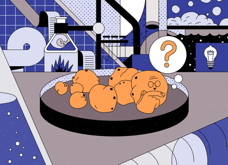
The Epigenetic Secrets Behind Dopamine, Drug Addiction and Depression

This is an interesting take on the "dopamine hypothesis" — that it isn't it's abundant access to the synaptic cleft once the amount is increased that causes habituation, but rather how prevalent it is to tag genes within cells.

By R. DOUGLAS FIELDS
October 27, 2020

New research links serotonin and dopamine not just to addiction and depression, but to the ability to control genes.

As I opened my copy of Science at home one night, an unfamiliar word in the title of a new study caught my eye: dopaminylation. The term refers to the brain chemical dopamine’s ability, in addition to transmitting signals across synapses, to enter a cell’s nucleus and control specific genes. As I read the paper, I realized that it completely upends our understanding of genetics and drug addiction. The intense craving for addictive drugs like alcohol and cocaine may be caused by dopamine controlling genes that alter the brain circuitry underlying addiction. Intriguingly, the results also suggest an answer to why drugs that treat major depression must typically be taken for weeks before they’re effective. I was shocked by the dramatic discovery, but to really understand it, I first had to unlearn some things.

“Half of what you learned in college is wrong,” my biology professor, David Lange, once said. “Problem is, we don’t know which half.” How right he was. I was taught to scoff at Jean-Baptiste Lamarck and his theory that traits acquired through life experience could be passed on to the next generation. The silly traditional example is the mama giraffe stretching her neck to reach food high in trees, resulting in baby giraffes with extra-long necks. Then biologists discovered we really can inherit traits our parents acquired in life, without any change to the DNA sequence of our genes. It’s all thanks to a process called epigenetics — a form of gene expression that can be inherited but isn’t actually part of the genetic code. This is where it turns out that brain chemicals like dopamine play a role.

All genetic information is encoded in the DNA sequence of our genes, and traits are passed on in the random swapping of genes between egg and sperm that sparks a new life. Genetic information and instructions are coded in a sequence of four different molecules (nucleotides abbreviated A, T, G and C) on the long double-helix strand of DNA. The linear code is quite lengthy (about 6 feet long per human cell), so it’s stored neatly wound around protein bobbins, similar to how magnetic tape is wound around spools in cassette tapes.

Inherited genes are activated or inactivated to build a unique individual from a fertilized egg, but cells also constantly turn specific genes on and off throughout life to make the proteins cells need to function. When a gene is activated, special proteins latch onto DNA, read the sequence of letters there and make a disposable copy of that sequence in the form of messenger RNA. The messenger RNA then shuttles the genetic instructions to the cell’s ribosomes, which decipher the code and make the protein specified by the gene.

But none of that works without access to the DNA. By analogy, if the magnetic tape remains tightly wound, you can’t read the information on the cassette. Epigenetics works by unspooling the tape, or not, to control which genetic instructions are carried out. In epigenetic inheritance, the DNA code is not altered, but access to it is.

“I was shocked by the dramatic discovery, but to really understand it, I first had to unlearn some things.”

This is why cells in our body can be so different even though every cell has identical DNA. If the DNA is not unwound from its various spools — proteins called histones — the cell’s machinery can’t read the hidden code. So the genes that would make red blood corpuscles, for example, are shut off in cells that become neurons.

How do cells know which genes to read? The histone spool that a specific gene’s DNA winds around is marked with a specific chemical tag, like a molecular Post-it note. That marker directs other proteins to “roll the tape” and unwind the relevant DNA from that histone (or not to roll it, depending on the tag).

It’s a fascinating process we’re still learning more about, but we never expected that a seemingly unrelated brain chemical might also play a role. Neurotransmitters are specialized molecules that transmit signals between neurons. This chemical signaling between neurons is what enables us to think, learn, experience different moods and, when neurotransmitter signaling goes awry, suffer cognitive difficulties or mental illness.

Serotonin and dopamine are famous examples. Both are monoamines, a class of neurotransmitters involved in psychological illnesses such as depression, anxiety disorders and addiction. Serotonin helps regulate mood, and drugs known as selective serotonin reuptake inhibitors are widely prescribed and effective for treating chronic depression. We think they work by increasing the level of serotonin in the brain, which boosts communication between neurons in the neural circuits controlling mood, motivation, anxiety and reward. That makes sense, sure, but it is curious that it usually takes a month or more before the drug relieves depression.

Dopamine, on the other hand, is the neurotransmitter at work in the brain’s reward circuits; it produces that “gimme-a-high-five!” spurt of euphoria that erupts when we hit a bingo. Nearly all addictive drugs, like cocaine and alcohol, increase dopamine levels, and the chemically induced dopamine reward leads to further drug cravings. A weakened reward circuitry could be a cause of depression, which would help explain why people with depression may self-medicate by taking illicit drugs that boost dopamine.

“In epigenetic inheritance, the DNA code is not altered, but access to it is.”

But (as I found out after reading that dopaminylation paper), research last year led by Ian Maze, a neuroscientist at the Icahn School of Medicine at Mount Sinai, showed that serotonin has another function: It can act as one of those molecular Post-it notes. Specifically, it can bind to a type of histone known as H3, which controls the genes responsible for transforming human stem cells (the forerunner of all kinds of cells) into serotonin neurons. When serotonin binds to the histone, the DNA unwinds, turning on the genes that dictate the development of a stem cell into a serotonin neuron, while turning off other genes by keeping their DNA tightly wound. (So stem cells that never see serotonin turn into other types of cells, since the genetic program to transform them into neurons is not activated.)

That finding inspired Maze’s team to wonder if dopamine might act in a similar way, regulating the genes involved in drug addiction and withdrawal. In the April Science paper that so surprised me, they showed that the same enzyme that attaches serotonin to H3 can also catalyze the attachment of dopamine to H3 — a process, I learned, called dopaminylation.

Together, these results represent a huge change in our understanding of these chemicals. By binding to the H3 histone, serotonin and dopamine can regulate transcription of DNA into RNA and, as a consequence, the synthesis of specific proteins from them. That turns these well-known characters in neuroscience into double agents, acting obviously as neurotransmitters, but also as clandestine masters of epigenetics.

Maze’s team naturally began exploring this new relationship. First they examined postmortem brain tissue from cocaine users. They found a decrease in the amount of dopaminylation of H3 in the cluster of dopamine neurons in a brain region known to be important in addiction: the ventral tegmental area, or VTA.

“That turns these well-known characters in neuroscience into double agents.”

That’s just an intriguing correlation, though, so to find out if cocaine use actually affects dopaminylation of H3 in these neurons, the researchers studied rats before and after they self-administered cocaine for 10 days. Just as in the human cocaine users’ brains, dopaminylation of H3 dropped within the neurons in the rats’ VTA. The researchers also found a rebound effect one month after withdrawing the rats from cocaine, with much higher dopaminylation of H3 found in these neurons than in control animals. That increase might be important in controlling which genes get turned on or off, rewiring the brain’s reward circuitry and causing an intense drug craving during withdrawal.

Ultimately, it looks as though dopaminylation — not just typical dopamine functioning in the brain — may control drug-seeking behavior. Long-term cocaine use modifies neural circuits in the brain’s reward pathway, making a steady intake of the drug necessary for the circuits to operate normally. That requires turning specific genes on and off to make the proteins for those changes, and this is an epigenetic mechanism driven by dopamine acting on H3, not a change in DNA sequence.

To test that hypothesis, the researchers genetically modified H3 histones in rats by replacing the amino acid that dopamine attaches to with a different one it doesn’t react with. This stops dopaminylation from occurring. Withdrawal from cocaine is associated with changes in the readout of hundreds of genes involved in rewiring neural circuits and altering synaptic connections, but in the rats whose dopaminylation was prevented, these changes were suppressed. Moreover, neural impulse firing in VTA neurons was reduced, and they released less dopamine, showing that these genetic changes were indeed affecting the brain’s reward circuit operation. This might account for why people with substance use disorder crave drugs that boost dopamine levels in the brain during withdrawal. Finally, in subsequent tests, the genetically modified rats exhibited much less cocaine-seeking behavior.

“It looks as though dopaminylation … may control drug-seeking behavior.”

To put it plainly, the discovery that monoamine neurotransmitters control epigenetic regulation of genes is transformative for basic science and medicine. These experiments show that the tagging of H3 by dopamine does indeed underlie drug-seeking behavior, by regulating the neural circuits operating in addiction.

And, equally exciting, the implications likely go well beyond addiction, given the crucial role of dopamine and serotonin signaling in other neurological and psychological illnesses. Indeed, Maze told me that his team’s latest research (not yet published) has also found this type of epigenetic marking in the brain tissues of people with major depressive disorder. Perhaps this connection even explains why antidepressant drugs take so long to be effective: If the drugs work by activating this epigenetic process, rather than just supplying the brain’s missing serotonin, it can take days or even weeks before these genetic changes become apparent.

Looking ahead, Maze wonders if such epigenetic changes might also occur in response to other addictive drugs, including heroin, alcohol and nicotine. If so, medicines based on this newly discovered epigenetic process could eventually lead to better treatments for many types of addiction and mental illnesses.

In a commentary accompanying the research, Jean-Antoine Girault of Sorbonne University in Paris made a final, intriguing observation. We know that typical neural impulse firing works by causing a ripple effect of dynamic changes in calcium concentration inside neurons that eventually reach the nucleus. But Girault noted that the enzyme that catalyzes the attachment of dopamine to H3 is also regulated by levels of intracellular calcium. In this way, electrical chatter between neurons is relayed to the nucleus, suggesting that neural activity — driven by a behavior — could attach the dopamine epigenetic marker to genes responsible for drug-seeking behavior. That’s how the experiences one has in life can select which genes get read out, and which do not. Lamarck would be proud.
 
It’s a really interesting article, I can definitely see what he’s getting at. I really do hate rat studies though, especially in regards to drugs as they are definitely manipulated in regards to what the study should show. I would be interested in some more post mortem brain results though.

Definitely something I will be researching and looking into. Great post!
 
Can lab-grown brains become conscious?

27 OCTOBER 2020
Sara Reardon

A handful of experiments are raising questions about whether clumps of cells and disembodied brains could be sentient, and how scientists would know if they were.

d41586-020-02986-y_18498284.jpg

Illustration by Fabio Buonocore

In Alysson Muotri’s laboratory, hundreds of miniature human brains, the size of sesame seeds, float in Petri dishes, sparking with electrical activity.

These tiny structures, known as brain organoids, are grown from human stem cells and have become a familiar fixture in many labs that study the properties of the brain. Muotri, a neuroscientist at the University of California, San Diego (UCSD), has found some unusual ways to deploy his. He has connected organoids to walking robots, modified their genomes with Neanderthal genes, launched them into orbit aboard the International Space Station, and used them as models to develop more human-like artificial-intelligence systems. Like many scientists, Muotri has temporarily pivoted to studying COVID-19, using brain organoids to test how drugs perform against the SARS-CoV-2 coronavirus.

But one experiment has drawn more scrutiny than the others. In August 2019, Muotri’s group published a paper in Cell Stem Cell reporting the creation of human brain organoids that produced coordinated waves of activity, resembling those seen in premature babies. The waves continued for months before the team shut the experiment down.

This type of brain-wide, coordinated electrical activity is one of the properties of a conscious brain. The team’s finding led ethicists and scientists to raise a host of moral and philosophical questions about whether organoids should be allowed to reach this level of advanced development, whether ‘conscious’ organoids might be entitled to special treatment and rights not afforded to other clumps of cells and the possibility that consciousness could be created from scratch.

The idea of bodiless, self-aware brains was already on the minds of many neuroscientists and bioethicists. Just a few months earlier, a team at Yale University in New Haven, Connecticut, announced that it had at least partially restored life to the brains of pigs that had been killed hours earlier. By removing the brains from the pigs’ skulls and infusing them with a chemical cocktail, the researchers revived the neurons’ cellular functions and their ability to transmit electrical signals.

Other experiments, such as efforts to add human neurons to mouse brains, are raising questions, with some scientists and ethicists arguing that these experiments should not be allowed.

The studies have set the stage for a debate between those who want to avoid the creation of consciousness and those who see complex organoids as a means to study devastating human diseases. Muotri and many other neuroscientists think that human brain organoids could be the key to understanding uniquely human conditions such as autism and schizophrenia, which are impossible to study in detail in mouse models. To achieve this goal, Muotri says, he and others might need to deliberately create consciousness.

Researchers are now calling for a set of guidelines, similar to those used in animal research, to guide the humane use of brain organoids and other experiments that could achieve consciousness. In June, the US National Academies of Sciences, Engineering, and Medicine began a study with the aim of outlining the potential legal and ethical issues associated with brain organoids and human–animal chimaeras.

The concerns over lab-grown brains have also highlighted a blind spot: neuroscientists have no agreed way to define and measure consciousness. Without a working definition, ethicists worry that it will be impossible to stop an experiment before it crosses a line.

The current crop of experiments could force the issue. If scientists become convinced that an organoid has gained consciousness, they might need to hurry up and agree on a theory of how that happened, says Anil Seth, a cognitive neuroscientist at the University of Sussex near Brighton, UK. But, he says, if one person’s favoured theory deems the organoid conscious whereas another’s doesn’t, any confidence that consciousness has been attained vanishes. “Confidence largely depends on what theory we believe in. It’s a circularity.”

Sentient states
Creating a conscious system might be a whole lot easier than defining it. Researchers and clinicians define consciousness in many different ways for various purposes, but it is hard to synthesize them into one neat operational definition that could be used to decide on the status of a lab-grown brain.

Physicians generally assess the level of consciousness in patients in a vegetative state on the basis of whether the person blinks or flinches in response to pain or other stimuli. Using electroencephalogram (EEG) readings, for instance, researchers can also measure how the brain responds when it is zapped with an electrical pulse. A conscious brain will display much more complex, unpredictable electrical activity than one that is unconscious, which responds with simple, regular patterns.

But such tests might not adequately probe whether a person lacks consciousness. In brain-imaging studies of people who are in a coma or vegetative state, scientists have shown that unresponsive individuals can display some brain activity reminiscent of consciousness — such as activity in motor areas when asked to think about walking.

d41586-020-02986-y_18520130.jpg

Micrograph of a developing brain organoid.
In developing human brain organoids, pre-neuronal cells (red) turn into neurons (green), which wire up into networks (white).

In any case, standard medical tests for consciousness are difficult to apply to brain cells grown in dishes, or disembodied animal brains. When Muotri suggested that his organoids’ firing patterns were just as complex as those seen in preterm infants, people were unsure what to make of that. Some researchers don’t consider the brain activity in a preterm infant to be complex enough to be classed as conscious. And organoids can’t blink or recoil from a painful stimulus, so they wouldn’t pass the clinical test for consciousness.

By contrast, it’s much more likely that an intact brain from a recently killed pig has the necessary structures for consciousness, as well as wiring created by memories and experiences the animal had while it was alive. “Thinking about a brain that has been filled with all this, it is hard to imagine that brain would be empty,” says Jeantine Lunshof, a philosopher and neuroethicist at Harvard University in Cambridge, Massachusetts. “What they can do in terms of thinking I don’t know, but it’s for sure not zero,” says Lunshof. Bringing a dead brain back to a semblance of life, as the Yale team did, might have the potential to restore a degree of consciousness, although the scientists took pains to avoid this by using chemical blocking agents that prevented brain-wide activity.

Researchers agree that they need to take the possibilities raised by these studies seriously. In October 2019, UCSD held a conference of about a dozen neuroscientists and philosophers, together with students and members of the public, with the intention of establishing and publishing an ethical framework for future experiments. But the paper has been delayed for months, partly because several of the authors could not agree on the basic requirements for consciousness.

Increasingly complex
Almost all scientists and ethicists agree that so far, nobody has created consciousness in the lab. But they are asking themselves what to watch out for, and which theories of consciousness might be most relevant. According to an idea called integrated information theory, for example, consciousness is a product of how densely neuronal networks are connected across the brain. The more neurons that interact with one another, the higher the degree of consciousness — a quantity known as phi. If phi is greater than zero, the organism is considered conscious.

Most animals reach this bar, according to the theory. Christof Koch, president of the Allen Institute for Brain Science in Seattle, Washington, doubts that any existing organoid could achieve this threshold, but concedes that a more advanced one might.

Other competing theories of consciousness require sensory input or coordinated electrical patterns across multiple brain regions. An idea known as global workspace theory, for instance, posits that the brain’s prefrontal cortex functions as a computer, processing sensory inputs and interpreting them to form a sense of being. Because organoids don’t have a prefrontal cortex and can’t receive input, they cannot become conscious. “Without input and output, the neurons may be talking with each other, but that doesn’t necessarily mean anything like human thought,” says Madeline Lancaster, a developmental biologist at the University of Cambridge, UK.

Connecting organoids to organs, however, could be a fairly simple task. In 2019, Lancaster’s team grew human brain organoids next to a mouse spinal column and back muscle. When nerves from the human organoid connected with the spinal column, the muscles began to spontaneously contract.

d41586-020-02986-y_18520132.jpg

Developmental biologist Madeline Lancaster works with organoids to study brain organization and disorders in her lab at the University of Cambridge, UK.

Most organoids are built to reproduce only one portion of the brain — the cortex. But if they develop long enough and with the right kinds of growth factor, human stem cells spontaneously recreate many different parts of the brain, which then begin coordinating their electrical activity. In a study published in 2017, molecular biologist Paola Arlotta at Harvard University coaxed stem cells to develop into brain organoids composed of many different cell types, including light-sensitive cells like those found in the retina5. When exposed to light, neurons in the organoids began firing. But the fact that these cells were active doesn’t mean the organoids could see and process visual information, Arlotta says. It simply means that they could form the necessary circuits.

Arlotta and Lancaster think their organoids are too primitive to be conscious, because they lack the anatomical structures necessary to create complex EEG patterns. Still, Lancaster admits that for advanced organoids, it depends on the definition. “If you thought a fly was conscious, it’s conceivable that an organoid could be,” she says.

However, Lancaster and most other researchers think that something like a revitalized pig brain would be much more likely to achieve consciousness than an organoid. The team that did the work on the pig brains, led by neuroscientist Nenad Sestan, was trying to find new ways to revitalize organs, not to create consciousness. The researchers were able to get individual neurons or groups to fire and were careful to try and avoid the creation of widespread brain waves. Still, when Sestan’s team saw what looked like coordinated EEG activity in one of the brains, they immediately halted the project. Even after a neurology specialist confirmed that the pattern was not consistent with consciousness, the group anaesthetized the brains as a precautionary measure.

Sestan also contacted the US National Institutes of Health (NIH) for guidance on how to proceed. The agency’s neuroethics panel, including Lunshof and Insoo Hyun, a bioethicist at Case Western University in Cleveland, Ohio, assessed the work and agreed that Sestan should continue to anaesthetize the brains. But the panel hasn’t settled on more general regulations, and doesn’t routinely require a bioethics assessment for organoid proposals because its members think that consciousness is unlikely to arise. The NIH hasn’t arrived at a definition of consciousness, either. “It’s so flexible, everyone claims their own meaning,” Hyun says. “If it’s not clear we’re talking about the same thing, it’s a big problem for discourse.”

d41586-020-02986-y_18520128.jpg

Neuroscientist Nenad Sestan used the BrainEx platform to restore neural activity in disembodied pig brains.

Fuzzy definitions
Some think it is futile to even try to identify consciousness in any sort of lab-maintained brain. “It’s just impossible to say meaningful things about what these bunches of brain cells could think or perceive, given we don’t understand consciousness,” says Steven Laureys, a neurologist at the University of Liège in Belgium, who pioneered some of the imaging-based measures of consciousness in people in a vegetative state. “We shouldn’t be too arrogant.” Further research should proceed very carefully, he says.

Laureys and others point out that the experience of an organoid is likely to be very different from that of a preterm infant, an adult human or a pig, and not directly comparable. Furthermore, the structures in an organoid might be too small to have their activity measured accurately, and similarities between the EEG patterns in organoids and preterm baby brains could be coincidental. Other scientists who work on brain organoids agree with Laureys that the question of whether a system is conscious could be unanswerable. Many avoid the idea entirely. “I don’t know why we would try to ask that question, because this system is not the human brain,” says Sergiu Pasça, a neuroscientist at Stanford University in California. “They’re made out of neurons, neurons have electrical activity, but we have to think carefully about how to compare them.”

Muotri wants his organoid systems to be comparable, in at least some ways, with human brains, so that he can study human disorders and find treatments. His motivation is personal: his 14-year-old son has epilepsy and autism. “He struggles hard in life,” Muotri says. Brain organoids are a promising avenue, because they recapitulate the earliest stages of brain wiring, which are impossible to study as a human embryo develops. But studying human brain disorders without a fully functioning brain, he says, is like studying a pancreas that doesn’t produce insulin. “To get there, I need a brain organoid model that really resembles a human brain. I might need an organoid that becomes conscious.”

Muotri says he is agnostic about which definition to use to decide whether an organoid reaches consciousness. At some point, he says, organoids might even be able to help researchers answer questions about how brains produce conscious states. For instance, mathematician Gabriel Silva at UCSD is studying neural activity in Muotri’s organoids to develop an algorithm that describes how the brain generates consciousness6. The goal of his project, which is partially funded by Microsoft, is to create an artificial system that works like human consciousness.

At the moment, there are no regulations in the United States or in Europe that would stop a researcher from creating consciousness. The National academies panel plans to release a report early next year, outlining the latest research and making a judgement on whether regulations are needed. Members plan to weigh in on questions such as whether to obtain people’s consent to develop their cells into brain organoids, and how to study and dispose of organoids humanely. The International Society for Stem Cell Research is also working on organoid guidelines, but is not addressing consciousness because it doesn’t think the science is there yet.

Hyun says that the NIH neuroethics panel has not yet seen any proposals to create complex, conscious organoids that would necessitate new guidelines. And Muotri says he doesn’t know of anyone else deliberately trying to create conscious organoids either, although a sufficiently complex organoid could, by some definitions, reach that status accidentally.

Still, Muotri and others say they would welcome some guidelines. These could include requiring scientists to justify the number of human brain organoids they use, to use them only for research that cannot be done in any other way, to restrict the amount of pain that can be inflicted on them, and to dispose of them humanely.

Having such advice in place ahead of time would help researchers weigh up the costs and benefits of creating conscious entities. And many researchers stress that such experiments have the potential to yield important insights. “There are truly conscious people out there with neurological disorders with no treatments,” Lancaster says. “If we did stop all of this research because of the philosophical thought experiment,” she adds, “that would be very detrimental to actual human beings who do need some new treatment.”

Treatments could still, however, be tested in brain organoids made using mouse stem cells , or in regular animal models. Such experiments could also inform discussions about the ethical use of human organoids. For instance, Hyun would like to see researchers compare the EEG patterns of mouse brain organoids with those of living mice, which might indicate how well human organoids recapitulate the human brain.

For his part, Muotri sees little difference between working on a human organoid or a lab mouse. “We work with animal models that are conscious and there are no problems,” he says. “We need to move forward and if it turns out they become conscious, to be honest I don’t see it as a big deal.”

Nature 586, 658-661 (2020)

doi: https://doi.org/10.1038/d41586-020-02986-y
 
2-(4-isobutylphenyl)propionatesodium.png


MOTRIN/ADVIL (ibuprofen)
2-(4-isobutylphenyl)propionate sodium

Teach A Man To Fish, And He Won't Go Hungry. Give A Man Some Stimulants And Unlimited Internet Access, And He May Not Bother You Again For Days, Weeks, Or Even Months.
 
Last edited:
It’s a really interesting article, …

Definitely something I will be researching and looking into. Great post!
I think so as well. There's always odd discrepancies between dopaminergic drugs and their ability to cause addictive response. There's been attempts to separate the DA transporter ligands between "closed–to–out" versus "open–to–out" confirmations as indicators but those don't seem to be the difference.

Also direct receptor agonists of the dopamine receptor don't seem to be reinforcing in the same way, it has to do with dopamine released from, or impeded re-entry to, the transporter.

I know there is CART (cocaine–amphetamine regulated transcript) that might have to do with addiction. I wonder how that relates to the above considerations of "dopaminylation"?
 
A peptide with stimulant effects. I can't remember whether I've posted this in some thread earlier.

Terence K. Y. Lai, Ping Su, Hailong Zhang & Fang Liu, "Development of a peptide targeting dopamine transporter to improve ADHD-like deficits", Molecular Brain volume 11, Article number: 66 (2018)

Attention-deficit hyperactivity disorder (ADHD) is a neurocognitive disorder characterized by hyperactivity, inattention, working memory deficits and impulsivity. Its worldwide prevalence is estimated to be 3–5% in children and adolescents. The mainstay treatment for ADHD is stimulant medications (e.g. methylphenidate), which increase synaptic dopamine by directly blocking dopamine transporter (DAT). Although these pharmacological agents are effective, they are often associated with various side effects including risks for future substance use disorders in ADHD patients. Here, we investigated an interaction between DAT and dopamine D2 receptor (D2R) as a novel target to develop potential therapeutics for the treatment of ADHD by using an interfering peptide (TAT-DATNT) to dissociate this protein complex. We found that TAT-DATNT promotes locomotor behavior in Sprague-Dawley rats. Furthermore, using in vivo microdialysis and high-performance liquid chromatography, we found that the disruption of D2R-DAT elevates extracellular dopamine level. More importantly, the interfering peptide, TAT-DATNT, attenuates hyperactivity and improves spontaneous alternation behavior in spontaneously hypertensive rats (SHR) ------ a common animal model of ADHD. This work presents a different means (i.e. other than direct blockade by a DAT inhibitor) to regulate the activity of DAT and dopaminergic neurotransmission, and a potential target site for future development of ADHD treatments.
 
Not that its recent, but apparently lanthanum ion acts as an analgesic via a mu-opioid based mechanism.[ref] The caveat is it needs to be directly piped into the brain, past the BBB, to have such an effect. There is the usual problem (very generally speaking, charged compounds/ions of any sort need some sort of pump system to move across the BBB's lipid membranes) but also a unique reactivity/precipitation issue... lanthanum phosphate has incredibly low solubility. In fact lanthanym carbonate is prescribed as an oral phosphate binder for people with kidney failure and hyperphospnatemia. Lanthanum chloride solution is used to condition the water in swimmig pools as well, by sequestering phosphate as something insoluble, a major nutrient for algae/scum is greatly reduced, meaning less growth, and less need for large amounts of chlorinating agents.
 
Not that its recent, but apparently lanthanum ion acts as an analgesic via a mu-opioid based mechanism.[ref] The caveat is it needs to be directly piped into the brain, past the BBB, to have such an effect. There is the usual problem (very generally speaking, charged compounds/ions of any sort need some sort of pump system to move across the BBB's lipid membranes) but also a unique reactivity/precipitation issue... lanthanum phosphate has incredibly low solubility. In fact lanthanym carbonate is prescribed as an oral phosphate binder for people with kidney failure and hyperphospnatemia. Lanthanum chloride solution is used to condition the water in swimmig pools as well, by sequestering phosphate as something insoluble, a major nutrient for algae/scum is greatly reduced, meaning less growth, and less need for large amounts of chlorinating agents.

Then have to use trimethyllanthanum or some BBB penetrating nanoparticle formulation? Probably not a good idea if it can be neurotoxic like mercury.

 
Screenshot-20201110-195424-2.png

S'il-Te-Plait, or please in French, aka STP, aka DOM

Funny thing is DOM means stupid in Dutch ..

FREE DOM STP!!
 
So if La binds phosphate to form insoluble precipitate, and the brain has at least some phosphate hanging around (from hydrolysis of nucleoside polyphosphates, etc).... seems like there is a possibility for things to go fucky there. deposits of LaPO4 crystallizing out in the subarachnoid space & loss of available phosphate reserves are two obvious possibilities as I see it

trimethyllanthanum has 1 paper on it and the title descries it as "elusive". probably not a drug candidate.

pumps that dose drugs to the CSF are quite doable - baclofen is one such drug dosed direct to CSF (for spasticity... I may have to get such a pump in future, depending on if my spasticity ggets worse :\)
 
Zinc ion concentration affects the dopamine transporter, but it's not clear whether this can be done with a non-toxic amount of zinc salts.


These results are the first to describe the involvement of synaptic Zn2+ in regulating DA neurotransmission as well as physiological and behavioral effects to cocaine and have important implications for both general DA-dependent behaviors and especially for the prevention and treatment of cocaine addiction. Specifically, our findings suggest that dietary Zn2+ intake, and potentially, impaired Zn2+ absorption or excretion mechanisms, are implicated in cocaine reward, seeking, and relapse.
 
Does baclofen really have that low bioavailability when taken orally? I coulda swore my friend have a bottle of pills of baclofen but i could be wrong. A pump makes sense I just never realized that they would do it for that drug
 
1-(2-carbomethoxyethyl)-2-methyl-3-phenyl-1-aza-4-oxocyclopentane.png


MANTEQUILLA
1-(2-carbomethoxyethyl)-2-methyl-3-phenyl-1-aza-4-oxocyclopentane

1-(4-carbomethoxyphenyl)-1-oxo-2-methylaminopropane.png


STANLEY STEEMER
1-(4-carbomethoxyphenyl)-1-oxo-2-methylaminopropane
 
Last edited:
Status
Not open for further replies.
Top