• N&PD Moderators: Skorpio | thegreenhand

News NEUROSCIENCE | Demystifying Psychedelics ->

brain-connections.jpg


Study uncovers psilocybin-induced changes in brain connectivity*

by Eric Dolan | PsyPost | Jan 20 2020

New research published in Biological Psychology sheds light on the neurophysiological underpinnings of the psychedelic experience. The study provides new details about how psilocybin — the active component in “magic” mushrooms — changes the communication patterns between regions of the brain.

Psychedelics are currently being investigated for the treatment of psychiatric disorders. However, it is still unclear how they change brain activity and connectivity to induce their unique effects,” explained study author Katrin Preller of the University of Zurich and Yale University.

We therefore conducted a study to investigate the time-dependent effects of psilocybin on brain connectivity and the association between changes in brain connectivity and receptor pharmacology.”

In the study, 23 healthy human participants underwent MRI brain scanning 20 minutes, 40 minutes, and 70 minutes after receiving either psilocybin or a placebo. The researchers observed reduced connectivity between brain areas involved in planning and decision-making but increased connectivity between areas involved in sensation and movement while the participants were under the influence of the psychedelic drug.

Preller and her colleagues conducted a similar study on LSD, and obtained “virtually identical” results.

Psilocybin – similar to LSD – induced a pattern of brain connectivity that is characterized by increased synchronization of sensory brain regions and decreased connectivity of associative networks,” she told PsyPost.

Increased sensory processing but altered integration of this sensory information may therefore underlie the psychedelic state and explain the symptoms induced by psilocybin. Furthermore, this pattern of changes in connectivity was closely associated with spatial expression of the serotonin 2A and 1A receptors – pinpointing these receptors as critical for the effects of psilocybin.”

Another drug called ketanserin, a serotonin 2A antagonist, prevented the effects of both psilocybin and LSD, suggesting that changes in brain connectivity caused by these psychedelic drugs are linked to stimulation of the serotonin 2A receptor.

In this study we investigated different time points from administration to subjective peak effects. Future studies need to investigate how psilocybin impacts brain connectivity during a later phase and post-acutely,” Preller noted.

*From the article here :
 
768px-Serotonin.svg.png


How serotonin modulates behavior

MIT | Neuroscience News

A new NIH funded study will use C. elegans to discover the secrets behind serotonin’s influence on behavior.

In popular experience the story of how serotonin modulates the brain might seem simple: pop an antidepressant, serotonin levels go up, mood improves. But neuroscientists acknowledge how little they know about how the neurotransmitter affects circuits and behavior in the incredibly complex human brain. To reveal the basics of how serotonin really works, scientists at MIT’s Picower Institute for Learning and Memory, funded by a new $1.16 million, four-year grant from the National Institutes of Health, will employ a far simpler model: the nematode worm C. elegans.

Though it is tiny, transparent and sports a nervous system with only 302 neurons, C. elegans is a powerful system for studying how serotonin modulates brain states, said the grant’s principal investigator Steven Flavell, Lister Brothers Career Development Professor in the Picower Institute and assistant professor in the Department of Brain and Cognitive Sciences. C. elegans and mammals share much of the same basic molecular machinery for emitting and receiving serotonin. But unlike in a mammal, all the neurons and their connectivity has been precisely mapped out in C. elegans and scientists can exert powerful genetic control over each cell, including those that express each of the worm’s five distinct serotonin receptors. Moreover, Flavell’s lab has developed an innovative imaging system that can reliably image the calcium activity of virtually every neuron in real time, even as a worm freely slithers and wriggles around in response to experimental manipulations.

c-elegans-QBI-worm-nerve-injury.jpg

The Flavell lab has developed the ability to image the activity of virtually every C. elegans neuron in real-time,
even in a freely moving and behaving worm.


Essentially, Flavell’s team can take nearly full control of the worm’s serotonergic system and simultaneously observe the response of virtually every neuron in the whole brain. This gives them needed capabilities that aren’t available in mammals to figure out how varying patterns of serotonin release can stimulate distinct receptors (or combinations of them) on a multitude of neurons in a variety of circuits to modulate different behaviors.

Focus on feeding

“By taking advantage of a well-defined paradigm for serotonergic function and cutting-edge imaging technologies, we are well positioned to examine how patterned serotonin release activates distinct receptor types throughout a circuit to change the large-scale activity patterns that give rise to behavior,” Flavell said.

In December 2018, Flavell’s lab published a paper in Cell showing how a particular C. elegans neuron called NSM senses when a worm has started feeding on bacteria and signals other neurons via serotonin to slow the worm down to savor the meal. Since then, his lab has studied how manipulating NSM’s serotonin release patterns affects the worm’s slowing behavior and has begun to map out which serotonin receptors on which neurons play a role in those effects, for instance by genetically knocking out individual receptors, or combinations of receptors, to see what changes.

With the new grant, the lab will expand on these studies and go well beyond to systematically achieve three aims: mapping out which combinations of serotonin receptors mediate serotonin’s effect on behavior and identifying the exact neurons where they function; analyzing how serotonin alters whole-brain activity; and determining how serotonin-responsive circuits and whole brain activity differs when worms must balance aversive stimuli with appetitive food cues. While the first two sets of experiments will elucidate how the brain deploys serotonin to modulate behavior, the third aim will show how those dynamics change in more complex environments.

Surprisingly, these fundamental issues related to serotonin signaling remain poorly understood,” Flavell said. “Resolving them would greatly enhance our understanding of the serotonergic system.”

 
lsd-against-depression@2x.png


Ketamine found to reverse neural changes that underlie depression-related behaviors*

Neuroscience News | April 11, 2019

The formation of prefrontal cortex dendritic spine formation sustains the remission of depressive related symptoms and behaviors following ketamine treatment by restoring lost spines.

Researchers have identified ketamine-induced brain-related changes that are responsible for maintaining the remission of behaviors related to depression in mice — findings that may help researchers develop interventions that promote lasting remission of depression in humans.

Major depression is one of the most common mental disorders in the United States, with approximately 17.3 million adults experienced a major depressive episode in 2017. However, many of the neural changes underlying the transitions between active depression, remission, and depression re-occurrence remain unknown. Ketamine, a fast-acting antidepressant which relieves depressive symptoms in hours instead of weeks or longer, provides an opportunity for researchers to investigate the short- and long-term biological changes underlying these transitions.

“Ketamine is a potentially transformative treatment for depression, but one of the major challenges associated with this drug is sustaining recovery after the initial treatment,” said study author Conor Liston, M.D., Ph.D., of Weill Cornell Medicine, New York City.

To understand mechanisms underlying the transition from active depression to remission in humans, the researchers examined behaviors related to depression in mice. Researchers took high-resolution images of dendritic spines in the prefrontal cortex of mice before and after they experienced a stressor. Dendritic spines are protrusions in the part of neurons that receive communication input from other neurons. The researchers found that mice displaying behaviors related to depression had increased elimination of, and decreased the formation of, dendritic spines in their prefrontal cortex compared with mice not exposed to a stressor. This finding replicates prior studies linking the emergence of behaviors related to depression in mice with dendritic spine loss.

In addition to the effects on dendritic spines, stress reduced the functional connectivity and simultaneous activity of neurons in the prefrontal cortex of mice. This reduction in connectivity and activity was associated with behaviors related to depression in response to stressors. Liston’s group then found that ketamine treatment rapidly restored functional connectivity and ensemble activity of neurons and eliminated behaviors related to depression. Twenty-four hours after receiving a single dose of ketamine, mice exposed to stress showed a reversal of behaviors related to depression and an increase in dendritic spine formation when compared to stressed mice that had not received ketamine. These new dendritic spines were functional, creating working connections with other neurons.

The researchers found that while behavioral changes and changes in neural activity in mice happened quickly (three hours after ketamine treatment), dendritic spine formation happened more slowly (12-24 after hours after ketamine treatment). While further research is needed, the authors suggest these findings might indicate that dendritic spine regrowth may be a consequence of ketamine-induced rescue of prefrontal cortex circuit activity.

Although dendritic spines were not found to underly the fast-acting effects of ketamine on behaviors related to depression in mice, they were found to play an important role in maintaining the remission of those behaviors. Using a new technology developed by Haruo Kasai, Ph.D., and Haruhiko Bito, Ph.D., collaborators at the University of Tokyo, the researchers found that selectively deleting these newly formed dendritic spines led to the re-emergence of behaviors related to depression.

“Ketamine is the first new anti-depressant medication with a novel mechanism of action since the 1980s. Its ability to rapidly decrease suicidal thoughts is already a fundamental breakthrough,” said Janine Simmons, M.D., Ph.D., chief of the NIMH Social and Affective Neuroscience Program. “Additional insights into ketamine’s longer-term effects on brain circuits could guide future advances in the management of mood disorders.”

*From the article here :
 
img.png


Non-psychedelic ibogaine compound TBG may revolutionize psychiatry

by Rebecca Coffey

Photographic and video evidence shows that a single dose of the non-psychedelic ibogaine compound TBG can repair stress-damaged neurons and brain circuitry in mice — all while reducing their anxious and depressive behaviors and curbing their addictions.

The compound is tabernanthalog (TBG), synthesized from the psychedelic ibogaine by UC Davis chemical neuroscientist David Olson. Ibogaine is an extract of the naturally-growing African plant, iboga. A drug for medicinal use in humans is in development; clinical trials are anticipated.

An article in the May 25, 2021, issue of Molecular Psychiatry depicts the work by Olson, UC Santa Cruz molecular biologist Yi Zuo, and other collaborators, and also spells out the rationale behind modeling TBG on ibogaine’s key structural elements. While ibogaine has demonstrated anti-anxiety and anti-depression effects and is used in some drug and alcohol rehab clinics around the world, it will probably never be licensed for use in the United States. This is because, at therapeutic doses, it can cause dangerous heart arrhythmias and fatal heart attacks. At high doses, it is neurotoxic. Enter Olson’s TBG. His hope for the new synthetic compound is that, in humans, it will prove to have all of ibogaine’s benefits and none of its toxicity or hallucinogenic potential. While the compound is still untested in humans, Zuo and Olson’s Molecular Psychiatry paper about it shows that sort of promise.

Anxiety, depression, addiction, and the psychedelic connection

Over the past several years, neuroscientists and mental health professionals have become intrigued with the potential of psychedelics to open people’s minds to healthy change and to combat mental illness. Ibogaine’s successes —as well as the successes of psilocybin and ketamine — suggest that psychedelics may one day be recognized as powerful medicines. Even so, hallucinations themselves can be dangerous, at least for people with a history of psychosis or mania. For other people, the prospect of hallucinating is daunting. Realizing all of this, in his lab at UC Davis Olson wondered whether the psychedelic effects of drugs like psilocybin and ketamine are necessary contributors to their efficacy as psychiatric medicines. Would treatment work just as well without the drugs’ “trippy” aspects?

TBG does not induce hallucinations, or at least that’s the impression that Olson, Zuo, and colleagues have drawn from their studies with mice. Typically, mice given a hallucinogen develop a characteristic side-to-side head movement referred to as a “head twitch response.” Mice given TBG don’t twitch their heads.

TBG reverses stress behaviors in mice

The new Molecular Psychiatry article was preceded by a paper published in January 2021 in Nature. Part of the purpose of that earlier study was to investigate whether TBG is safer than ibogaine. Working with young zebrafish, Olson and colleagues found that ibogaine caused disabling changes in shape, while TBG did not. The study was also designed to determine whether TBG, like ibogaine, is useful in treating mental illness. They found that, in mice, it reduced alcohol- and heroin-seeking behavior and it reversed post-stress behaviors associated with depression.

Five months after the publication of that Nature article, the study described in Molecular Psychiatry demonstrated anti-anxiety effects of TBG and revealed the breakthrough neuronal and brain circuitry repair benefits of the compound.

In the newer study, Zuo, Olson, and colleagues worked with two-month-old lab mice, exposing them for several days to unpredictable mild stress such as illumination changes, forced swimming, and extra handling. At the end of the period of stressful days, the mice were put through challenges meant to reveal whether stress had changed behavior, sensory processing abilities, or cognitive flexibility.

Indeed, it had. The consecutive days of stress had made mice reluctant to move about in high places and had reduced their ability to learn novel stimulus-response associations. When stressed-out mice rubbed their whiskers on textures that were new to them, the level of excitation in the sensory processing area of the brain was unusually low.

Then the mice were given TBG. Within one day, the behavioral, learning, and sensory processing changes wrought by stress were reversed, and no toxic effects on heart or brain were evident.

Looking inside the living mice’s brains, the researchers also saw that damage to neurons’ dendrites and to neuronal communication had been righted. (Dendrites are the spined branches extending out of neurons. Electrical impulses flow through them into and out of the neuronal network.)

Looking inside a living mouse’s brain

One month before the experiment began, the researchers had created a single “cranial window” at the top of each mouse’s skull. Having anesthetized the mice, they drilled a 2.3 mm diameter hole into the head, and then replaced the missing bone and tissue with glass glued in place with dental cement. This is a common procedure in labs studying neurology in live rodents. It’s considered humane because skulls have few nerves and, for any given rodent, the large mass of remaining bone plus the tiny cranial window create a stable skull.

Using high-powered microscopes and special lights, Olson’s team shot photos and video through the cranial windows. They saw evidence of damaged dendritic spines, which they speculated lay at the root of behavior, sensory, and cognitive deficits. They also noted a lack of calcium bursts within the neural networks. This indicated that electrical communication among neurons had ebbed. After giving mice a single dose of TBG, they saw improvements in spine growth and density. Calcium bursts indicated that communication was back at par.

The possibility of a permanent cure

How does TBG repair neurons and neural circuits? Could it be that psilocybin, ketamine, and other psychedelics can also facilitate brain repair?

In a Zoom interview, Olson explained his original interest in creating a synthetic counterpart to ibogaine, and also spoke of what the future might hold.

"I’d learned from the work of others that sometimes a single administration of a psychedelic substance can produce long-lasting effects. For example, studies conducted by other researchers had suggested that an initial dose of ibogaine can keep heroin addicts drug free for up to six months."

Olson also spoke of wondering whether stress or addiction had produced neuronal changes that the drugs were reversing.

“The idea that a substance can quickly rescue structural and functional deficits in neural networks is a new concept in pharmacological research and intervention,” he explained. “Such substances are called ‘psychoplastogens.’ Ketamine, ibogaine, and psilocybin are all psychoplastogens. The non-hallucinogenic analog TBG is also, but it repairs damage without creating hallucinations or injuring hearts.”

The concept of psychoplastogenesis seems beyond revolutionary. For, if some drugs are capable of truly repairing what has gone wrong in a brain, they may not just treat mental illness. They may cure it.

In conversation, Olson speculated that safe, non-hallucinogenic psychoplastogens might even one day be approved for weekly, monthly, or annual home use.

“In which case,” he said, “people could keep ’cures’ in their medicine cabinets. Psychiatry would never be the same.”

To date, there are no data suggesting efficacy or safety for TBG in humans. Olson’s research has led to the founding of Delix Therapeutics, which is advancing TBG through preclinical and clinical development.​
 
Last edited:
tingri-everest-base-camp-trek.jpg


Neuronal and neurotransmitter systems dynamic coupling explains the effects of psilocybin

UPF Barcelona | Neuroscience News

Computational simulations reveal the integration of both neuronal and neurotransmitter systems at a whole-brain level is vital to fully understand the effects of psilocybin on brain activity.

Interest in research into the effects of psilocybin has increased significantly in recent years due to its promising therapeutic effects in neuropsychiatric disorders such as depression, anxiety and addiction.

It is widely accepted that the brain’s powerful ability to adapt its behaviour flexibly is largely due to the neurotransmitter system. Psilocybin is the substance responsible for the psychoactive effect caused by nearly 200 species of fungi. This substance has a particular affinity for serotonin receptors found primarily in the brain, but also in other parts of the body such as the stomach. Upon consumption, psilocybin selectively changes the function of serotonin receptors, thus generating an altered state of consciousness characterized by the dissolution of the ego, changes in the quality and attribution of thoughts, impaired visual and sensory perception, and greater awareness of repressed memories.

An international team of researchers has developed a computational biophysical model of the entire brain that integrates real data about its anatomical structural connectivity, the functional dynamics of neurons and a map which shows the concentration of serotonin receptors in various brain regions.

This international research, published on 13 April in the journal Proceedings of the National Academy of Sciences, was led by Gustavo Deco, ICREA research professor and director of the Center for Brain and Cognition at the Department of Information and Communication Technologies (DTIC) at UPF, with the participation of Josephine Cruzat, a member of his team, Morten Kringelbach, a neuroscientist at the University of Oxford (UK), and other scientists from research centres in Germany, Denmark, the US, Portugal and the UK.

This work has shown that the new dynamic model of the whole brain is capable of addressing one of the major challenges in neuroscience: to explain the paradoxical flexibility of brain function despite having a fixed anatomical structure

Study of psilocybin’s mechanisms of action in humans

This theoretical and experimental study modelled the interaction between neuronal and neurotransmitter systems throughout the brain to explain how psilocybin affects brain activity. To study the drug’s mechanisms of action, Morten Kringelbach, Josephine Cruzat and Gustavo Deco analysed data from functional magnetic resonance imaging (fMRI) in 16 healthy subjects. In the experiment, participants were given small doses of psilocybin intravenously or saline solution (placebo effect) while in the scanner, to measure their brain function. The experimental part of the study was carried out at Imperial College London under the direction of Robin Carthart-Harris, co-author of the study.

The functional data acquired under the conditions of placebo and psilocybin were then combined with data from diffusion magnetic resonance imaging (dMRI) capturing brain structure by describing the anatomical connections between the different brain regions; and with data on the density of serotonin receptors estimated by positron emission tomography (PET).

The integration of neuronal and neurotransmitter systems at whole-brain level is important to fully explain the effects of psilocybin on brain activity.

psilocybin-neurotransmission-neuroscienews.jpg

Interest in research into the effects of psilocybin has increased significantly in recent years due to its
promising therapeutic effects in neuropsychiatric disorders such as depression, anxiety and addiction.


As explained by by Deco and Cruzat, co-authors of the work, “the computational simulations performed in this study revealed that the integration of neuronal and neurotransmitter systems at the whole-brain level is important to fully explain the effects of psilocybin on brain activity, specifically through the stimulation of serotonin receptors 5 -HT2A, involved in psychoactive modulation.”

Overall, the remarkable flexibility of human brain function depends crucially on the bidirectional dynamic participation of neuronal and neurotransmission systems. According to the authors, this new approach provides a better and deeper understanding of the effects of psilocybin on the brain and may lead to the development of new treatments for neuropsychiatric diseases such as depression, anxiety and addiction.

 
shutterstock_580332481.jpg


Brain morphology may help explain differing psilocybin experiences

by Barb Bauer | Psychedelic Science Review | 17 April 2020

Understanding brain morphology differences may prove essential for creating effective psychedelic drug formulations.

Scientists have some understanding of the receptor mechanisms involved in the psychedelic experience. The current theory is that the binding of an agonist or partial agonist to the serotonin 5-HT2A receptor elicits a psychedelic effect via signaling pathways. Some experts hypothesize that the 5-HT2C receptor may also be involved.

The general effects of psilocybin (actually, its active metabolite psilocin) are documented, and they cover a range of emotional, cognitive, and perceptual changes. Set and setting (aka context) is known to play a critical role in the subjective effects of psychedelics. Despite this understanding, there is still considerable unexplained variability in the individual experience from ingesting psilocybin.

Now, research is showing that the morphology of certain parts of the brain may be a predictor of the magnitude of the psychedelic experience from psilocybin.

In February 2020, a research team, including Franz Vollenweider, published a randomized, double-blind, placebo-controlled study in Biomedicines. In this study, the authors built on previous research supporting the hypothesis that “Individual brain morphology measures can be used to predict various pharmacological challenges and behavior.” Specifically, the team measured the thickness of areas in the cingulate cortex of people’s brains and compared it to their subjective ratings from ingesting psilocybin.

Study design

The study recruited 55 people from local universities (33 men and 22 women) with a mean age of 25 years. They were randomly divided into three groups. The low and high dose groups received 0.16 mg/kg and 0.215 mg/kg psilocybin, respectively, in two sessions at least ten days apart. The control group received maltose as a placebo.

One hour after each psilocybin dose, the participants underwent a magnetic resonance imaging (MRI) of their brain. The areas of the brain the researchers measured the thickness of were in the right hemisphere. They were the rostral anterior cingulate, caudal anterior cingulate, and the posterior cingulate (Figure 1). The authors stated that they chose these areas because they have a high expression of 5-HT2A receptors compared to other regions of the limbic system. For the control area, they chose a part of the right hemisphere called the post central, where there is low expression of 5-HT2A receptors.

Lewis-et-al.-Figure-1-2.jpg

Figure 1: Schematic showing the brain regions used in the analyses. (A) Right hemisphere cingulate cortex region used in primary analyses. Dark purple = rostral anterior cingulate; medium purple = caudal anterior cingulate; light purple = posterior cingulate. (B) Red = right hemisphere post central region used as the control region analysis.

The Five-Dimensional Altered State of Consciousness (5D-ASC) questionnaire was administered to each participant six hours after each dose. Of the 11 sub-scales measured in the 5D-ASC, the researchers focused on the data from four of them: experience of unity, spiritual experience, blissful state, and insightfulness.

Results

The data revealed that there were no significant differences in the four 5D-ASC sub-scale ratings based on dose. The authors theorized this might be because the doses reflected the high and low ends of the medium dose range for psilocybin based on the literature. They recognized two doses being a strength of the study, but also a limitation because they were not sufficiently different to detect any dose effects. Therefore, the authors then looked at all the data together, regardless of the dose.

Analyzing the data in this way, the researchers observed that the thickness of the right hemisphere rostral anterior cingulate predicted ratings for the four 5D-ASC sub-scales (i.e., the thicker the region, the higher the person’s subjective experience ratings). Conversely, the thickness of the caudal and posterior cingulate areas was not predictive of the sub-scale ratings. The authors noted that these results correlate with previous studies in the literature.

The authors stated, “This is the first study to evaluate brain morphology as a predictor of the emotional subjective experience of psilocybin in healthy controls.” They hypothesized from the data that,

"...morphology metrics such as cortical thickness may reflect differences in brain substrates that mediate drug effects. Investigating the relationship between psilocybin modulation and cingulate structure could provide insight into mechanisms underlying psilocybin’s profound emotional effects and provides a theoretical framework for brain-based predictors of 5HT2A psychedelic modulation."

 
psychedelics-promote-plasticity_orig.png


Psychedelics Promote Structural and Functional Neural Plasticity

Calvin Ly, Alexandra C. Greb, Lindsay P. Cameron, Jonathan M. Wong, Eden V. Barragan, Paige C. Wilson, Kyle F. Burbach, Sina Soltanzadeh Zarandi, Alexander Sood, Michael R. Paddy, Whitney C. Duim, Megan Y. Dennis, A. Kimberley McAllister, Kassandra M. Ori-McKenney, John A. Gray, David E. Olson

Atrophy of neurons in the prefrontal cortex (PFC) plays a key role in the pathophysiology of depression and related disorders. The ability to promote both structural and functional plasticity in the PFC has been hypothesized to underlie the fast-acting antidepressant properties of the dissociative anesthetic ketamine. Here, we report that, like ketamine, serotonergic psychedelics are capable of robustly increasing neuritogenesis and/or spinogenesis both in vitro and in vivo. These changes in neuronal structure are accompanied by increased synapse number and function, as measured by fluorescence microscopy and electrophysiology. The structural changes induced by psychedelics appear to result from stimulation of the TrkB, mTOR, and 5-HT2A signaling pathways and could possibly explain the clinical effectiveness of these compounds. Our results underscore the therapeutic potential of psychedelics and, importantly, identify several lead scaffolds for medicinal chemistry efforts focused on developing plasticity-promoting compounds as safe, effective, and fast-acting treatments for depression and related disorders.

Discussion

Classical serotonergic psychedelics are known to cause changes in mood and brain function that persist long after the acute effects of the drugs have subsided. Moreover, several psychedelics elevate glutamate levels in the cortex and increase gene expression in vivo of the neurotrophin BDNF as well as immediate-early genes associated with plasticity. This indirect evidence has led to the reasonable hypothesis that psychedelics promote structural and functional neural plasticity, although this assumption had never been rigorously tested. The data presented here provide direct evidence for this hypothesis, demonstrating that psychedelics cause both structural and functional changes in cortical neurons.

Prior to this study, two reports suggested that psychedelics might be able to produce changes in neuronal structure. Jones et al. (2009) demonstrated that DOI was capable of transiently increasing the size of dendritic spines on cortical neurons, but no change in spine density was observed. The second study showed that DOI promoted neurite extension in a cell line of neuronal lineage. Both of these reports utilized DOI, a psychedelic of the amphetamine class. Here we demonstrate that the ability to change neuronal structure is not a unique property of amphetamines like DOI because psychedelics from the ergoline, tryptamine, and iboga classes of compounds also promote structural plasticity. Additionally, D-amphetamine does not increase the complexity of cortical dendritic arbors in culture, and therefore, these morphological changes cannot be simply attributed to an increase in monoamine neurotransmission.

The identification of psychoplastogens belonging to distinct chemical families is an important aspect of this work because it suggests that ketamine is not unique in its ability to promote structural and functional plasticity. In addition to ketamine, the prototypical psychoplastogen, only a relatively small number of plasticity-promoting small molecules have been identified previously. We observe that hallucinogens from four distinct structural classes (i.e., tryptamine, amphetamine, ergoline, and iboga) are also potent psychoplastogens, providing additional lead scaffolds for medicinal chemistry efforts aimed at identifying neurotherapeutics. Furthermore, our cellular assays revealed that several of these compounds were more efficacious (e.g., MDMA) or more potent (e.g., LSD) than ketamine. In fact, the plasticity-promoting properties of psychedelics and entactogens rivaled that of BDNF. The extreme potency of LSD in particular might be due to slow off kinetics, as recently proposed following the disclosure of the LSD-bound 5-HT2B crystal structure.

Importantly, the psychoplastogenic effects of psychedelics in cortical cultures were also observed in vivo using both vertebrate and invertebrate models, demonstrating that they act through an evolutionarily conserved mechanism. Furthermore, the concentrations of psychedelics utilized in our in vitro cell culture assays were consistent with those reached in the brain following systemic administration of therapeutic doses in rodents. This suggests that neuritogenesis, spinogenesis, and/or synaptogenesis assays performed using cortical cultures might have value for identifying psychoplastogens and fast-acting antidepressants. It should be noted that our structural plasticity studies performed in vitro utilized neurons exposed to psychedelics for extended periods of time. Because brain exposure to these compounds is often of short duration due to rapid metabolism, it will be interesting to assess the kinetics of psychedelic-induced plasticity.

A key question in the field of psychedelic medicine has been whether or not psychedelics promote changes in the density of dendritic spines. Using super-resolution SIM, we clearly demonstrate that psychedelics do, in fact, increase the density of dendritic spines on cortical neurons, an effect that is not restricted to a particular structural class of compounds. Using DMT, we verified that cortical neuron spine density increases in vivo and that these changes in structural plasticity are accompanied by functional effects such as increased amplitude and frequency of spontaneous EPSCs. We specifically designed these experiments to mimic previous studies of ketamine so that we might directly compare these two compounds, and, to a first approximation, they appear to be remarkably similar. Not only do they both increase spine density and neuronal excitability in the cortex, they seem to have similar behavioral effects. We have shown previously that, like ketamine, DMT promotes fear extinction learning and has antidepressant effects in the forced swim test. These results, coupled with the fact that ayahuasca, a DMT-containing concoction, has potent antidepressant effects in humans, suggest that classical psychedelics and ketamine might share a related therapeutic mechanism.

Although the molecular targets of ketamine and psychedelics are different (NMDA and 5-HT2A receptors, respectively), they appear to cause similar downstream effects on structural plasticity by activating mTOR. This finding is significant because ketamine is known to be addictive whereas many classical psychedelics are not. The exact mechanisms by which these compounds stimulate mTOR is still not entirely understood, but our data suggest that, at least for classical psychedelics, TrkB and 5-HT2A receptors are involved. Although most classical psychedelics are not considered to be addictive, there are still significant safety concerns with their use in medicine because they cause profound perceptual disturbances and still have the potential to be abused. Therefore, the identification of non-psychedelic analogs capable of promoting plasticity in the PFC could facilitate a paradigm shift in our approach to treating neuropsychiatric diseases. Moreover, such compounds could be critical to resolving the long-standing debate in the field concerning whether the subjective effects of psychedelics are necessary for their therapeutic effects. Although our group is actively investigating the psychoplastogenic properties of non-psychedelic analogs, others have reported the therapeutic potential of safer structural and functional analogs of ketamine.

Our data demonstrate that classical psychedelics from several distinct chemical classes are capable of robustly promoting the growth of both neurites and dendritic spines in vitro, in vivo, and across species. Importantly, our studies highlight the similarities between the effects of ketamine and those of classical serotonergic psychedelics, supporting the hypothesis that the clinical antidepressant and anxiolytic effects of these molecules might result from their ability to promote structural and functional plasticity in prefrontal cortical neurons. We have demonstrated that the plasticity-promoting properties of psychedelics require TrkB, mTOR, and 5-HT2A signaling, suggesting that these key signaling hubs may serve as potential targets for the development of psychoplastogens, fast-acting antidepressants, and anxiolytics. Taken together, our results suggest that psychedelics may be used as lead structures to identify next-generation neurotherapeutics with improved efficacy and safety profiles.

*See the entire study here :
 
594ad6b14df31.png


High doses of ketamine found to temporarily 'switch off' the brain

University of Cambridge | 11 Jun 2020

Researchers have identified two brain phenomena that may explain some of the side-effects of ketamine. Their measurements of the brain waves of sheep sedated by the drug may explain the out-of-body experience and state of complete oblivion it can cause.

Researchers have identified two brain phenomena that may explain some of the side-effects of ketamine. Their measurements of the brain waves of sheep sedated by the drug may explain the out-of-body experience and state of complete oblivion it can cause.

In a study aimed at understanding the effect of therapeutic drugs on the brains of people living with Huntington's disease, researchers used electroencephalography (EEG) to measure immediate changes in the animals' brain waves once ketamine -- an anaesthetic and pain relief drug -- was administered. Low frequency activity dominated while the sheep were asleep. When the drug wore off and the sheep regained consciousness, the researchers were surprised to see the brain activity start switching between high and low frequency oscillations. The bursts of different frequency were irregular at first, but became regular within a few minutes.

"As the sheep came around from the ketamine, their brain activity was really unusual," said Professor Jenny Morton at the University of Cambridge's Department of Physiology, Development and Neuroscience, who led the research. "The timing of the unusual patterns of sheep brain activity corresponded to the time when human users report feeling their brain has disconnected from their body."

She added: "It's likely that the brain oscillations caused by the drug may prevent information from the outside world being processed normally."

The findings arose as part of a larger research project into Huntington's disease, a condition that stops the brain working properly. The team want to understand why human patients respond differently to various drugs if they carry the gene for this disease. Sheep were used because they are recognised as a suitable pre-clinical model of disorders of the human nervous system, including Huntington's disease.

Six of the sheep were given a single higher dose of ketamine, 24mg/kg. This is at the high end of the anaesthetic range. Initially, the same response was seen as with a lower dose. But within two minutes of administering the drug, the brain activity of five of these six sheep stopped completely, one of them for several minutes -- a phenomenon that has never been seen before.

"This wasn't just reduced brain activity. After the high dose of ketamine the brains of these sheep completely stopped. We've never seen that before," said Morton. Although the anaesthetised sheep looked as though they were asleep, their brains had switched off. "A few minutes later their brains were functioning normally again -- it was as though they had just been switched off and on."

The researchers think that this pause in brain activity may correspond to what ketamine abusers describe as the 'K-hole' -- a state of oblivion likened to a near-death experience, which is followed by a feeling of great serenity. The study was published in the journal Scientific Reports.

Ketamine abusers are known to take doses many times higher than those given to the sheep in this research. It is also likely that progressively higher doses have to be taken to get the same effect. The researchers say that such high doses can cause liver damage, may stop the heart, and be fatal.

To conduct the experiment sheep were put into veterinary slings, which are commonly used to keep animals safe during veterinary procedures. Different doses of ketamine were given to 12 sheep and their brain activity recorded with EEG.

Ketamine was chosen for the study because it is widely used as a safe anaesthetic and pain-relief drug for treating large animals including dogs, horses and sheep. It is also used medically, and is known as a 'dissociative anaesthetic' because patients can appear awake and move around, but they don't feel pain or process information normally -- many report feeling as though their mind has separated from their body.

At lower doses ketamine has a pain-relieving effect, and its use in adult humans is mainly restricted to field situations such as frontline pain-relief for injured soldiers or victims of road traffic accidents.

"Our purpose wasn't really to look at the effects of ketamine, but to use it as a tool to probe the brain activity in sheep with and without the Huntington's disease gene," said Morton. "But our surprising findings could help explain how ketamine works. If it disrupts the networks between different regions of the brain, this could make it a useful tool to study how brain networks function -- both in the healthy brain and in neurological diseases like Huntington's disease and schizophrenia."

Ketamine has recently been proposed as a new treatment for depression and post-traumatic stress disorder. Beyond its anaesthetic actions, however, very little is known about its effects on brain function.

"We think of anaesthetic drugs as just slowing everything down. That's what it looks like from the outside: the animals basically go to sleep and are unresponsive, and then they wake up very quickly. But when we looked at the brain activity, it seems to be a much more dynamic process," said Morton.

 
baboonbrains.jpg

MDMA neurotoxicity/brain damage

Is MDMA neurotoxic?

by Aimee Sarmiento, PharmD 2021 and Benjamin Malcolm, PharmD, MPH, BCPP | 10 June 2020

Neurotoxicity resulting in low mood, anxiety, insomnia and problems with cognition has been reported and well documented in literature relating to use of ‘Ecstasy’, but what about pure MDMA?

MDMA-induced neurotoxicity use has been the subject of intense controversy and even scandal within the medical community over decades. As MDMA assisted psychotherapy progresses toward approval as a legal therapeutic entity, the questions regarding neurotoxicity of MDMA will be clinically relevant as patients and providers consider risks and benefits of use. Moreover, clinical trials are shedding some light on thresholds for neurotoxic effects and providing high quality data to help guide safe use.

In this post we’ll give some background information on what neurotoxicity is, how MDMA may act as a neurotoxin, summarize research findings on MDMA-induced neurotoxicity, and explain use parameters that are likely to prevent development of significant neurotoxic effects.

“Recommendations” for harm reduction in persons that choose to use MDMA or ‘Ecstasy’ are not intended to condone the use of illicit substances or recommending their use. This article is for information, education, and harm-reduction purposes. The authors recommend you do not break the law.

First of all, what IS neurotoxicity?

Neurotoxicity involves damage to neurons of the central or peripheral nervous system. It can be caused by several things, including drugs. One common misconception is that neurotoxicity exists as an all or nothing event - that is your neurons are either healthy or destroyed, although it is best understood as a symptom spectrum that can be reversible in some cases and irreversible in others. It can begin with small functional deficits, which gradually progress to a larger-scale functional decline or symptoms.

In the case of neurotoxicity as it relates to MDMA, this progression may present itself in an insidious manner, whether it be gradual loss of memory, behavioral and mood issues, or decreased cognitive function over repeated exposures. Of course, large overdoses can produce severe neurotoxicity rapidly. Drugs can produce neurotoxicity through many different types of mechanisms, too many to recount here, although it can be noted that a drug may have a dosing window in which no neurotoxicity is observed due to the dose being modest enough to not push a biological system to such extreme points that toxic responses occur. Even substances we consider completely benign and healthful, such as water, can become neurotoxic under particular circumstances (e.g. drinking 2 gallons in a single hour).

Therefore, our question of MDMA-induced neurotoxicity needs more nuance than ‘is it neurotoxic or not?’ The answer is “yes it is”, but it’s misleading. This is because it inevitably depends on the circumstances of administration. Instead, the better question to ask is ‘under what conditions does MDMA cause neurotoxicity?’

How does MDMA lead to neurotoxicity?

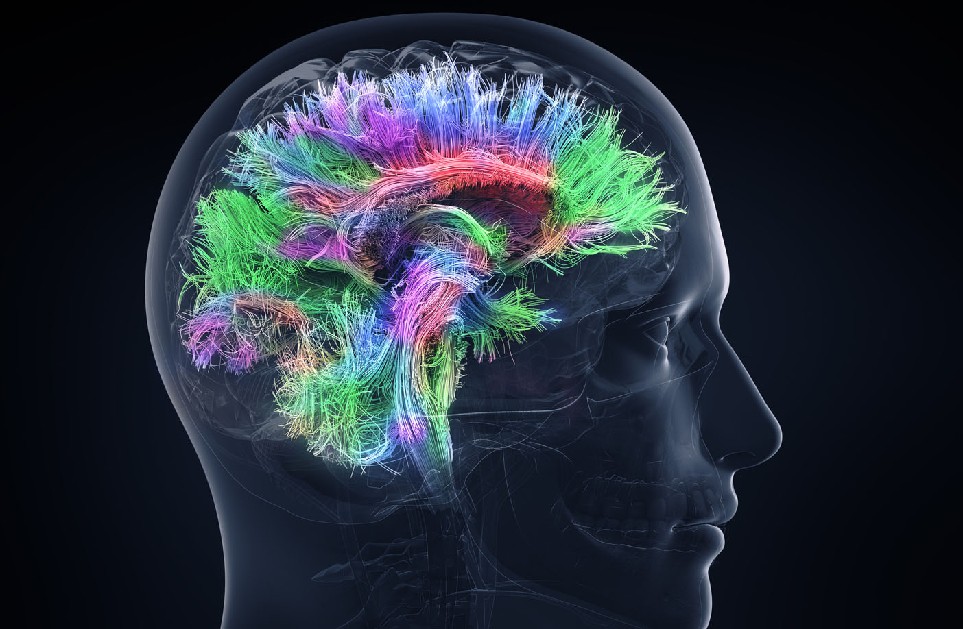
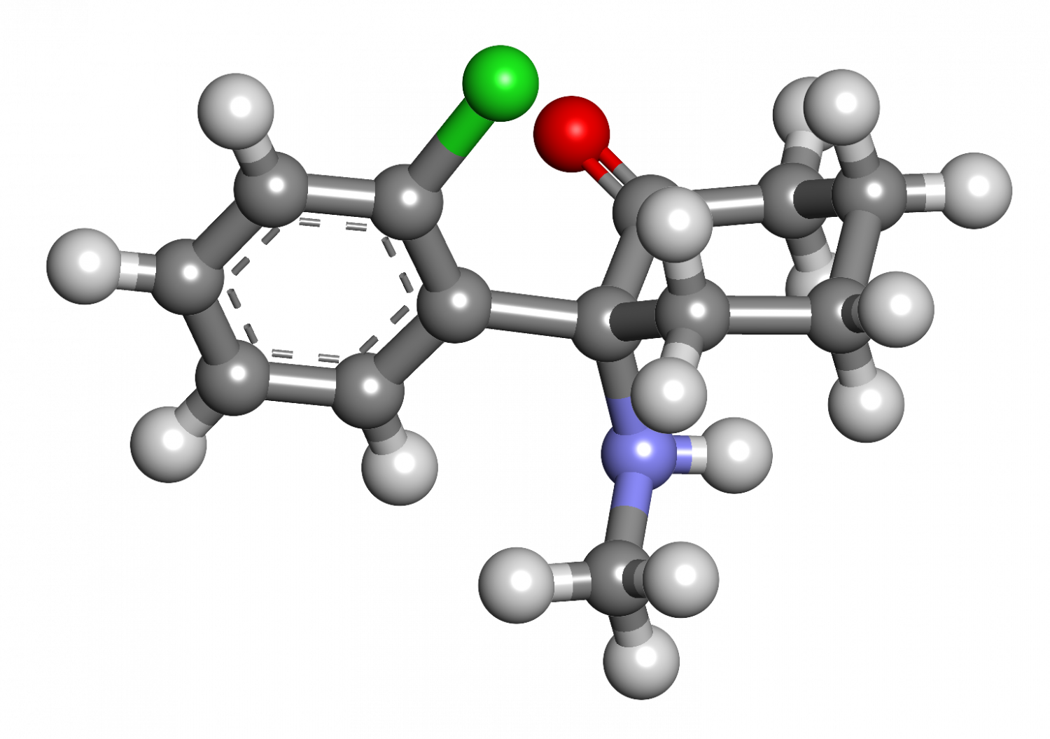
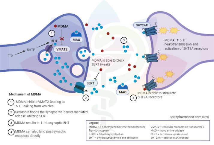
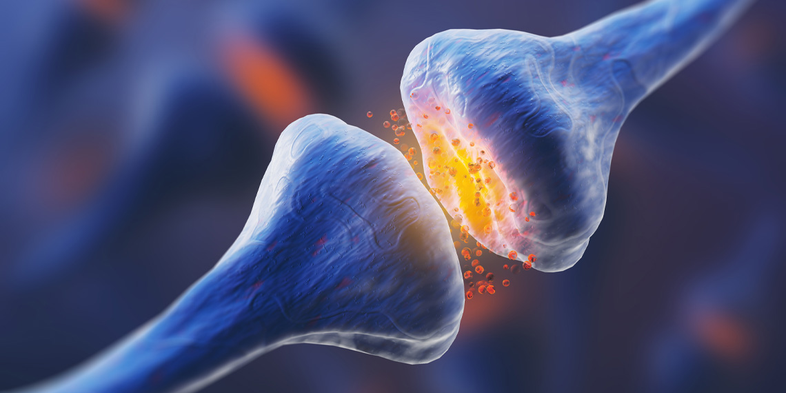
Before discussing how MDMA leads to neurotoxicity, it may be helpful to review how MDMA works. MDMA acts on several neurotransmitter systems, although primarily releases serotonin from presynaptic nerve terminals, resulting in acute depletion of serotonin stores and synaptic flooding of serotonin. This has led researchers to believe neurotoxicity is a consequence of damage to serotonergic neurocircuitry. Subsequently, evidence of neurotoxicity for MDMA is mainly quantified through 5-HT (serotonin) concentrations, activity or presence of enzymes involved with serotonin synthesis or transport (TH and SERT), and visualization of axons immunoreactive for 5-HT or SERT through imaging.

5HT+Synapse+MDMA+MOA.png


Mechanisms of MDMA-induced neurotoxicity

Sometimes it may be as simple as the dose determining if poisonous or not. With higher doses there is a greater chance of profound serotonin depletion. Frequency of use is also probably consequential as dosing on a weekly basis (as some may be if frequenting raves often) is not enough time to allow for recovery from prior use, even in some taking doses in clinical environments. This is rather simple in that your serotonin neuron simply has a capacity for how high the concentrations can be without damage and how fast it can return to balance after use. The two may go hand in hand as frequent use can produce tolerance and result in increased doses when used.

Other explanations are more complicated. For example, MDMA is metabolized to reactive metabolites such as HHMA or HHA, which can cause cellular damage. The serotonin excess may also lead to lasting reduction in gene expression resulting in lowered expressed of 5HT or SERT. The latter hypothesis has challenges the ‘neurodegenerative’ theory of MDMA induce neurotoxicity which argues for direct damage to serotonin neurocircuits and argues instead for a genetic reason for changes in serotonin function.

It is crucial to note that MDMA’s neurotoxic potential is increased by other substances such as alcohol or amphetamine, which are commonly taken in conjunction with MDMA at nightlife events. Concurrent substances can be ingested either advertently or inadvertently as many Ecstasy tablets contain adulterants or misrepresented substances. For example, one analysis found almost half of ecstasy tablets continued < 67% MDMA and that caffeine and amphetamine were common adulterants. Other common adulterants include novel psychoactive substances such as other phenethylamine (amphetamine) based drugs. Many events sell alcohol or may limit water availability. In combination, both alcohol and MDMA exert neurotoxic effects by impairing the survival of neuronal precursors in the hippocampal dentate gyrus, an area of the brain important for neuronal generation, learning, and memory. Beyond alcohol or amphetamine(s), rave scenes can expose users to myriad other substances, such as GHB (gamma-hydroxybutyrate) or ketamine, which may also increase risk. Recently and tragically, other adulterants from diverse drug classes have began to be found on ecstasy tablets such as fentanyl. Ecstasy is recommended to be tested for presence of desired agent (MDMA) and absence of deadly adulterants (fentanyl) by harm reduction organizations. Further information and testing kits for purchase can be found at dancesafe.org

Other aspects of MDMA use that may contribute to neurotoxicity is the recreational environment and timing of administration. Users may stay up all night on MDMA and related stimulants, which could trigger a sleep deprived state. Sleep deprivation is well known to impair cognition and lower mood, thus plausibly leads to some of the observed week after effects. This issue may be minimized by timing MDMA administration such that the user can fall asleep at a regular time or with minimal disruption to habitual circadian patterns. Another factor that may play a role in toxicity is the bioenergetic stress (namely thermal stress) secondary to MDMA being used in hot environments, raising core body temperatures, and being associated with high output physical activity.

Though the jury is still out on exactly how MDMA exerts its neurotoxic effects, the bottom line is that MDMA has several underlying explanations of why it could be neurotoxic as well as evidence from recreational use environments that it can be neurotoxic under certain circumstances. There circumstances tend to work in conjunction within subcultures that use Ecstasy heavily.

At what point does MDMA become neurotoxic?

The potential for MDMA to act as a neurotoxin is determined by the dose, frequency of exposure, timeline of exposure, concurrent substance use, and overall setting or context in which it is administered.

Doses of 1.7mg/kg (~125mg in a ~160lb adult) have been evaluated in clinical laboratory conditions without evidence of damage to serotonin neurocircuits or functional deficits. In the six phase II trials of MDMA-AP for PTSD conducted thus far, doses have ranged between 75-187.5mg and participants have undergone 2-3 experimental sessions spaced 3-5 weeks apart. In phase III trials, the maximum dose in any session will be 120mg as an initial dose and 60mg as a booster dose (180mg total) and all participants will undergo 3 sessions. In phase II, there were no detected changes in neurocognitive function over a 2-month period following the second and third experimental sessions. During MDMA-AP sessions participants reported a range of short-lived side effects characteristic of amphetamine use and mild serotonin excess. In the week after use fatigue peaked on day 1 and decreased towards day 6, the need for more sleep peaked on day 2 and decreased towards day 6, and low mood peaked between days 2-4 and decreased towards day 6. Notably, in 4 of the 6 phase II studies, depression scores were measured and showed greater improvements than placebo over the course of the trial. In summary, it’s clear that a course of 2-3 MDMA sessions spaced at least a month apart utilizing doses of 75-125mg followed by a booster dose of 37.5mg-75mg 2-3 hours later does not produce persistent neurotoxic effects.

In the previously mentioned study of MDMA samples, the ones that contained >67% MDMA averaged 205mg per dose, higher than the initial and booster doses in MDMA-AP combined (however weights include fillers thus is likely an overestimate of MDMA per tablet). Consumption of multiple tablets (stacking) can bring doses up to 0.5g or more throughout an evening for some users; similar to amounts consumed in all 3 MDMA-AP sessions combined. Stacking was investigated in laboratory conditions by giving 100mg of MDMA followed by another 100mg of MDMA 4 hours later. Results demonstrated that blood concentrations, cardiovascular stress, and temperature were all increased, however subjective intensity of effect was similar due to development of rapid tolerance to pleasurable effects. This may cause users to underestimate the physical stress they are placing on their body with repeated dosing.

So, what does the research tell us so far?

There are two converging streams of evidence here. One from recreation literature and Ecstasy use suggesting potential for neurotoxicity. The other from clinical environments in which MDMA is predominantly therapeutic, has a promising safety profile, and lacks evidence of clinically significant neurotoxicity despite using measures that could reasonably detect such problems. Therefore, models of dosing from clinical trials could be extrapolated and generalized into a harm reduction framework that could be applied broadly to persons who use MDMA.

While phase II trials did detect several cases of persons experiencing side effects of MDMA in the week after their session, the changes were transient and resolved after a week in almost all cases. This suggests that transient changes to mood, sleep, or cognition in the week after use is a side effect that persons should be educated about, while neurotoxicity is an adverse drug reaction associated with ‘incorrect’ or harmful administration patterns.
How can MDMA-induced neurotoxicity be avoided?

The good news is that avoiding or severely limiting the potential for significant neurotoxicity occurring with MDMA is straight-forward, not difficult, and preserves the ability for profound benefits.

- Limit MDMA doses to the moderate range 75-125mg initially
- Limit dosing in a single session to a single booster of 50% the original dose
- Space MDMA sessions at least one month apart
- Limit dosing to 3-4 times per calendar year
- Avoid using other drugs and alcohol with MDMA
- Test ‘ecstasy’ tablets for presence of MDMA prior to use (as well as absence of other drugs such as fentanyl)
- Limit sleep disruption due to MDMA use
- Plan regular breaks and sip water if exposed to hot environments (do not drink excessive amounts of water)

If you notice a pattern of low mood or cognitive problems in the days after MDMA use that seems to worsen upon repeated administration or is persistent beyond a few days post-use then take an extended break from using MDMA or similar substances

Do antidepressants or 5-HTP prevent MDMA-induced neurotoxicity?

Serotonin blocking antidepressants such as Selective Serotonin Reuptake Inhibitors (SSRIs e.g. Prozac or fluoxetine) can diminish neurotoxic potential of MDMA. However, this is a poor strategy overall because serotonin blocking antidepressants also greatly diminish the subjective experience of MDMA.

Supplements such as L-tryptophan or 5-HTP are able to boost serotonin synthesis. It may not be wise to take large doses prior to MDMA use as it could increase the risk of physical side effects of serotonin excess such as nausea, vomiting, or diarrhea. Conversely, it may be reasonable to use L-tryptophan or 5-HTP for a few days after MDMA use to aid in restoring serotonin levels.

Neither antidepressants nor serotonin precursor supplementation has adequate data suggesting benefits to recommend use. While further research and testing of interventions to reduce either week after side effects or prevent neurotoxic effects is encouraged, current clinical trial data do not support such measures as necessary.

Summary & conclusions

Neurotoxicity can manifest in many forms and severity levels and MDMA appears to be neurotoxic under certain circumstances. High doses or several doses throughout the period of ingestion, frequent use (e.g. weekly use at nightlife events), aduleration, and substance mixing all are implicated in the adverse event of MDMA neurotoxicity. Data from phase II trials of MDMA-AP do not support risks of clinically significant neurotoxicity from MDMA use as side effects were observed to be mild and limited to the week after use, while participants also saw profound improvements in clinical symptoms for PTSD. In summary, MDMA induced neurotoxicity is easy to avoid with moderate doses, adequate time between use, and pure substances without combination.

 
180618-Conversatoin-mind-molding-psychedelic-drugs-could-treat-depression-hero_red1cq


Psychedelic drugs affect how the brain is wired

by David Olson | The Conversation | The Daily Beast

Researchers know that mind-altering drugs including LSD, DMT, and MDMA affect brain function, but new findings show they can alter the structure of the brain as well.

It seems that psychedelics do more than simply alter perception. According to the latest research from my colleagues and me, they change the structures of neurons themselves.

My research group has been studying the effects of psychedelics on neuronal structure and function, and we found that these compounds cause neurons to grow. A lot. Many of these compounds are well-known and include lysergic acid diethylamide (LSD), psilocin (from magic mushrooms), N,N-dimethyltryptamine (DMT, from ayahuasca) and 3,4-methylenedioxymethamphetamine (MDMA, aka ecstasy).

These are among the most powerful drugs known to affect brain function, and our research shows that they can alter the structure of the brain as well. Changes in neuronal structure are important because they can impact how the brain is wired, and consequently, how we feel, think and behave.

Prior to our study, few compounds were known to have such drastic and rapid effects on neuronal structure. One of those was ketamine—a dissociative anesthetic and quite possibly the best fast-acting antidepressant that we have available to us at the moment.

If you think of a neuron like a tree, then its dendrites would be the large branches, and its dendritic spines—which receive signals from other neurons—would be the small branches. Some of these small branches might have leaves, or synapses in the case of a neuron. In fact, neuroscientists often use terms like “arbor” and “pruning” much like a horticulturist would.

“When we grew neurons in a dish—which is not unlike growing a plant in a pot—and fed them psychedelic compounds, the neurons sprouted more dendritic branches, grew more dendritic spines, and formed more connections with neighboring neurons.”

Thanks to studies on ketamine, slow-acting antidepressants and chronic stress models of depression, scientists now know that depression is not simply the result of a “chemical imbalance,” as pharmaceutical companies like to suggest. It is far more complicated and involves structural changes in key neural circuits that regulate emotion, anxiety, memory and reward.

Rethinking depression

One of the hallmarks of depression is the atrophy of neurons in the prefrontal cortex—a region of the brain that controls anxiety and regulates mood among other things. Basically, these branches and spines shrivel up, disconnecting from other neurons in the brain. One hypothesis for why ketamine is so effective is because it can rapidly regrow the arbors and spines of these critical neurons.

Like ketamine, psychedelics have shown promise in the clinic for treating neuropsychiatric diseases. The DMT-containing herbal tea known as ayahuasca produces fast-acting antidepressant effects within a day, psilocybin eases the anxiety of terminally ill cancer patients and MDMA can reduce fear in those suffering from post-traumatic stress disorder(PTSD). Our recent papers suggest the intriguing possibility that psychedelic compounds and ketamine might share a common therapeutic mechanism.

Strictly speaking, a psychedelic is a “mind-manifesting” drug—a definition that’s open to interpretation. They tend to produce perceptual distortions or hallucinations by activating 5-HT2A receptors. Our research group has found that compounds typically regarded as psychedelics, like LSD and DMT, as well as those that are sometimes called psychedelics, like MDMA, and those that are not usually called psychedelics, like ketamine, are all capable of profoundly impacting neuronal structure.

Psychedelics vs. Psychoplastogens

Our group has coined the term “psychoplastogen” to refer to such compounds, and we believe that these molecules may hold the key to treating a wide variety of brain diseases.

Our studies on neurons grown in dishes, as well as experiments performed using fruit flies and rodents, have demonstrated that several psychoplastogens, including psychedelics and ketamine, encourage neurons to grow more branches and spines. It seems that all of these compounds work by activating mTOR—a key protein involved in cell growth.

The biochemical machinery that regulates mTOR activity is intricate. As we tease apart how psychedelics and other psychoplastogens turn on mTOR signaling, we might be able to engineer compounds that only produce the therapeutic effects on neuronal growth while bypassing pathways that lead to undesired hallucinations.

The field has known for some time now that psychedelics can produce lasting positive effects on brain function, and it’s possible that these long-lasting changes result from the psychoplastogenic effects of these drugs. If true, this would suggest that psychoplastogens might be used to repair circuits that are damaged in mood and anxiety disorders.

Many diseases, such as depression and anxiety disorders, are characterized by atrophy of dendritic branches and spines. Therefore, compounds capable of rapidly promoting dendritic growth, like psychedelics, have broad therapeutic potential. The number of papers demonstrating that psychedelics can produce therapeutic effects continues to grow every year.

Panacea or poison?

However, we should temper our enthusiasm because we do not yet know all of the risks associated with using these drugs. For example, it’s possible that promoting neuronal growth during development could have negative consequences by interfering with the normal processes by which neural circuits are refined. We just don’t know, yet.

Similarly, it is unclear what effects psychoplastogens will have on the aging brain. It’s important to keep in mind that excessive mTOR activation is also associated with a number of diseases including autism spectrum disorder (ASD) and Alzheimer’s disease.

To me, it’s obvious that we need to understand how these powerful compounds affect the brain, in both positive and negative ways, if we hope to fully comprehend the fundamental laws governing how the nervous system works and how to fix it when it doesn’t.

By David E. Olson, assistant Professor, Department of Chemistry; Department of Biochemistry & Molecular Medicine; Center for Neuroscience, University of California, Davis

 
shutterstock_236914165-2048x1707.jpg


Examining the cognitive neuroscience of psychedelics

by Joel Ng, MA | Psychedelic Science Review | 4 Aug 2020

Much research has been conducted into the use of psychedelics in concurrence with therapy as novel treatments for a host of mental disorders, from OCD (obsessive-compulsive disorder) to depression to substance abuse. Such research has only just started to regain popularity after the widespread ban on psychedelic substances in the 1970s. However, less is known about how psychedelics work on a granular level. A deeper understanding of psychedelics, and being able to closely tie neurochemical changes caused by psychedelics with subjective experiences, could expand our understanding of the brain and advance mental health care greatly.

A recent paper published by Drs. Robin Carhart-Harris and Karl Friston in the Journal Pharmacological Reviews suggests a new way of explaining how psychedelics affect the brain’s way of understanding one’s environment, and by extension, provides a potential explanation for how psychedelics work in treating mental disorders. Their model is known by the acronym REBUS, which stands for “relaxed beliefs under psychedelics.”

The REBUS model and prior beliefs

The authors view the brain as an engine that generates mental models of the world with the purpose of predicting future sensory data. These predictions are called ‘priors’, meaning ‘prior beliefs’. Much of human behaviour and cognition are based on these priors. For example, there is the mental model of how a washroom sink functions – turn this knob, water comes out. This can be extended beyond beliefs about the physical nature of the world, also including more abstract beliefs. For example, drug addiction could be viewed through the lens of a prior: “taking this drug leads to large amounts of reward.”

Usually, priors that are inaccurate reflections of the world are updated by incoming sensory data. New information from the senses updates the model to better reflect reality. Sometimes, however, new sensory information is ignored by the brain, due to priors being too rigid to be updated. An example of this is drug addicts who keep abusing their drug of choice even after the negative aspects of drug addiction cause significant damage to their lives or depressed individuals with otherwise comfortable lives. Carhart-Harris and Friston suggest that psychedelics decrease the rigidity of priors – making priors more malleable to incoming sensory data. Likening this process to heating a metal to increase its plasticity, psychedelics allow new information to better mould the prior into something more reflective of reality. This model explains how psychedelic trips can often result in long-term changes in individuals even after the trip ends by promoting a reorganization of the brain’s way of perceiving the world.

What does the dissolution of priors feel like on the individual level? One of the most accepted models of personality, the five-factor model of personality, describes five domains of personality – Openness, neuroticism, extraversion, agreeableness, and conscientiousness. Openness, the factor of interest in this case, broadly describes an individual’s level of affinity for new experiences, people, and viewpoints. Personality analyses of subjects who ingested psilocybin showed significant increases in their openness domains over a year after the psilocybin dose. Viewing these results, broadly, this dissolution of priors feels like an imposed sort of openness of mind, where the psychedelic compound forces one to be more open-minded regarding incoming information. As information about the world enters the consciousness – information that might otherwise have been previously ignored to retain prior cognitive structures – psychedelic mind-states are less able to discount this information, which subjectively feels like being more open-minded.

Learning from the brains of children

Psychedelics’ effect on the serotonin 5-HT2 receptors in the brain has been theorized as the main neurochemical process by which psychedelics exert an effect on subjective experience. Interestingly, psychedelics induce certain biological events that are similar to childhood. First, serotonin receptors, which are the primary receptors that psychedelics act on, are more numerous in children than in adults. Second, neurogenesis and brain plasticity, both traits that are more pronounced in childhood brains, are induced by psychedelics. All in all, the evidence indicates that psychedelic states of mind and childhood states of mind bear great similarities. Learning about the world and the individual’s relationship with it takes place at a critical period in one’s childhood, one that psychedelics might be able to reactivate in adults.

With this implication in mind, one can examine further the interesting mental abilities of children, abilities that fade away as they grow up. For example, enhanced learning potential and enhanced memory are just two of the things that children are capable of that adults are not. Children with an eidetic memory often lose this ability as they grow. This is an ability that has, anecdotally, been retriggered in psychedelic mind-states. However, more rigorous research is required to corroborate these accounts.

Other advantages of the REBUS model

One additional benefit of the REBUS model of viewing psychedelic effects on cognition is that it can explain a host of other subjective effects that psychedelics produce, from ego dissolution, altered time perception, geometric hallucinations, and magical thinking. All of the previously stated subjective effects can be seen as the result of new information altering previously rigid cognitive structures like the ego, subjective experiences of time units, visual recognition and classification of objects, and large-scale worldviews on the nature of reality.

Lastly, and perhaps most importantly, this model allows the psychiatric community to better explain exactly why and how psychedelics seem to be extremely effective at treating certain types of mental illnesses. Conditions including anxiety, depression, PTSD (post-traumatic stress disorder), viewed as maladaptive priors that are immune to sensory information updates in sober mind-states, are able to be integrated with new information in the psychedelic mind-state due to the reduced rigidity of the priors. Depressed individuals remaining depressed after positive events in their lives can be viewed as the prior “I am not worth anything” resisting integration with the new information. This resistance is reduced in the psychedelic mind-state, allowing for psychedelic-enhanced psychotherapy to be more effective than regular psychotherapy.

The impact on psychedelic therapy

In conclusion, authors Carhart-Harris and Friston posit a new model of viewing the brain, one that also explains the subjective and neurochemical effects of psychedelics on the brain. This model sheds more light on many facets of psychedelics, from the recreational, subjective effects of psychedelics to more serious medical effects that might one day pave the way to greater, more effective mental health treatments for a variety of individuals.

 
RS-Dennis-McKenna-1200x630-1-1-800x400.jpg


The neuroscience of psychedelic visuals

by Dr. James Cooke | Reality Sandwich | 30 Aug 2020

‘Psychedelic’ can be applied not only to a class of chemical, but to a whole style of visual art, inspired by the visions that these chemicals produce. Why do these substances produce similar visual experiences across different individuals and where do these experiences come from? From breathing walls to melting hands, from symbolic imagery to entity encounters, psychedelic visions can tell us something about who and what we are.

During everyday waking consciousness we typically perceive a stable world of objects. This feels like a completely passive process–just open your eyes and there’s the world, no effort required. In reality, the felt simplicity of this experience masks the highly complex processing going on in your brain that allows you to see. Your retina is not a clear window through which your soul looks out onto the world. Instead it is a fleshly surface similar to the rest of your body, a piece of meat that blocks the light from the outside world, rather than letting it in.

When we see, what actually happens is that the patterns of light are transformed into electrochemical signals that are sent down the optic nerve to the brain. The brain, sitting in your pitch-black skull, learns to actively build models of the objects out there in the world that these electrochemical clues relate to. Your normal perception can be understood as a controlled hallucination, kept in check by the data coming in through your senses and by your expectations of the world around you. However, this balance between the internal creativity of the models in your brain and the extent to which they are kept in check can be altered.

Psychedelics interact with brain cells to alter their activity in ways that disrupt the normal process of perception. At low doses of a psychedelic, the visual world becomes distorted in reliable ways. Perhaps you experience trails of light following your hands as you move them, or you perceive the walls to be breathing. This can be understood as the result of a temporary impairment in the Default Mode Network (DMN).

The DMN is a group of brain areas that builds up expectations about the world and uses them to keep our perception in check. When its ability to do this is reduced, our internal models of the visual world are free to make guesses about what we’re seeing. We may overestimate the distance of the wall then correct for our error based on the sensory data. Give the newfound flexibility offered to the brain areas involved in this process, they may underestimate, then overestimate. This process can continue on and on, resulting in a pendulating interpretation of the wall’s distance and the perception of the walls breathing. The same goes for the precise position of one’s hand in space over time.

We’ve all seen shapes in the clouds or perhaps faces in the bark of a tree. Here we are using ambiguous sensory input and are finding internal models in our brains that roughly match the pattern. When on a psychedelic this process goes into overdrive and can result in us mistaking a flower for a lizard or a bowl of spaghetti for a bowl of worms. Why do we tend to see natural shapes like animals and not artificial ones? Why lizards and worms and not buildings and airplanes?

Our visual system evolved to recognize the patterns of the natural world that are relevant to survival and so, when given to opportunity to play, our visual systems show us these natural forms that we were built to detect. We have templates for snakes and spiders deeply programmed into us for example, something that simply isn’t the case for modern dangerous objects like cars and guns.

Our hallucinations can be thought of as what our brains expect to see, something that may also account for why we perceive colors to be highly saturated in the psychedelic state, as we’re experiencing the more pure template of the color than we typically experience in daily life. Understanding the basis of hallucination in our evolutionarily programmed expectation of the visual world also offers a way of understanding why eyes, serpents, and insects are so common in higher-dose psychedelic experiences, as these are highly biologically relevant patterns for us as a species.

The models in our brain that underlie perception are constructed by networks of brain cells. The vast computational power of such networks has resulted in their being imitated in modern Artificial Intelligence systems. Artificial Neural Networks such as Google’s Deep Dream can be trained to recognize different images, resulting in the construction of models in the neworks in a way that approximates learning in the brain. When we dream or hallucinate, the contents of these models become active with no sensory input to keep them in check. The same can be done with the artificial Neural Networks. When it is tasked with generating rather than recognizing images, they produce distinctly psychedelic visuals. The fact that this is the case provides striking evidence that certain psychedelic visuals are generated by the models in your brain being pushed into “create” mode.

At higher doses geometric patterns can be observed, especially when one closes one’s eyes, thereby excluding the sensory data that would otherwise keep one’s models in check. The patterns of the natural word are geometric in essence, all patterns are. Geometric hallucinations can reveal certain common patterns that are fundamental to our experience of the world. Psychedelics are not the only way to shift the visual brain into a mode where it displays their core patterns. When one “sees stars” after being hit on the head, the visuals being perceived are called phosphenes. Phosphenes can also be seen when pressure is placed on the eyeball. These geometric patterns are routinely experienced in the psychedelic state and have been observed in ancient cave art, leading to the suggestion that early cave art represents the earliest attempts of our species to carry back the experiences of the psychedelic state.

At high doses of classical psychedelics, people routinely experience ancient imagery exemplified in the art of cultures as found in ancient Egypt and Mesoamerica. Mesoamerican cultures are known to have ingested psychedelic mushrooms and it has been suggested that religion in ancient Egypt for a time revolved around the consumption of such mushroom, based on the similarity of depictions of Egyptian crowns to different stages of the development of this type of mushroom. The psychoactive blue lotus is also believed to have been consumed ritually in ancient Egypt. From our contemporary perspective, the styles depicted in the artworks feel as if they originated in these cultures. In reality the imagery of the psychedelic experience may have come first and the art later.

Another experience that can be had at high doses, especially with DMT, is the experience of visiting another “dimension”. In such an experience the person typically feels as if they have left their body behind and have been transported to another world. Understanding that our perception of the world around us is not the passive experience of a true picture of the world but instead is a controlled hallucination generated by our minds allows us to explain such experiences. As in a dream, the brain is pushed into a creative mode where it is especially decoupled from the sensory data coming from the world around. Certain contents, such as snakes and eyes, can be readily explained by the idea that they reflect deeply programmed visual patterns that are relevant to survival.

Not all of the contents fit neatly into this interpretation however, with visions of technology being particularly difficult to account for. One speculative explanation for such visions is that they reflect geometric hallucinations varying in three dimensions of space and in time.

When having the experience of moving to another dimension it is common to experience passage down a tunnel. It has been suggested by “the Godfather of LSD”, Stan Grof, that the tunnel experience is a memory of the experience of birth, although the fact that those born by cesarean section can still experience tunnels seems to rule this explanation out. Neurobiologist Jack Cowan has argued that spontaneous activity moving across the visual cortex might translate into the experience of concentric rings, as a result of the way that the retina maps onto the surface of the cortex.

Dramatic psychedelic experiences can feel like they reflect actual experiences of pre-existing phenomena that exist outside oneself, rather than being generated from within. This shouldn’t be a surprise as our perception always presents us with the feeling of an objective reality outside of ourselves, even though it is a controlled hallucination created inside us.

The nature of these experiences has led some scientists and philosophers to suggest that the material world is secondary to another reality that we interact with in these states, this ultimate reality varying from a spiritual realm, other dimensions, baseline reality in which our current world is merely a simulation, or a universal consciousness that underpins reality itself. The challenge of these perspectives, other than overturning the current prevailing paradigm, is to explain why low doses produce effects that are so readily explained by our understanding of how normal perception functions in the brain, and at high doses why these other worlds are show patterns that are biologically relevant to us, such as human eyes and natural imagery.

Psychedelic experiences can allow us to delve into our deepest personal programming and beyond into our deepest evolutionary programming, bequeathed to us by countless ancestors. Understanding the origin of psychedelic visions as coming from within does not reduce their meaning. We get the opportunity to explore ancient patterns on which our sense of meaning itself is scaffolded, we may confront aspects of Jung’s collective unconscious or realize how our visions connect us in time to our unimaginably long past as an evolved creature. Understanding how such experiences are generated, while interesting, is not necessary to derive pleasure and benefit from them. The value really is in the experience itself, whatever is going on behind the scenes.

 
shutterstock_1304852701-1536x945.jpg


The Nuances of Ketamine’s Neurochemistry

Melody Pezeshkian, BS | Psychedelic Science Review | 15 Feb 2021

What's similar and different about the R and S isomers of ketamine?

Ketamine, an N-methyl-D-aspartate (NMDA) antagonist, can produce antidepressant effects more rapidly than current first-line treatments for depression. Ketamine is a racemic (50-50 mixture of the R and S isomers) drug known for its dissociative, anesthetic, and antidepressant effects. Ketamine’s novel mechanism of action, contrasting to that of typical SSRI’s, has driven its popularity in the field of psychiatry and clinical psychology. The drug has been clinically approved for treatment-resistant depression (TRD) and is being studied for the treatment of other mental illnesses.

Ketamine’s novel mechanisms of action

Ketamine acts as an antagonist at the NMDA receptor, a glutamatergic, ligand-gated ion channel. As a quick refresher, gamma-aminobutyric acid (GABA) is an inhibitory neurotransmitter that works to stabilize the levels of glutamate released in the brain. Glutamate is an excitatory neurotransmitter. Ketamine binds to the NMDA receptors found on GABA-eric interneurons and inhibits GABA release. By Inhibiting the release of GABA, more excitatory glutamate molecules become available in the cortex, via disinhibition, which may contribute to ketamine’s antidepressant effects.

Zanos-and-Gould.png

Figure 1: Ketamine’s proposed novel mechanisms of action as compiled by Zanos and Gould.3 Note that ketamine binds to NMDA receptors on GABAergic interneurons to disinhibit glutamate release (section ‘a’ in the upper left of the image). Evoked release of glutamate binds to AMPA receptors, in turn releasing BDNF. This figure also illustrates the proposed release of ketamine metabolites.

Researchers are discovering that ketamine’s antidepressant mechanism of action is not solely dependent on its activity at the NMDA site.3 Ketamine activates α-amino-3-hydroxy-5-methyl-4-isoxazole-propionic acid receptors (AMPARs) which are another major ionotropic, glutamate receptor (Figure 1). AMPARs are activated via glutamate release and subsequently induce the production of brain-derived neurotrophic factor (BDNF), a growth factor that possesses anti-depressant effects.

BDNF is also a factor in synaptic plasticity and its production can lead to resilience to chronic stress. The metabolites of ketamine also have antidepressant effects which are NMDA-independent. Ketamine’s metabolites (2R,6R)-HNK and (2S,6S)-HNK are released by the breakdown of ketamine and promote AMPAR-mediated synaptic potentiation.

Ketamine can be isolated into ‘R’ and ‘S’ Isomers

The racemic (50-50) mixture of ketamine can be consumed via different routes of administration—orally, nasally, intravenously. However, racemic mixtures can also be filtered to isolate one isomer.

Isomers are molecular structures with the same chemical formula but with a different molecular geometry. Some isomers, known as structural isomers, have atoms that are connected in different ways (consider butane and isobutane). However, in the case of ketamine’s R and S isomers, the atoms are connected in the same structural way but differ in their spatial orientation. Isomers differing in spatial orientation but maintaining structural similarity are known as stereoisomers.

While the R and S stereoisomers of ketamine look nearly identical, they are chiral molecules–meaning that they are non-superimposable mirror images (Figure 2). The preceding letters R and S are used to refer to the ‘handedness’ of the isomer. R indicates right-handed, or clockwise geometry, whereas S indicates left-handed, or counterclockwise geometry. Take for example human hands; when facing each-other they are mirror images. However, when rotated they cannot be indistinguishably placed over each other. Chiral molecules are also known in the field of chemistry as enantiomers. Though this difference between the enantiomers is seemingly small, each isomer of ketamine has unique neurochemical effects.

Muller-et-al..png

Figure 2: The optical isomers of ketamine.

R and S Ketamine have different therapeutic ratios, meaning that isomers vary in the ratio needed to induce different therapeutic effects. Isomers may differ in their affinity for certain biological targets, which enables them to produce significantly different therapeutic effects from one another. Similarly, one isomer may have certain adverse effects compared to another. Studying isomer differences can shed light on the benefits of one isomer over a racemic mixture, or over the other.

Clinical differences between the effects of the R and S Isomers


Neither isomer has been studied extensively in human trials however, some research elucidates differences between them.

The S-isomer (also known as esketamine) is currently isolated and distributed for a higher cost than the racemic mixture. The isomer withstood a non-inferiority trial when compared to the racemic mixture, indicating equivalence between s-ketamine and racemic in treating depression. S-ketamine was FDA approved in 2019 for the treatment of TRD and is administered nasally as the drug Spravato.

Studies show that S-ketamine has a fourfold greater potency when it comes to inhibiting the NMDA receptor when compared to R-ketamine (also called arketamine).3 Its greater affinity at the NMDA receptor likely contributes to its selection by pharmaceutical companies in developing the drug. S-ketamine also has a greater analgesic potency; its analgesic potency is twice that of the racemate. For this reason, there is a preferential use of s-ketamine when it comes to anesthesia.

Surprisingly, the R-isomer shows longer-lasting anti-depressant effects compared to S-ketamine. A study on mice showed that R-ketamine increased prefrontal 5-HT (serotonin) at significantly greater levels than S-ketamine.7 Another study on mice suggests that R-ketamine produces less psychomimetic side-effects related to mobility, resulting in less distorting side-effects typically associated with ketamine.

Summary and conclusion

Ketamine acts as an NMDA receptor antagonist to produce novel antidepressant effects with both R and S isomers as well as the racemic displaying comparable anti-depressant effects. While esketamine is currently being touted as the major clinical resource for TRD, it is important to call attention to the varying effects of isomers.

Most studies that suggest evidence of differences between the isomers have been conducted using mice. However, some results suggest similar findings in humans. Scientists continue to study the mechanisms and effects of the ketamine R and S isomers. Understanding the complete pharmacology and effects of the racemic and other mixtures is a wide-open area in the field of psychedelic research. Much remains unknown about the effects of ketamine and the mechanisms of action of its metabolites, necessitating further research on the drug and its potential benefits.

 
neurogenesis-dmt-neurosncience.jpg


Ayahuasca stimulates the formation of new neurons

Complutense University of Madrid | Neuroscience News | 6 Nov 2020

DMT, a natural component of ayahuasca tea, promotes neurogenesis, a new study reports. Researchers found DMT was capable of activating neural stem cells and promoted the formation of new neurons.

One of the main natural components of ayahuasca tea, dimethyltryptamine (DMT), promotes neurogenesis (the formation of new neurons) according to research led by the Complutense University of Madrid (UCM).

In addition to neurons, the infusion used for shamanic purposes also induces the formation of other neural cells such as astrocytes and oligodendrocytes.

“This capacity to modulate brain plasticity suggests that it has great therapeutic potential for a wide range of psychiatric and neurological disorders, including neurodegenerative diseases”, explained José Ángel Morales, a researcher in the UCM and CIBERNED Department of Cellular Biology.

The study, published in Translational Psychiatry, reports the results of four years of in vitro and in vivo experimentation on mice, demonstrating they exhibit “a greater cognitive capacity when treated with this substance”, according to José Antonio López, a researcher in the Faculty of Psychology at the UCM and co-author of the study.

Changing the receptor eliminates the psychedelic effect

Ayahuasca is produced by mixing two plants from the Amazon: the ayahuasca vine (Banisteriopsis caapi) and the chacruna shrub (Psychotria viridis).

The DMT in ayahuasca tea binds to a type-2A serotonergic brain receptor, which enhances its psychedelic effect. In this study, the receptor was changed to a sigma type receptor that does not have this effect, thus “greatly facilitating its future administration to patients.”

In neurodegenerative diseases, it is the death of certain types of neuron that causes the symptoms of pathologies such as Alzheimer’s and Parkinson’s. Although humans have the capacity to generate new neuronal cells, this depends on several factors and is not always possible.

“The challenge is to activate our dormant capacity to form neurons and thus replace the neurons that die as a result of the disease. This study shows that DMT is capable of activating neural stem cells and forming new neurons,” concluded Morales.

 
shutterstock_1691666992-1536x878.jpg


Insights into the neurophysiology of tryptamine-derived psychedelics*

by Justin R. Kulchycki, MS | Psychedelic Science Review | 14 Dec 2020

Brain imaging research reveals where psychedelic compounds such as psilocybin and LSD manifest their effects.

Given the recent clinical interest in psychedelic compounds, many researchers have sought to elucidate their mechanism of action from a neurophysiological perspective. These research teams are seeking to understand changes in brain activity between the psychedelic state and normal waking consciousness. More specifically, the goal of these studies is to characterize the networks and regions of the brain affected by psychedelic ingestion. This can be accomplished with functional magnetic resonance imaging (fMRI), which accurately produces real-time depictions of brain activity.​

Effects of psilocybin on the Default Mode Network

A seminal 2012 study at Imperial College London was the first to use fMRI to characterize brain activity between psychedelic and non-psychedelic states. In this work, Carhart-Harris et al. used fMRI to record alterations in brain activity before and after injections of placebo and psilocybin. The paramount discovery was that psilocybin appeared to reduce overall cerebral blood flow. The brain regions which demonstrated the most consistent deactivation were the posterior cingulate cortex and the medial prefrontal cortex. These two regions reside in the default mode network (DMN), which refers to specific areas of the brain involved in various domains of cognitive and social processing, as well as autobiographical memory recollection.

In normal waking consciousness, activity in the posterior cingulate cortex and medial prefrontal cortex is relatively high compared with other regions of the brain. Interestingly, the degree to which activity in these regions was decreased was proportional to the intensity of the subjective effects reported by the volunteers. There is speculation that the posterior cingulate cortex is involved in consciousness, formulation of the self, and ego. The “ego-dissolution” phenomenon is a commonly reported experience following the administration of high doses of psychedelics. While this could be related to decreased posterior cingulate cortex activity, no direct quantitative relationship has been observed between the disintegration of the DMN and ego-dissolution.

A study published earlier this year from the University of Zurich also examined psilocybin-induced neurophysiological changes in volunteers. Like the previously mentioned study, this group used fMRI to capture changes in brain activity as the psilocybin-induced psychedelic state began to manifest in their volunteers. Their results supported the findings of the group at Imperial College London, in that associative regions become disintegrated with the rest of the brain upon psilocybin ingestion. Additionally, this study found that sensory areas of the brain, which are related to motor function, sensation, and visual perception become more integrated with the rest of the brain in the psychedelic state.​

LSD demonstrates similar effects on the brain

The team at the University of Zurich also conducted a similar study with LSD, another tryptamine-derived hallucinogen. Observations regarding the neurophysiological effects of LSD are consistent with the two previously mentioned studies using psilocybin. LSD ingestion results in a decreased connectivity of associative regions of the brain, and increased integration of sensory and somatomotor regions. Moreover, participants who showed relatively higher connectivity in the somatomotor network also reported a higher intensity of subjective effects.​

How do tryptamine psychedelics affect the whole brain?

A study from the Institute for Scientific Interchange in Turin, Italy indicates that psilocybin induces more persistent connections throughout the brain as a whole compared with normal waking consciousness. This work was conducted using the original fMRI data collected at Imperial College London. The researchers say psilocybin induces a more intercommunicative mode of brain function. This may mean there is more sharing of information between brain regions that don’t associate with each other in normal waking consciousness. Furthermore, the researchers speculate that this intercommunicative mode of brain function may be the root of the synesthesia phenomenon associated with the psychedelic experience.​

Summary

Collectively, these studies help describe the neurobiological mechanisms of tryptamine-derived hallucinogens. The general observation is higher-level functional networks involved in various cognitives processes become less integrated, and sensory and somatomotor networks become more integrated. What’s more, there seem to be more persistent connections formed throughout the whole brain during the psychedelic state relative to normal waking consciousness. These studies add to the psychedelic knowledge base which other researchers can tap into for further investigation.

*From the article here :
 
1ead8c783f7af89917e20bf15262716184-03-Microdosing-lede.rsquare.w1200.jpg


The emerging revival of psychedelics in neuroscience

by Cami Rosso | Psychology Today | 13 Jan 2021

There’s a common saying that goes, “Everything old is new again.” Does this pithy maxim apply to psychedelics? Psychedelic drugs, also known as hallucinogens or psychotomimetic drugs, may be in the early stages of a neuroscience renaissance in the form of legal medication since its research heyday in the 1950s and early 1960s. On Monday, Culver-City-based Kernel, a brain-computer interface (BCI) pioneer, announced a partnership with Toronto-based biotech Cybin to apply Kernel’s Flow for real-time quantification of brain activity to conduct research on psychedelics for treatment of mental health disorders.

“The ability to collect quantitative data from our sponsored drug development programs with Kernel’s Flow is potentially game-changing in terms of our ability to measure where psychedelics work in the brain in real-time, and how we ultimately design our future therapeutics,” stated Doug Drysdale, CEO of Cybin, in a statement released on Monday.

In 2016, visionary entrepreneur, venture capitalist (OS Fund), and author Bryan Johnson founded Kernel with his own investment and is the current CEO. Johnson also founded Braintree, which acquired Venmo in 2012, and was later sold for USD 800 million in 2013 to PayPal Holdings, Inc., which was then a wholly owned subsidiary of eBay.

Recently Kernel made a brain-computer interface breakthrough with its non-invasive Neuroscience as a Service (NaaS) brain recording technologies called Flux and Flow. Flux catches the magnetic fields produced by brain activity, and Flow tracks cortical hemodynamics, or the brain’s blood flow. In July 2020, Kernel announced USD 53 million funding from General Catalyst, Khosla Ventures, Eldridge, Manta Ray, and Tiny Blue Dot.

Psychedelics are an emerging area of treatment for a wide range of disorders such as major depressive disorder (MDD), post-traumatic stress disorder (PTSD), narcolepsy, treatment-resistant depression, and more mental health issues. The Cybin partnership with Kernel is just one example.

Last week, the Icahn School of Medicine at Mount Sinai in New York announced the launch of its Center for Psychedelic Psychotherapy and Trauma Research. The Center plans to research psychedelic compounds such as psilocybin and MDMA in efforts to discover novel therapeutics to help veterans and civilians who are struggling with depression, anxiety, PTSD, and similar stress conditions and mental health disorders.

Last month, psychedelic medicine startup Beckley Psytech raised GBP 14 million in equity funding from Jim Mellon, the British billionaire entrepreneur and founder of the longevity biopharma Juvenescence, Richard Reed, the co-founder of Innocent Drinks, and other investors. Beckley Psytech is researching a psychedelic agent called 5-MeO-DMT (5-methoxy-N,N-dimethyltryptamine) that is structurally similar to other indoleamine hallucinogens like LSD. 5-MeO-DMT is found in nature from a variety of plants such as the calcium tree that is native to the Caribbean and South America, as well as certain species of toads, such as the Sonoran Desert toad, which is also known as the Colorado River Toad (Bufo alvarius).

In November 2020, billionaire Peter Thiel invested EUR 10 million (~USD 12 million) via Thiel Capital in Atai Life Sciences, a biotech accelerator with headquarters in Berlin that is focused on providing a platform for companies developing novel treatments for depression, mild traumatic brain injury, generalized anxiety disorder, PTSD, anxiety, and addiction using lead compounds that include psilocybin, MDMA derivatives, N, N-dimethyltryptamine, noribogaine, deu-mitragynine, N-acetlycysteine, deu-Etifoxine, arketamine, and ibogaine. Thiel was an early investor in Facebook, and the entrepreneur who co-founded PayPal, and Palantir Technologies, among other companies.

Johns Hopkins University’s psychedelics research center received USD 17 million in funding in 2019 from investors that include Tim Ferriss, the famed podcaster and author of The 4-Hour Workweek, WordPress co-founder Matt Mullenweg, TOMS founder and former Navy SEAL Blake Mycoskie, angel investor Craig Nerenberg, and the Steven & Alexandra Cohen Foundation. The Center for Psychedelic and Consciousness Research at Johns Hopkins Medicine aims to identify new treatments using psychedelics for various diseases such as addiction, PTSD, post-treatment Lyme disease syndrome, anorexia nervosa, alcohol use with patients with major depression, and Alzheimer’s disease.

In the spring of 2019, the U.S. Food and Drug Administration (FDA) approved Spravato (esketamine), a nasal spray by Johnson & Johnson that is a chemical similar to anesthetic ketamine, and used for treatment-resistant major depressive disorder. The fast-acting antidepressant is a glutamate NMDA (N-methyl-D-aspartate) receptor modulator, which are associated with synaptic plasticity.

There is a global need for mental health therapeutics. Over 264 million people globally suffer from depression according to a 2017 Global Health Metrics study that was funded by the Bill & Melinda Gates Foundation and later published in The Lancet. An estimated eight million adult Americans have PTSD during a given year according to the U.S. Department of Veterans Affairs. In 2017, roughly 792 million people worldwide suffer from a mental health disorder according to Our World in Data.

With new investments and neuroscience breakthroughs, visionary entrepreneurs, cutting-edge biotech startups, research scientists, and pharmaceutical companies are trailblazing with novel therapeutics and rapidly thawing the psychedelic winter.

 
shutterstockRF_644108704.jpg

University of Glasgow

Neuroscience students launch Psychedelic Society

by Carmen Blaque | Glasgow Guardian } 2 Feb 2021

The society hopes to promote positive research into psychedelic drugs.

Illegal hallucinogenic drugs such as LSD, magic mushrooms and DMT have become increasingly researched amongst the scientific community in the past decade. Government grants have been issued in the UK to study such compounds, in hope to ease the mental health burden in the UK. Traditional pharmaceutical antidepressants, such as SSRI’s, fail to work for up to half of individuals. For the individuals who do respond, various side effects are common. With antidepressant medications seeing a 48% increase in the past decade in Scotland, the need for innovative treatments is paramount.

Dr Adrian James from The Royal College of Psychiatrists has recently stated that coronavirus will worsen the UK's mental health problems: “It is probably the biggest hit to mental health since the second world war.” Neuroscience students at the University of Glasgow have launched a Psychedelic Society to raise awareness about the potential for psychedelics to treat various mental health conditions such as depression, PTSD, anorexia, and addiction.

The Psychedelic Society are inspired by previous acts of social change that have occurred at the University of Glasgow. The University’s Climate Action Society contributed to the University’s plan of divestment from fossil fuels in 2013, with full divestment estimated to occur by 2024. Students at the University of Glasgow Psychedelic Society are hopeful that the University will also see the urgency to study psychedelic medicine and hope that they can make social change through providing students and staff with general education surrounding the therapeutic potential of psychedelics, in addition to the scheduling status of these substances.

Psychedelics are Schedule 1 controlled substances, which not only makes them illegal but makes scientific research extremely difficult. The University of Glasgow's Psychedelic Society is hopeful that they can make changes to the current scheduling which would make research easier and help those who are in desperate need. With ecological scientists such as Dr Sam Gandy showing that psychedelics can also increase nature-relatedness - which could, in turn, increase positive environmental behaviour - psychedelics offer hope for the current climate crisis.

The society is inspired by political scientist Erica Chenoweth from Harvard University, who found that past historical acts of change occur when around 3.5% of the population are in active participation. The society is hopeful that others will engage to help lower the scheduling of psychedelics, which could help research and ultimately save the lives of those who have not benefited from other mental health treatments.

Professor Davit Nutt, a former advisory committee of the misuse of drugs was famously sacked for stating that ecstasy and LSD are less harmful than drugs such as alcohol, has been invited to talk as part of the society's psychedelic science event. Professor David Nutt, who is now chair of Drug Science and deputy head of the Imperial College London's Centre for Psychedelic Research, is also hopeful that psychedelics could help those suffering from mental health illness, which has been proven from clinical trials. Professor Nutt stated: “The results were quite remarkable, possibly the most powerful single-intervention impact in depression there’s ever been, half participants were depression-free at one week and about a third were depression-free at three months.”

Psychedelic Research Centers are launching around the world, and UK leading institutions such as Imperial College London, have a dedicated Centre for Psychedelic Research and have made a significant contribution to psychedelic science and mental health in recent years. Students at the University of Glasgow's Psychedelic Society are hopeful that Scotland's universities will follow, to pave the way for the healing potential of psychedelics.

 
190301-rat-full-1440x810.jpg


Microdoses of DMT produce positive effects on mood and anxiety in rodents*

Lindsay Cameron, Charlie Benson, Brian DeFelice, Oliver Fiehn, and David Olson

Drugs capable of ameliorating symptoms of depression and anxiety while also improving cognitive function and sociability are highly desirable. Anecdotal reports have suggested that serotonergic psychedelics administered in low doses on a chronic, intermittent schedule, so-called “microdosing”, might produce beneficial effects on mood, anxiety, cognition, and social interaction. Here, we test this hypothesis by subjecting male and female Sprague Dawley rats to behavioral testing following the chronic, intermittent administration of low doses of the psychedelic N,N-dimethyltryptamine (DMT). The behavioral and cellular effects of this dosing regimen were distinct from those induced following a single high dose of the drug. We found that chronic, intermittent, low doses of DMT produced an antidepressant-like phenotype and enhanced fear extinction learning without impacting working memory or social interaction. Additionally, male rats treated with DMT on this schedule gained a significant amount of body weight during the course of the study. Taken together, our results suggest that psychedelic microdosing may alleviate symptoms of mood and anxiety disorders, though the potential hazards of this practice warrant further investigation.

Drugs capable of ameliorating symptoms of depression and anxiety while also improving cognitive function and sociability are highly desirable. Anecdotal reports have suggested that serotonergic psychedelics administered in low doses on a chronic, intermittent schedule, so-called “microdosing”, might produce beneficial effects on mood, anxiety, cognition, and social interaction. Here, we test this hypothesis by subjecting male and female Sprague Dawley rats to behavioral testing following the chronic, intermittent administration of low doses of the psychedelic N,N-dimethyltryptamine (DMT). The behavioral and cellular effects of this dosing regimen were distinct from those induced following a single high dose of the drug. We found that chronic, intermittent, low doses of DMT produced an antidepressant-like phenotype and enhanced fear extinction learning without impacting working memory or social interaction. Additionally, male rats treated with DMT on this schedule gained a significant amount of body weight during the course of the study. Taken together, our results suggest that psychedelic microdosing may alleviate symptoms of mood and anxiety disorders, though the potential hazards of this practice warrant further investigation.

*See the entire article here :
 
Neuroplasticity+-+IG.png


New evidence that a single dose of psilocybin can boost brain connections*

by Eric Dolan | PsyPost | 18 Mar 2021

Scientists in Denmark believe the psychedelic substance psilocybin might produce rapid and lasting antidepressant effects in part because it enhances neuroplasticity in the brain. Their new research, published in the International Journal of Molecular Sciences, has found evidence that psilocybin increases the number of neuronal connections in the prefrontal cortex and hippocampus of pig brains.

Psilocybin — the active component in so-called “magic” mushrooms — has been shown to have profound and long-lasting effects on personality and mood. But the mechanisms behind these effects remain unclear. Researchers at Copenhagen University were interested in whether changes in neuroplasticity in brain regions associated with emotional processing could help explain psilocybin’s antidepressant effects.

“Both post-mortem human brain and in vivo studies in depressed individuals have shown a loss of synapses through the down-regulation of synaptic proteins and genes,” the authors of the study wrote. “Hence, upregulation of presynaptic proteins and an increase in synaptic density may be associated with the potential antidepressive effects of psychedelics.”

The researchers had previously conducted tests to establish the proper dose of psilocybin needed to produce psychoactive effects in pigs, who were examined because their brains are anatomically similar to the brains of humans.

A group of 12 pigs received a psychoactive dose of psilocybin, while a separate group of 12 pigs received inert saline injections. Half of the pigs were euthanized one day after the administration of psilocybin, while the rest were euthanized one week later.

An examination of brain tissue from the hippocampus and prefrontal cortex revealed increases in the protein SV2A in pigs who had received psilocybin. SV2A, or synaptic vesicle glycoprotein 2A, is commonly used as a marker of the density of synaptic nerve endings in the brain. SV2A is typically reduced in patients with major depressive disorder.

“We find that a single dose of psilocybin increases the presynaptic marker SV2A already after one day and that it remains higher seven days after,” the researchers said, adding that the “increased levels of SV2A after intervention with a psychedelic drug adds to the scientific evidence that psychedelics enhance neuroplasticity, which may explain the mechanism of action of its antidepressant properties.”


hofmann_1405878c.jpg

Albert Hofmann

Study reveals how LSD leads to greater brain flexibility

by Kristi Pahr | LUCID NEWS | 12 Mar 2021

Research into consciousness has been gaining ground lately, but the majority of the studies have revolved around the loss of consciousness: sleep, anesthesia, and coma. A recent paper delves into LSD’s impact on consciousness in the waking brain – and the results are unexpected.

Research recently published in the journal NeuroImage shows that LSD allows the normally strict neuronal pathways in the brain to become more flexible, allowing the brain to explore and make connections separate from those that are typically allowed by brain structure.

“In general, integration and segregation of information are fundamental properties of brain function: we found that LSD has specific effects on each,” says Andrea Luppi, Ph.D. candidate at the University of Cambridge and lead author of the study. “We also know that brain structure has a large influence on brain function under normal conditions. Our research shows that under the effects of LSD, this relationship becomes weaker: function is less constrained by structure."

"This is largely the opposite of what happens during anesthesia.”


Luppi’s team examined data collected by Dr. Robin Carhart-Harris and Dr. Leor Roseman from the Centre for Psychedelic Research at Imperial College London for a separate study. The data included functional magnetic resonance imaging (fMRI) that examined brain activity in 20 volunteers. Volunteers underwent two MRI sessions, one after taking a placebo and one after taking 75 micrograms of LSD.

MRI results showed marked changes in the typical segregation and integration of stimuli in areas of the brain that normally do not interact. “Reduced similarity between structural and functional connectivity indicates that under the effects of LSD, brain regions interact functionally in a way that is less constrained than usual by the presence or absence of an underlying anatomical connection,” researchers wrote in the study.

These new interactions explain the dramatic altered consciousness one experiences after consuming LSD. “Being less constrained by pre-existing priors due to the effects of LSD, the brain is free to explore a variety of functional connectivity patterns that go beyond those dictated by anatomy – presumably resulting in the unusual beliefs and experiences reported during the psychedelic state, and reflected by increased functional complexity,” reads the study.

“Studying psychoactive substances offers a unique opportunity for neuroscience: we can study their effects in terms of brain chemistry, but also at the level of brain function and subjective experience,” said Dr. Luppi.

“In particular, the mind is never static, and neither is the brain: we are increasingly discovering that when it comes to brain function and its evolution over time, the journey matters just as much as the destination. A more thorough characterization of how psychedelics influence the brain may also shed light on potential clinical applications – such as the ongoing research at the new Centre for Psychedelic Research in London.”

This research was a collaborative effort between Dr. Luppi and Dr. Robin Carhart-Harris and Dr. Leor Roseman from the Centre for Psychedelic Research at Imperial College London, who collected and shared the data and was supervised by Dr. Emmanuel Stamatakis from the Cognition and Consciousness Imaging Group at the University of Cambridge.

 
Top