

djsim
Bluelight Crew
- Joined
- Mar 18, 2007
- Messages
- 3,220
I just wanted to draw attention to the latest Case Study titled Posterior retinal hole secondary to a candida retinitis added on July 27th (which would benefit from someone who can translate the Spanish text better than Google has done).
I want to make it clear IV drug users should NEVER use citric acid from lemons to prep heroin (usually brown heroin). Only sterile citric acid should ever be used, or failing that, acetic acid (vinegar). Heating the shot will NOT kill the Candida fungus if it is in the juice of the lemon... hell, they've found fungus which have survived re-entry on the outside of the space shuttle, so a lighter ain't going to do shit
This fungus can cause blindness within a month. Fungus introduced via IV are very, very difficult to kill, so if there is fungus in the juice, chances are almost certain the fungus will thrive in your body... and preferentially on your retina . Within 30 days vision can be totally occluded. And if you're really unlucky, it could be both eyes.
*******
Posterior retinal hole secondary to a candida retinitis
Alvarez-Suarez ML, Sanchez-Tabar L et al, 2005, The Spanish Ophthalmology society files (Archivos de la Sociedad Espanola de Oftalmologia) 80(7): 421-4.
http://scielo.isciii.es/scielo.php?script=sci_arttext&pid=S0365-66912005000700008&lng=es&nrm=iso&tlng=es&skpa=on
ABSTRACT:
CASE REPORT: We describe the case of a 36-year-old man with a history of intravenous heroin use, who was HIV negative. Left ocular examination disclosed a focal candida retinitis in the posterior pole associated with vitritis and moderate iritis. Treatment with fluconazole inactived the chorio-retinal lesion and resolved the vitritis, but developed an inner limiting membrane contraction over the macula. Two years later, vitreous traction produced a retinal hole that needed argon laser photocoagulation. DISCUSSION: Candida retinitis which penetrates into the vitreous cavity can produce retinal holes by vitreous traction over the lesion.
CLINICAL CASE (rough translation)
Male 30 age that attends emergency for loss of vision, redness and pain in left eye (OS) of 3 days of evolution. The patient also concerns fever approximately 20 days ago and is heroinómano sporadic recognizing a last episode of several Brown heroin injections approximately 25 days ago.
Exploration of the right eye (OD) was normal, while in the left eye (OS) there was an accompanied by a most focus of retinitis with localized vitritis previous Uveitis. The clinical exploration only detected febrícula and a few small bilateral latero-cervicales adenopathy.
Eye injuries were typical of candidiasis started hospital treatment with intravenous, fluconazole at dose of 200 mg / 24 h, and an eye pattern of corticosteroids and midriáticos topics. Complementary studies showed a Leukocytosis with normal formula (no neutropenia), a proteinograma with discrete increase in the alpha-2-globulins and some negative hepatitis B and HIV markers. The blood cultures, Lues, toxoplasma, CMV, serology and BK sputum and urine samples were also negative. Puncture a cervical Lymphadenopathy showed a reactive lymphadenitis with little activity blastic.
The box improved gradually with fever, the cilium-conjunctival reaction and inflammation of anterior chamber disappearance. The back of eye could see a good scarring of the prerretiniana injury and treatment was completed with oral fluconazole other 4 weeks.
At the end of treatment visual acuity in OI was 0.6, with a vitreous clear and with the focus of totally scar retinitis but associated vitreous traction which produced a marked contraction of the internal level macular limiting membrane ( fig. 1 ). 2 Years occurred a retinal hole where griped retino-vitreous scar focus, that the patient detected by sudden clinic in metamorfopsia. The tear was accompanied by a small detachment surrounded with two barriers of laser argon ( figs. 2 and 3 ) and was followed by a marked visual improvement until 0.9 release of traction on limiting internal.
DISCUSSION
Eye thrush is a pathology that typically appears in drug addicts injecting, in immunosuppressed patients, and when there is a long-term use of catheter. These last two risk factors seems to have moved to the use of heroin, the main cause of endophthalmitis Candida in the 1980s and early 90 (2). They are also predisposing causes hemodialysis, corticosteroids, a prolonged (especially associated to abdominal surgery) antibioterapia, diabetes, pregnancy, alcoholism, liver failure, the post-partum, prematurity, manipulation genitourinaria e even described in people healthy (2).
The diagnosis is set by the special characteristics of risk in these patients, the morphological aspect of the retino-vitreous lesions and often, by the coexistence of other associated manifestations of systemic candidiasis (3). Retinitis Candidal appears as an injury blanco-amarillentas coriorretinianas located mainly in later pole. The retinitis progresses and crosses the internal limiting membrane to spread itself over ... to the vitreous ... and can (cause major) complications including retinal detachment, retraction of the limiting internal, membranes fibrovasculares and ptisis bulbi (1.3). An intense iritis and sinequiante is also typical and translates a sterile in anterior chamber inflammatory reaction.
Most of these patients with eye affectation typically present other clinical signs as inflammation ... The treatment of patients ... relies on fluconazole po or iv, at a dose of 100-400mg/day for 4-8 weeks (1,2). The vitrectomy is done in case of poor response to the fluconazole, presence of severe vitritis and for the management of vítreo-retinianas complications as grout persistent vitreous haze, retinal detachment, membrane epirretiniana macular and neovascularization (4). A significant visual loss by contraction of the internal limiting membrane would also be an indication of peeled of the constraint with the help of the biological stains. At the time of the vitrectomy amphotericin B intravítrea, can be associated corticoids intraocular and complemented by oral or intravenous fluconazole (2).
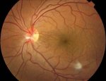
fig. 1 Folds macular in limiting membrane internal due to the vitreous traction on the focus scar of retinitis.
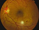
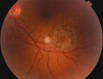
fig. 2 & 3 Retinal hole in secondary later pole the focus of candidiasis (before and after fotocoagular).
I want to make it clear IV drug users should NEVER use citric acid from lemons to prep heroin (usually brown heroin). Only sterile citric acid should ever be used, or failing that, acetic acid (vinegar). Heating the shot will NOT kill the Candida fungus if it is in the juice of the lemon... hell, they've found fungus which have survived re-entry on the outside of the space shuttle, so a lighter ain't going to do shit
This fungus can cause blindness within a month. Fungus introduced via IV are very, very difficult to kill, so if there is fungus in the juice, chances are almost certain the fungus will thrive in your body... and preferentially on your retina
. Within 30 days vision can be totally occluded. And if you're really unlucky, it could be both eyes.*******
Posterior retinal hole secondary to a candida retinitis
Alvarez-Suarez ML, Sanchez-Tabar L et al, 2005, The Spanish Ophthalmology society files (Archivos de la Sociedad Espanola de Oftalmologia) 80(7): 421-4.
http://scielo.isciii.es/scielo.php?script=sci_arttext&pid=S0365-66912005000700008&lng=es&nrm=iso&tlng=es&skpa=on
ABSTRACT:
CASE REPORT: We describe the case of a 36-year-old man with a history of intravenous heroin use, who was HIV negative. Left ocular examination disclosed a focal candida retinitis in the posterior pole associated with vitritis and moderate iritis. Treatment with fluconazole inactived the chorio-retinal lesion and resolved the vitritis, but developed an inner limiting membrane contraction over the macula. Two years later, vitreous traction produced a retinal hole that needed argon laser photocoagulation. DISCUSSION: Candida retinitis which penetrates into the vitreous cavity can produce retinal holes by vitreous traction over the lesion.
CLINICAL CASE (rough translation)
Male 30 age that attends emergency for loss of vision, redness and pain in left eye (OS) of 3 days of evolution. The patient also concerns fever approximately 20 days ago and is heroinómano sporadic recognizing a last episode of several Brown heroin injections approximately 25 days ago.
Exploration of the right eye (OD) was normal, while in the left eye (OS) there was an accompanied by a most focus of retinitis with localized vitritis previous Uveitis. The clinical exploration only detected febrícula and a few small bilateral latero-cervicales adenopathy.
Eye injuries were typical of candidiasis started hospital treatment with intravenous, fluconazole at dose of 200 mg / 24 h, and an eye pattern of corticosteroids and midriáticos topics. Complementary studies showed a Leukocytosis with normal formula (no neutropenia), a proteinograma with discrete increase in the alpha-2-globulins and some negative hepatitis B and HIV markers. The blood cultures, Lues, toxoplasma, CMV, serology and BK sputum and urine samples were also negative. Puncture a cervical Lymphadenopathy showed a reactive lymphadenitis with little activity blastic.
The box improved gradually with fever, the cilium-conjunctival reaction and inflammation of anterior chamber disappearance. The back of eye could see a good scarring of the prerretiniana injury and treatment was completed with oral fluconazole other 4 weeks.
At the end of treatment visual acuity in OI was 0.6, with a vitreous clear and with the focus of totally scar retinitis but associated vitreous traction which produced a marked contraction of the internal level macular limiting membrane ( fig. 1 ). 2 Years occurred a retinal hole where griped retino-vitreous scar focus, that the patient detected by sudden clinic in metamorfopsia. The tear was accompanied by a small detachment surrounded with two barriers of laser argon ( figs. 2 and 3 ) and was followed by a marked visual improvement until 0.9 release of traction on limiting internal.
DISCUSSION
NSFW:
Eye thrush is a pathology that typically appears in drug addicts injecting, in immunosuppressed patients, and when there is a long-term use of catheter. These last two risk factors seems to have moved to the use of heroin, the main cause of endophthalmitis Candida in the 1980s and early 90 (2). They are also predisposing causes hemodialysis, corticosteroids, a prolonged (especially associated to abdominal surgery) antibioterapia, diabetes, pregnancy, alcoholism, liver failure, the post-partum, prematurity, manipulation genitourinaria e even described in people healthy (2).
The diagnosis is set by the special characteristics of risk in these patients, the morphological aspect of the retino-vitreous lesions and often, by the coexistence of other associated manifestations of systemic candidiasis (3). Retinitis Candidal appears as an injury blanco-amarillentas coriorretinianas located mainly in later pole. The retinitis progresses and crosses the internal limiting membrane to spread itself over ... to the vitreous ... and can (cause major) complications including retinal detachment, retraction of the limiting internal, membranes fibrovasculares and ptisis bulbi (1.3). An intense iritis and sinequiante is also typical and translates a sterile in anterior chamber inflammatory reaction.
Most of these patients with eye affectation typically present other clinical signs as inflammation ... The treatment of patients ... relies on fluconazole po or iv, at a dose of 100-400mg/day for 4-8 weeks (1,2). The vitrectomy is done in case of poor response to the fluconazole, presence of severe vitritis and for the management of vítreo-retinianas complications as grout persistent vitreous haze, retinal detachment, membrane epirretiniana macular and neovascularization (4). A significant visual loss by contraction of the internal limiting membrane would also be an indication of peeled of the constraint with the help of the biological stains. At the time of the vitrectomy amphotericin B intravítrea, can be associated corticoids intraocular and complemented by oral or intravenous fluconazole (2).
fig. 1 Folds macular in limiting membrane internal due to the vitreous traction on the focus scar of retinitis.
fig. 2 & 3 Retinal hole in secondary later pole the focus of candidiasis (before and after fotocoagular).
Attachments
Last edited:
