• N&PD Moderators: Skorpio | thegreenhand

News NEUROSCIENCE | Demystifying Psychedelics ->

mr peabody

Bluelight Crew
Joined
Aug 31, 2016
Messages
5,714
41009784_m-848x424.jpg


Ketamine may relieve depression by repairing damaged brain circuits

by Jon Hamilton | NPR

The anesthetic ketamine can relieve depression in hours and keep it at bay for a week or more. Now scientists have found hints about how ketamine works in the brain.

In mice, the drug appears to quickly improve the functioning of certain brain circuits involved in mood. Then, hours later, it begins to restore faulty connections between cells in these circuits.

The finding comes after the Food and Drug Administration in March approved Spravato, a nasal spray that is the first antidepressant based on ketamine.

The anesthetic version of ketamine has already been used to treat thousands of people with depression. But scientists have known relatively little about how ketamine and similar drugs affect brain circuits.

The study offers "a substantial breakthrough" in scientists' understanding, says Anna Beyeler, a neuroscientist at INSERM, the French equivalent of the National Institutes of Health, who wasn't involved in the research. But there are still many remaining questions, she says.

Research has found evidence that ketamine was creating new synapses, the connections between brain cells. But the new study appears to add important details about how and when these new synapses affect brain circuits, says Ronald Duman, a professor of psychiatry and neuroscience at Yale University.

Studying ketamine's antidepressant effects in mice presented a challenge. "There's probably no such thing as a depressed mouse," says Dr. Conor Liston, a neuroscientist and psychiatrist at Weill Cornell Medicine in New York and an author of the Science paper.

Liston and his team of scientists gave mice a stress hormone that caused them to act depressed. For example, the animals lost interest in favorite activities like eating sugar and exploring a maze.

Then the team used a special laser microscope to study the animals' brains. The researchers were looking for changes to synapses.

"Stress is associated with a loss of synapses in this region of the brain that we think is important in depression," Liston says. "And sure enough, the stressed-out mice lost a lot of synapses."

Next, the scientists gave the animals a dose of ketamine. And Liston says that's when they noticed something surprising. "Ketamine was actually restoring many of the exact same synapses in their exact same configuration that existed before the animal was exposed to chronic stress," he says.

In other words, the drug seemed to be repairing brain circuits that had been damaged by stress.

That finding suggested one way that ketamine could be relieving depression in people. But it didn't explain how ketamine could work so quickly.

Was the drug really creating all these new synapses in just a couple of hours?

To find out, the team used a technology that makes living brain cells glow under a microscope. "You can kind of imagine Van Gogh's Starry Night," Liston says. "The brain cells light up when they become active and become dimmer when they become inactive."

That allowed the team to identify brain circuits by looking for groups of brain cells that lit up together.

And that's when the scientists got another surprise.

After the mice got ketamine, it took less than six hours for the brain circuits damaged by stress to begin working better. The mice also stopped acting depressed in this time period.

But both of these changes took place long before the drug was able to restore many synapses.

"It wasn't until 12 hours after ketamine treatment that we really saw a big increase in the formation of new connections between neurons," Liston says.

The research suggests that ketamine triggers a two-step process that relieves depression.

First, the drug somehow coaxes faulty brain circuits to function better temporarily. Then it provides a longer-term fix by restoring the synaptic connections between cells in a circuit.

"One possibility is that the synapses are restored spontaneously once the cells in a circuit begin firing in a synchronized fashion," says INSERM'S Beyeler, who wrote a commentary accompanying the study.

"The new study suggests not only how ketamine works but also why its effects typically wear off after a few days or weeks", she says. "What we can imagine is that ketamine always has this short-term antidepressant effect, but then if the synaptic changes are not maintained, you will have relapse, and if that's true," she says, "scientists' next challenge is to find a way to maintain the brain circuits that ketamine has restored."

 
Last edited:
pcblsd_withskull.jpg


How psilocybin works on the brain

Frederick Barrett, Samuel Krimmel, Roland Griffiths, David Seminowicz, Brian Mathur | Johns Hopkins Medicine | 5 Jun 2020

To see how psychedelics impact the claustrum, a mysterious region of the brain believed to control the ego, researchers compared the brain scans of people after they took psilocybin with their scans after taking a placebo.

Perhaps no region of the brain is more fittingly named than the claustrum, taken from the Latin word for "hidden or shut away." The claustrum is an extremely thin sheet of neurons deep within the cortex, yet it reaches out to every other region of the brain. Its true purpose remains "hidden away" as well, with researchers speculating about many functions. For example, Francis Crick of DNA-discovery fame believed that the claustrum is the seat of consciousness, responsible for awareness and sense of self.

What is known is that this region contains a large number of receptors targeted by psychedelic drugs such as LSD or psilocybin ¾ the hallucinogenic chemical found in certain mushrooms. To see what happens in the claustrum when people are on psychedelics, Johns Hopkins Medicine researchers compared the brain scans of people after they took psilocybin with their scans after taking a placebo.

Their findings were published online on May 23, 2020, in the journal NeuroImage.

The scans after psilocybin use showed that the claustrum was less active, meaning the area of the brain believed responsible for setting attention and switching tasks is turned down when on the drug. The researchers say that this ties in with what people report as typical effects of psychedelic drugs, including feelings of being connected to everything and reduced senses of self or ego.

"Our findings move us one step closer to understanding mechanisms underlying how psilocybin works in the brain," says Frederick Barrett, Ph.D., assistant professor of psychiatry and behavioral sciences at the Johns Hopkins University School of Medicine and a member of the school's Center for Psychedelic and Consciousness Research.

"This will hopefully enable us to better understand why it's an effective therapy for certain psychiatric disorders, which might help us tailor therapies to help people more."
Because of its deep-rooted location in the brain, the claustrum has been difficult to access and study. Last year, Barrett and his colleagues at the University of Maryland, Baltimore, developed a method to detect brain activity in the claustrum using functional magnetic resonance imaging (fMRI).

For this new study, the researchers used fMRI with 15 people and observed the claustrum brain region after the participants took either psilocybin or a placebo. They found that psilocybin reduced neural activity in the claustrum by 15% to 30%. This lowered activity also appeared to be associated with stronger subjective effects of the drug, such as emotional and mystical experiences. The researchers also found that psilocybin changed the way that the claustrum communicated with brain regions involved in hearing, attention, decision-making and remembering.

With the highly detailed imaging of the claustrum provided by fMRI, the researchers next hope to look at the mysterious brain region in people with certain psychiatric disorders such as depression and substance use disorder. The goal of these experiments will be to see what roles, if any, the claustrum plays in these conditions. The researchers also plan to observe the claustrum's activity when under the influence of other psychedelics, such as salvinorin A, a psychedelic derived from a Mexican plant.

 
dendrite-connections.png

This figure shows the effects of three psychedelics, DMT, LSD, amphetamines (DOI) and one control
(VEH) on neurons in the prefrontal cortex (Ly et al)


LSD and magic mushrooms could heal brain cells damaged by depression, study

by Alex Matthews-King | The Independent

Psychedelics could be 'next generation' of safer treatments for mental health.

Psychedelic drugs like LSD and ecstasy ingredient MDMA have been shown to stimulate the growth of new branches and connections between brain cells which could help address conditions like depression and addiction.

Researchers in California have demonstrated these substances, banned as illicit drugs in many countries, are capable of rewiring parts of the brain in a way that lasts well beyond the drugs' effects.

This means psychedelics could be the "next generation" of treatments for mental health disorders which could be more effective and safer than existing options, the study's authors from the University of California.

In previous studies by the same team, a single dose of DMT, the key ingredient in ayahuasca medicinal brews of Amazonian tribes, has been shown to help rats overcome a fear of electric shock meant to emulate post-traumatic stress disorder (PTSD).

Now they have shown this dose increases the number of branch-like dendrites sprouting from nerve cells in the rat's brain.

These dendrites end at synapses where their electrical impulses are passed on to other nerve cells and underpin all brain activity. But they can atrophy and draw back in people with mental health conditions.

“One of the hallmarks of depression is that the neurites in the prefrontal cortex – a key brain region that regulates emotion, mood, and anxiety – those neurites tend to shrivel up,” says Dr David Olson, who lead the research team.

These brain changes also appear in cases of anxiety, addiction, and post-traumatic stress disorder and stimulating them to reconnect could help to address this.

The research, published in the journal Cell Reports today, looked at drugs in several classes including tryptamines, DMT and magic mushrooms; amphetamines, including MDMA; and ergolines, like LSD.

In tests on human brain cells in the lab, flies and rats, it found these substances consistently boosted brain connections.

Dr Olson compared the effects to ketamine, another illicit drug which represents one of the most important new treatments for depression in a generation, and found many psychedelics have equal or greater effects.

A ketamine nasal spray is being fast-tracked through clinical trials after it was shown to rapidly relieve major depression and suicidal thoughts in people who cannot be helped by other treatments.

However its use has to be weighed against its potential for abuse, and its ability to cause a form of drug-induced psychosis.

“The rapid effects of ketamine on mood and plasticity are truly astounding,” said Dr Olson. “The big question we were trying to answer was whether or not other compounds are capable of doing what ketamine does.”

“People have long assumed that psychedelics are capable of altering neuronal structure, but this is the first study that clearly and unambiguously supports that hypothesis."


The fact that many of these drugs seem to mimic the groundbreaking benefits of ketamine opens up an array of new treatment options, which may be less open to abuse, if these drugs can make it to clinical trials.

Dr Olson said: “Ketamine is no longer our only option. Our work demonstrates that there are a number of distinct chemical scaffolds capable of promoting plasticity like ketamine, providing additional opportunities for medicinal chemists to develop safer and more effective alternatives.”

The news that yet more banned substances could help tackle serious and debilitating disease comes as the UK Home Office is embroiled in a row over medicinal cannabis in treating epilepsy.

After months seizure-free, 12-year-old Billy Caldwell had a seizure last night after airport customs officials confiscated his prescription from Canada.

Billy had previously had the UK’s only NHS medical cannabis prescription, for an oil which banished seizures that used to strike 100 times a day, but the Home Office intervened to block his GP from prescribing it.

 
rutger-hauer-blade-runner-1.jpg


Psychedelic drugs change the structure of neurons*

by Peter Hess | INVERSE

The brain is continually reorganizing itself by forming new neural connections throughout life. This phenomenon is known as Neuroplasticity.

Psychedelic drugs - LSD, DMT, and psilocybin - have shaken off a lot of their stigma and reputation as party drugs in the past few years, as scientists begin to investigate their significant healing potential for people with mental illness. Similarly, the drug ketamine, best known as a rave drug, has also shown promise in rapidly treating medication-resistant depression, and like psychedelics, its effects persist after treatment has ended.

New research shows that the way psychedelics repair the brain is very similar to ketamine’s action, which could pave the way for a future class of fast-acting drugs to treat conditions like depression, post-traumatic stress disorder, and substance use disorders. In a paper published Tuesday in the journal Cell Reports, a team of researchers showed evidence that psychedelic drugs can induce structural changes in nerve cells — a trait called Neuroplasticity — that could, in turn, help repair brain dysfunction in people with mood and anxiety disorders.

“Psychedelics are some of the most powerful compounds known to impact brain function so I was very interested to know what their mechanisms of action are,” David Olson, Ph.D., an assistant professor of biochemistry and molecular medicine at UC Davis and the corresponding author on the study, tells Inverse. "This paper adds to the on growing body of psychedelic neuroscience research by showing some of the changes induced by psychedelics."

Through experiments conducted on cultured rat neurons, as well as the actual brains of fruit flies and rats, Olson and his colleagues found that LSD, DMT, and DOI (2,5-dimethoxy-4-iodoamphetamine, a potent psychedelic amphetamine) increased the number of dendrites (branches) in nerve cells, increased the density of dendritic spines (protrusions on dendrites that help the neurons receive input from other cells), and increased number of synapses (functional connections between neurons). Altogether, these findings suggest that psychedelics induce structural changes to the brain, which Olson says can help treat mental illness.

“The structure of neurons affects their function, and in the case of a lot of neuropsychiatric diseases, particularly mood and anxiety disorders, these are characterized by an atrophy of neurons in the prefrontal cortex, a key brain region that regulates emotion, fear, and reward,” says Olson. “Finding compounds that promote growth of those neurons we might enable us to repair the circuits are damaged in those diseases.”

Since prefrontal cortex helps control other areas of the brain involved in fear, anxiety, and reward, says Olson, it’s a critical region for the treatment of depression, PTSD, and substance use disorders.

But Olson and his co-authors aren’t just interested in using psychedelics to treat patients. They hope to use psychedelic compounds as tools to dig down into the biochemical signaling pathways that lead to the Neuroplasticity observed in this study. By identifying the specific ways in which psychedelics act on the nervous system, Olson and his colleagues hope that they can develop a new generation of drugs that will replicate — or improve upon — the rapid, long-lasting healing effects of ketamine and psychedelics, but without the potential for abuse or challenging experiences.

“That’s the ultimate goal: to use psychedelics as inspiration for better medicine,” Olson says.

Of course, this is just one snapshot of the neuronal changes induced by psychedelics, so further research will be necessary to find out long-term effects on brain function.

“Plasticity is not universally a good thing. We were hoping to induce plasticity in prefrontal cortex, which can be potentially useful for treating mood and anxiety disorders, but promoting plasticity in other parts of the brain, like the amygdala can induce anxiety,” says Olson. “It’s very unclear what the risks are right now.”

 
iu


Serotonin and brain function: A tale of two receptors*

Carhart-Harris, Nutt (2017)

Previous attempts to identify a unified theory of brain serotonin function have largely failed to achieve consensus. In this present synthesis, we integrate previous perspectives with new and older data to create a novel bipartite model centred on the view that serotonin neurotransmission enhances two distinct adaptive responses to adversity, mediated in large part by its two most prevalent and researched brain receptors: the 5-HT1A and 5-HT2A receptors.

We propose that passive coping is mediated by postsynaptic 5-HT1AR signalling and characterised by stress moderation. Conversely, we argue that active coping is mediated by 5-HT2AR signalling and characterised by enhanced plasticity (capacity for change). We propose that 5-HT1AR-mediated stress moderation may be the brain’s default response to adversity but that an improved ability to change one’s situation and/or relationship to it via 5-HT2AR-mediated plasticity may also be important – and increasingly so as the level of adversity reaches a critical point. We propose that the 5-HT1AR pathway is enhanced by conventional 5-HT reuptake blocking antidepressants such as the selective serotonin reuptake inhibitors (SSRIs), whereas the 5-HT2AR pathway is enhanced by 5-HT2AR-agonist psychedelics. This bipartite model purports to explain how SSRIs and psychedelics that modulate the serotonergic system in different ways, can achieve complementary adaptive and potentially therapeutic outcomes.

The function of brain serotonin

Here we suggest that the principal function of brain serotonin is to enhance adaptive responses to adverse conditions via two distinct pathways: (1) a passive coping pathway which improves stress tolerability; and (2) an active coping pathway associated with heightened plasticity, which, with support, can improve an organism’s ability to identify and overcome source(s) of stress by changing outlook and/or behaviour. Crucially, we propose that these two functions are mediated by signalling at postsynaptic 5-HT1A and 5-HT2A receptors respectively, with 5-HT1AR signalling dominating under ordinary conditions but 5-HT2AR signalling becoming increasingly operative as the level of adversity reaches a critical point.

We suggest that the two functions of interest (5-HT1AR-mediated stress relief and 5-HT2AR-mediated plasticity) are sufficiently distinct – and may even be mutually oppositional in certain contexts, evoking dilemmas over whether it is better to passively endure or actively approach, and in so doing, initiate some sort of fundamental change – with the potential for major resolution. This rule may not be absolute however - the two functions may also be complementary, e.g. in the case of enhanced serotonin functioning with chronic SSRI use – or indeed with normal basal 5-HT functioning, facilitating improved endurance and plasticity.

Despite this complementarity, we do anticipate that conventional serotonergic antidepressants such as the SSRIs and classic psychedelics such as psilocybin may become competitive options for the treatments of certain disorders such as depression; most fundamentally because they work via distinct pathways, but also because they cannot easily be taken in combination, i.e. conventional antidepressants attenuate the characteristic psychological effects of psychedelics. SSRIs are established evidence-based treatments for anxiety and major depression, whereas psychedelics are experimental medicines in an early phase of development. However, if evidence supporting the therapeutic value of psychedelics accrues – as we anticipate, and it is increasingly shown that their therapeutic mechanisms are significantly distinct from those of conventional medications, then this will open-up new and potentially empowering options for patients and clinicians. For the brave new psychiatry of the future – that many would like to see – decisions about whether to passively endure or actively address, may become increasingly pertinent.

*From the article here: https://journals.sagepub.com/doi/10.1177/0269881117725915
 
psychedelics-and-neurogenesis-via-bdnf-and-mtor.jpg

This figure shows the effects of three psychedelics and one control (VEH) on cortical neurons.


Sprucing up your brain with potent psychedelics

A new study, published June 12 in the journal Cell Reports, found psychedelics, specifically DOI, DMT, and LSD, can change brain cells in rats and flies, making neurons more likely to branch out and connect with one another. The work supports the theory that psychedelics could help to fight depression, anxiety, addiction, and post-traumatic stress disorder.

“One of the hallmarks of depression is that the neurites in the prefrontal cortex — a key brain region that regulates emotion, mood, and anxiety — those neurites tend to shrivel up,” says Olson. These brain changes also appear in cases of anxiety, addiction, and post-traumatic stress disorder.

Psychedelics are not the most popular drugs for treating depression, but as we better understand how they promote the growth of new dendrites and synapses, we should be better able to develop safer and more effective antidepressants to accomplish the same effect.

More from Cell 12 June 2018:

Neuropsychiatric diseases, including mood and anxiety disorders, are some of the leading causes of disability worldwide and place an enormous economic burden on society. Approximately one-third of patients will not respond to current antidepressant drugs, and those who do will usually require at least 2–4 weeks of treatment before they experience any beneficial effects (Rush et al., 2006). Depression, post-traumatic stress disorder (PTSD), and addiction share common neural circuitry and have high comorbidity.

Atrophy of neurons in the prefrontal cortex (PFC) plays a key role in the pathophysiology of depression and related disorders. The ability to promote both structural and functional plasticity in the PFC has been hypothesized to underlie the fast-acting antidepressant properties of the dissociative anesthetic ketamine. Here, we report that, like ketamine, serotonergic psychedelics are capable of robustly increasing neuritogenesis and/or spinogenesis both in vitro and in vivo. These changes in neuronal structure are accompanied by increased synapse number and function, as measured by fluorescence microscopy and electrophysiology. The structural changes induced by psychedelics appear to result from stimulation of the TrkB, mTOR, and 5-HT2A signaling pathways and could possibly explain the clinical effectiveness of these compounds. Our results underscore the therapeutic potential of psychedelics and, importantly, identify several lead scaffolds for medicinal chemistry efforts focused on developing plasticity-promoting compounds as safe, effective, and fast-acting treatments for depression and related disorders.

Psychedelics promote structural and functional neural plasticity

The neuroplasticity described in the 12 June 2018 paper in Cell consists of “neuritogenesis,” or the growth of dendrites and synaptic buttons — providing a denser connectivity between neurons.

Another form of neuroplasticity which may take place under some conditions is “neurogenesis,” or the growth of stem cells which develop into neurons. In adults, this may occur in the hippocampus and along the ventricular lining of the brain.

The hippocampus is the region of the medial temporal lobes thought to play a prominent role in the retention of long term memories. More on hippocampal neurogenesis:

Here we assessed whole autopsy hippocampi from healthy human individuals ranging from 14 to 79 years of age. We found similar numbers of intermediate neural progenitors and thousands of immature neurons in the DG, comparable numbers of glia and mature granule neurons, and equivalent DG volume across ages. Nevertheless, older individuals have less angiogenesis and neuroplasticity and a smaller quiescent progenitor pool in anterior-mid DG, with no changes in posterior DG. Thus, healthy older subjects without cognitive impairment, neuropsychiatric disease, or treatment display preserved neurogenesis.

It is possible that some psychedelic mixtures may stimulate the transition of neural stem cells into functioning neurons:

When grown with ayahuasca compounds, the stem cells in the neurospheres also began to differentiate (change their properties) to resemble neurons more effectively than under control conditions. This means that the stem cells started to lose their stem-cell-like properties, and started making proteins that are found in adult neurons.

Overall, it seems that exposing neural stem cells to harmine, THH, and harmaline encourages them to grow and change into new neurons more effectively than under control conditions.

This is all very controversial

Many neuroscientists still doubt that adult human brains can generate new neurons past adolescence. Proving that psychedelics can promote the growth of new neurons in the brain (e.g. hippocampus) will take some time, and perhaps a somewhat different perspective on neuropharmaceutical ethics than is dominant at this time.

It is less controversial to pursue the use of ketamine derivatives to promote new “branching” of existing neurons. But ketamine does not seem to be as effective at neuritogenesis as LSD, DMT, or psilocybin. In other words, some old prejudices may need to fall in order to achieve a better solution to an age-old scourge of the human spirit.

Here at the Al Fin Institutes of Neuroscience, we prefer the use of occasional treatments for chronic ailments over the everyday medication of persons. As people age, their ailments may tend to accumulate — as do the number of treatments required. Many persons may take as many as 10 to 20 medication doses per day. The use of psychedelics or psychedelic analogs for treating depression would likely be of an intermittent nature, rather than daily, and would help to cut down on the total daily dosage regimen.

We actually prefer to avoid medication altogether

If possible, the avoidance of pharmaceuticals is best. Electromagnetic or photic approaches to treating depression are preferred, as are other alternatives such as advanced neurofeedback, mindfulness, and cognitive behavioural therapies.

Still, some people have experienced relief from depression lasting 6 months or longer from a single psychedelic experience, if well designed and overseen.

We have barely begun to understand all the productive approaches to functional neuroplasticity in the treatment of neurologic and neuropsychiatric disorders.

Renewed scientific interest in the use of psychedelics to treat disorders of the spirit can be seen as a positive sign.

https://alfinnextlevel.wordpress.com/2018/06/15/sprucing-up-your-brain-with-potent-psychedelics/
 
sleepingbrain_wide-e40290d47221863e13990f78f86b983781d5673e-s800-c85.jpeg


Waves of fluid bathe the sleeping brain*

Neuroscience News | Boston University | Nov 1 2019

New research from Boston University suggests that tonight while you sleep, something amazing will happen within your brain. Your neurons will go quiet. A few seconds later, blood will flow out of your head. Then, a watery liquid called cerebrospinal fluid (CSF) will flow in, washing through your brain in rhythmic, pulsing waves.

The study, published on October 31 in Science, is the first to illustrate that the brain’s CSF pulses during sleep, and that these motions are closely tied with brain wave activity and blood flow.

“We’ve known for a while that there are these electrical waves of activity in the neurons,” says study coauthor Laura Lewis, a BU College of Engineering assistant professor of biomedical engineering and a Center for Systems Neuroscience faculty member. “But before now, we didn’t realize that there are actually waves in the CSF, too.”

This research may also be the first-ever study to take images of CSF during sleep. And Lewis hopes that it will one day lead to insights about a variety of neurological and psychological disorders that are frequently associated with disrupted sleep patterns, including autism and Alzheimer’s disease.

The coupling of brain waves with the flow of blood and CSF could provide insights about normal age-related impairments as well. Earlier studies have suggested that CSF flow and slow-wave activity both help flush toxic, memory-impairing proteins from the brain. As people age, their brains often generate fewer slow waves. In turn, this could affect the blood flow in the brain and reduce the pulsing of CSF during sleep, leading to a buildup of toxic proteins and a decline in memory abilities. Although researchers have tended to evaluate these processes separately, it now appears that they are very closely linked.

To further explore how aging might affect sleep’s flow of blood and CSF in the brain, Lewis and her team plan to recruit older adults for their next study, as the 13 subjects in the current study were all between the ages of 23 and 33. Lewis says they also hope to come up with a more sleep-conducive method of imaging CSF. Wearing EEG caps to measure their brain waves, these initial 13 subjects were tasked with dozing off inside an extremely noisy MRI machine, which, as anyone who has had an MRI can imagine, is no easy feat.

“We have so many people who are really excited to participate because they want to get paid to sleep,” Lewis says with a laugh. “But it turns out that their job is actually–secretly–almost the hardest part of our study. We have all this fancy equipment and complicated technologies, and often a big problem is that people can’t fall asleep because they’re in a really loud metal tube, and it’s just a weird environment.”

But for now, she is glad to have the opportunity to take images of CSF at all. "One of the most fascinating yields of this research," Lewis says, "is that they can tell if a person is sleeping simply by examining a little bit of CSF on a brain scan."

“It’s such a dramatic effect,”
she says.

As their research continues to move forward, Lewis’ team has another puzzle they want to solve: How exactly are our brain waves, blood flow, and CSF coordinating so perfectly with one another? “We do see that the neural change always seems to happen first, and then it’s followed by a flow of blood out of the head, and then a wave of CSF into the head,” says Lewis.

cerebrospinal-fluid-sleep-neurosciencenews.jpg

This shows the CSF in the brain during sleep. During sleep, the brain exhibits large-scale waves:
waves of blood oxygenation (red) are followed by waves of cerebrospinal fluid (blue).

"One explanation may be that when the neurons shut off, they don’t require as much oxygen, so blood leaves the area. As the blood leaves, pressure in the brain drops, and CSF quickly flows in to maintain pressure at a safe level."

“But that’s just one possibility,”
Lewis says. “What are the causal links? Is one of these processes causing the others? Or is there some hidden force that is driving all of them?”

*From the article here :
 
28220245083_0e79aa1921_b.jpg

Cambridge

Scientists discover key neural circuit regulating alcohol consumption

University of North Carolina | Neuroscience News

Specific neurons in the central nucleus of the amygdala regulate alcohol consumption.

Scientists have known that a region of the brain called the central nucleus of the amygdala (CeA) plays a role in behaviors related to alcohol use and consumption in general. It’s been less known which precise populations of brain cells and their projections to other brain regions mediate these behaviors. Now, UNC School of Medicine scientists discovered that specific neurons in the CeA contribute to reward-like behaviors, alcohol consumption in particular.

Published in the Journal of Neuroscience, this research pinpoints a specific neural circuit that when altered caused animal models to drink less alcohol.

“The fact that these neurons promote reward-like behavior, that extremely low levels of alcohol consumption activate these cells, and that activation of these neurons drive alcohol drinking in animals without extensive prior drinking experience suggests that they may be important for early alcohol use and reward,” said senior author Zoe McElligott, PhD, assistant professor of psychiatry and pharmacology.

“It’s our hope that by understanding the function of this circuit, we can better predict what happens in the brains of people who transition from casual alcohol use to subsequent abuse of alcohol, and the development of alcohol use disorders.”

McElligott, who is also a member of the UNC Bowles Center for Alcohol Studies, set out to investigate if a population of neurons that express a specific neuropeptide (neurotensin or NTS) contributes to reward-like behaviors and alcohol drinking. She was especially interested in these neurons in the context of inexperienced alcohol use, such as when a person first begins to drink alcohol. Also, NTS neurons are a subpopulation of other neurons in this CeA brain region that have been implicated in anxiety and fear – known as the somatostatin and corticotropin releasing factor neurons.

Using modern genetic and viral technologies in male mice, McElligott and colleagues found that selectively lesioning or ablating the NTS neurons in the CeA, while maintaining other types of CeA neurons, would cause the animals to drink less alcohol. This manipulation did not alter anxiety-like behavior. It also did not affect the consumption of other palatable liquids such as sucrose, saccharin, and bitter quinine solutions.

“We found that these NTS neurons in the CeA send a strong projection to the hindbrain, where they inhibit the parabrachial nucleus, near the brainstem,” McElligott said.

Using optogenetics – a technique where light activates these neurons – the researchers stimulated the terminal projections of the CeA-NTS neurons in the parabrachial and found that this stimulation inhibited the neurons in the parabrachial. When the scientists stimulated this projection with a laser in one half of the animal’s box, animals would spend more time where the stimulation would occur.

Animals also learned to perform a task to get the laser stimulation to turn on, and they would do this repeatedly, suggesting that they found this stimulation to be rewarding.

“Furthermore, when we stimulated this projection, animals would drink more alcohol as compared to when they had an opportunity to drink alcohol without laser stimulation,” McElligott said. “In contrast to our study where we ablated the NTS neurons, laser stimulation of this parabrachial pathway also caused the animals to consume caloric and non-caloric sweetened beverages. When the animals were presented with regular food and a sweet food, however, laser stimulation did not enhance the consumption regardless of the mouse’s hunger state. This suggests that different circuits may regulate the consumption of rewarding fluids and solids.”

McElligott and her graduate student María Luisa Torruella Suarez, the first author of this study, hope to explore how alcohol experience may change these neurons over time.

“Would these cells respond differently after animals have been drinking high quantities of alcohol over time?” McElligott said. “We also want to discover which populations of neurons in the parabrachial are receiving inputs from these neurons. Fully understanding this circuit could be the key to developing therapeutics to help people with alcohol use disorders.”

 
Stocksy-Womna-double-exposure-Lauren-Naefe.jpg


LSD and psilocybin could heal damaged brain cells in people suffering from depression, study

by Alex Matthews-King | The Independent

Psychedelics could be 'next generation' of safer treatments for mental health.

Psychedelic drugs like LSD and ecstasy ingredient MDMA have been shown to stimulate the growth of new branches and connections between brain cells which could help address conditions like depression and addiction.

Researchers in California have demonstrated these substances, banned as illicit drugs in many countries, are capable of rewiring parts of the brain in a way that lasts well beyond the drugs' effects.

This means psychedelics could be the "next generation" of treatments for mental health disorders which could be more effective and safer than existing options, the study's authors from the University of California.

In previous studies by the same team, a single dose of DMT, the key ingredient in ayahuasca medicinal brews of Amazonian tribes, has been shown to help rats overcome a fear of electric shock meant to emulate post-traumatic stress disorder (PTSD).

Now they have shown this dose increases the number of branch-like dendrites sprouting from nerve cells in the rat's brain.

These dendrites end at synapses where their electrical impulses are passed on to other nerve cells and underpin all brain activity. But they can atrophy and draw back in people with mental health conditions.

“One of the hallmarks of depression is that the neurites in the prefrontal cortex – a key brain region that regulates emotion, mood, and anxiety – those neurites tend to shrivel up,” says Dr David Olson, who lead the research team.

These brain changes also appear in cases of anxiety, addiction, and post-traumatic stress disorder and stimulating them to reconnect could help to address this.

The research, published in the journal Cell Reports today, looked at drugs in several classes including tryptamines, DMT and magic mushrooms; amphetamines, including MDMA; and ergolines, like LSD.

In tests on human brain cells in the lab, flies and rats, it found these substances consistently boosted brain connections.

Dr Olson compared the effects to ketamine, another illicit drug which represents one of the most important new treatments for depression in a generation, and found many psychedelics have equal or greater effects.

A ketamine nasal spray is being fast-tracked through clinical trials after it was shown to rapidly relieve major depression and suicidal thoughts in people who cannot be helped by other treatments.

However its use has to be weighed against its potential for abuse, and its ability to cause a form of drug-induced psychosis.

“The rapid effects of ketamine on mood and plasticity are truly astounding,” said Dr Olson. “The big question we were trying to answer was whether or not other compounds are capable of doing what ketamine does.”

“People have long assumed that psychedelics are capable of altering neuronal structure, but this is the first study that clearly and unambiguously supports that hypothesis."


The fact that many of these drugs seem to mimic the groundbreaking benefits of ketamine opens up an array of new treatment options, which may be less open to abuse, if these drugs can make it to clinical trials.

Dr Olson said: “Ketamine is no longer our only option. Our work demonstrates that there are a number of distinct chemical scaffolds capable of promoting plasticity like ketamine, providing additional opportunities for medicinal chemists to develop safer and more effective alternatives.”

The news that yet more banned substances could help tackle serious and debilitating disease comes as the UK Home Office is embroiled in a row over medicinal cannabis in treating epilepsy.

After months seizure-free, 12-year-old Billy Caldwell had a seizure last night after airport customs officials confiscated his prescription from Canada.

Billy had previously had the UK’s only NHS medical cannabis prescription, for an oil which banished seizures that used to strike 100 times a day, but the Home Office intervened to block his GP from prescribing it.

https://www.independent.co.uk/news/h...-a8395511.html
 
shutterstock_391979371-scaled-e1605639148596.jpg


Psychedelics may help the brain repair itself, study

Calvin Ly, Alexandra Greb, Lindsay Cameron, Kassandra Ori-McKenney, John Gray, David Olson

A new study in Cell Reports has found that psychedelics promote structural and functional neural plasticity.

In recent years, psychedelic party drugs such as LSD and MDMA have been studied by scientists for their potential ability to treat mental health problems like depression and anxiety—often in microdoses much smaller than the what a person would take to trip. But while the research into these drugs is promising, there’s still a lot we don’t understand about how they affect the brain. A new study, published Tuesday in Cell Reports, seems to offer the strongest evidence yet that they can actually help repair the brain’s circuitry and function.

The researchers, primarily from the University of California, Davis, exposed lab-cultured neurons from humans, rats, and other animals to various psychedelics. Drugs from different classes were used, including the amphetamine MDMA, the tryptamine psilocin, and the ergoline LSD. The neurons were taken from the prefrontal cortex, an area of the brain thought to be crucial in the development of certain mental illnesses.

Most of the psychedelics tested, the researchers found, promoted the growth of new dendrites from a neuron cell, which help transmit information from other neurons to the cell, as well as increased the density of small protrusions on these dendrites, known as dendritic spines. They also jumpstarted the growth of new connections, or synapses, between individual neurons. Similar effects were also seen in the brains of living test animals.

The net result of these changes, the authors say, is that they improve the brain’s plasticity, which includes its ability to repair itself from damage caused by things like stress or trauma. These changes, the researchers noted, are the reverse of what seems to happen in the brains of people living with chronic depression, post-traumatic stress disorder, or addiction. And they resemble the changes seen in people who take ketamine, an anesthetic and recreational drug that has been retooled in recent years as a fast-acting, if still experimental, antidepressant that some research has found can quickly tamp down suicidal thoughts.

“People have long assumed that psychedelics are capable of altering neuronal structure, but this is the first study that clearly and unambiguously supports that hypothesis,” said lead author David Olson, an assistant professor in the Departments of Chemistry and of Biochemistry and Molecular Medicine, in a statement. “What is really exciting is that psychedelics seem to mirror the effects produced by ketamine.”

It’s exciting, the authors say, because it means there’s more than one way for drugs to quickly improve a person’s brain plasticity. And the more options available, the better the chances someone can benefit from treatment, especially if other current drugs haven’t worked. Ultimately, it also provides researchers like Olson that many more avenues to pursue in developing more palatable versions of the psychedelic drugs we have available (i.e., versions that don’t cause long, mind-bending trips). The team even wants to rebrand these drugs as “psychoplastogens.”

“Ketamine is no longer our only option,”
Olson said. “Our work demonstrates that there are a number of distinct chemical scaffolds capable of promoting plasticity like ketamine, providing additional opportunities for medicinal chemists to develop safer and more effective alternatives.”

Olson’s team is already studying whether non-hallucinogenic analogs of these psychedelics can still improve brain plasticity, and they note that researchers elsewhere are in the middle of developing safer analogs of ketamine, which has some potential for addiction and abuse.

https://www.cell.com/cell-reports/fulltext/
 
tingri-everest-base-camp-trek.jpg


Can 5-MeO-DMT unlock the mystery of neurogenesis?

by Troy Farah | Psychedelic Times

2018 has been an incredible year for advances in psychedelic research, including psilocybin mushrooms, ketamine and more, not to mention a landmark year for MDMA and cannabis. Meanwhile, some research on a much lesser known psychedelic, 5-MeO-DMT, has slipped past national attention.

Known as 5-methoxy-N,N-dimethyltryptamine, this potent little molecule is most famous for occurring in the venom of a desert toad species, but it’s also present in many plant species or can be made synthetically. It’s in a class of chemicals called tryptamines, which includes DMT, psilocybin and even serotonin and melatonin.

Like many psychedelics, 5-MeO-DMT is highly active at serotonin receptors, but also at acetylcholine, glutamate and dopamine receptors as well, although at much lower potency. This has led some scientists to theorize that many DMT-related drugs could help with mood disorders. Some case studies have shown tryptamines to help with depression, anxiety, PTSD and more—but scientists still aren’t sure how exactly it all works.

The answer may lie in neurogenesis. In a newly published study in Frontiers in Molecular Neuroscience, Brazilian researchers looked at whether 5-MeO-DMT can stimulate neurogenesis in mice. After a single injection of 100 micrograms, the rodents showed significant cell proliferation compared to placebo.

Neurogenesis is exactly what it sounds like—the creation of new brain cells. When you’re young, the brain is constantly making new neurons. This activity slows down in adulthood, except in a few key places in the brain, such as a part of the hippocampus called the dentate gyrus. By stimulating this process, we can potentially fight diseases like Alzheimer’s.

The DG may be small, but it is thought to help form new memories, modulate mood disorders, and may even play a role in addiction. The debate is still raging over this, of course, with some studies saying the DG makes hundreds of new neurons per day, and others showing little change. But one theory suggests neurogenesis may improve a range of cognitive processes, especially in older people.

Perhaps 5-MeO-DMT can help us answer those questions. In the Brazilian study, the mice were injected with a drug called BrdU that helps scientists look at dividing cells in living tissue. Then, the mice were sacrificed by making them overdose on ketamine and xylazin. Unfortunately, it’s only possible to really look at these cell changes by cutting open the brains of mice—part of the reason this experiment hasn’t been done in humans—but at least they were euthanized peacefully.

The mice brains were washed and preserved, and the researchers found that 5-MeO-DMT not only produced more new neurons compared to controls, but they matured faster and survived better.

“Cells from animals submitted to a single 5-MeO-DMT injection showed dendrites with more branches and intersections,” the researchers wrote. Dendrites are the long branches of nerve cells that play an important role in brain chemistry. Like a tree, healthy branches means healthy brain cells. “Interestingly, chronic antidepressant therapy also accelerates the maturation of dendrites… To our knowledge, this work was the first to demonstrate a direct effect of a naturally occurring psychoactive compound in adult neurogenesis.”

There are some limitations to this research. For one, mice aren’t humans, so the conclusions that can be drawn are limited. Second, the researchers aren’t entirely sure how 5-MeO-DMT could be fostering neurogenesis, so what this means for the future of mental health remains to be seen.

With a few exceptions, there isn’t much new research on 5-MeO-DMT or related drugs. Last year, a different Brazilian team gave lab-grown mini-brains 5-MeO-DMT and found it had anti-inflammatory properties, and also promoted the growth of dendrites. Another rodent study shed some light on how 5-MeO-DMT works at serotonin receptors, but most studies are few and far between. There was also a first-of-its-kind epidemiological study that looked at spiritual and recreational use of 5-MeO-DMT, and concluded it has, “low potential for addiction, and might have psychotherapeutic effects.” Otherwise, that’s about it.

There may be a reason why 5-MeO-DMT research is largely ignored in the press. The chemical name is complex and difficult to remember, and most of this science is in very early stages.

There is still far too little research on 5-MeO-DMT to draw any strict conclusions on how it may benefit—or harm—humans; however, the positives seem stronger than the negatives. But this is exactly the point. While so much attention is centered on drugs like LSD and magic mushrooms, this compound is overlooked, and clearly could use more academic interest.

It would be wrong not to mention that federal scheduling has played a role in dampening this research. While 5-MeO-DMT has been used for centuries in some cultures, the United States only placed it in the highly illegal Schedule I category in 2011. Studying Schedule I drugs like marijuana, LSD and mescaline is notoriously difficult, so many institutions don’t even bother. If we really want to unlock the mysteries of this chemical—and what it can teach us about consciousness and the human mind—we must loosen restrictions on psychedelic research. The potential benefits are too important to ignore.

 
Beach-Tripping-709x473.jpg


Psychedelic drugs change the structure of neurons

by Andy Fell | UC Davis | Jun 12 2018

A team of scientists at the University of California, Davis, is exploring how psychedelic drugs impact the structure and function of neurons — research that could lead to new treatments for depression, anxiety and related disorders. In a paper published on June 12 in the journal Cell Reports, they demonstrate that a wide range of psychedelic drugs, including well-known compounds such as LSD and MDMA, increase the number of neuronal branches (dendrites), the density of small protrusions on these branches (dendritic spines), and the number of connections between neurons (synapses). These structural changes could suggest that psychedelics are capable of repairing the circuits that are malfunctioning in mood and anxiety disorders.

“People have long assumed that psychedelics are capable of altering neuronal structure, but this is the first study that clearly and unambiguously supports that hypothesis. What is really exciting is that psychedelics seem to mirror the effects produced by ketamine,” said David Olson, assistant professor in the departments of Chemistry and of Biochemistry and Molecular Medicine, who leads the research team.

Ketamine, an anesthetic, has been receiving a lot of attention lately because it produces rapid antidepressant effects in treatment-resistant populations, leading the U.S. Food and Drug Administration to fast-track clinical trials of two antidepressant drugs based on ketamine. The antidepressant properties of ketamine may stem from its tendency to promote neural plasticity — the ability of neurons to rewire their connections.

“The rapid effects of ketamine on mood and plasticity are truly astounding. The big question we were trying to answer was whether or not other compounds are capable of doing what ketamine does,” Olson said.

Psychedelics that show effects similar to ketamine

Olson’s group has demonstrated that other psychedelics mimic the effects of ketamine on neurons grown in a dish, and that these results extend to structural and electrical properties of neurons in animals. Rats treated with a single dose of DMT — a psychedelic compound found in the Amazonian herbal tea known as ayahuasca — showed an increase in the number of dendritic spines, similar to that seen with ketamine treatment. DMT itself is very short-lived in the rat: Most of the drug is eliminated within an hour. But the “rewiring” effects on the brain could be seen 24 hours later, demonstrating that these effects last for some time.

Behavioral studies also hint at the similarities between other psychedelics and ketamine. In another recent paper published in ACS Chemical Neuroscience, Olson’s group showed that DMT treatment enabled rats to overcome a “fear response” to the memory of a mild electric shock. This test is considered to be a model of post-traumatic stress disorder, or PTSD, and interestingly, ketamine produces the same effect. Recent clinical trials have shown that like ketamine, DMT-containing ayahuasca might have fast-acting effects in people with recurrent depression, Olson said.

These discoveries potentially open doors for the development of novel drugs to treat mood and anxiety disorders, Olson said. His team has proposed the term “psychoplastogen” to describe this new class of “plasticity-promoting” compounds.

“Ketamine is no longer our only option. Our work demonstrates that there are a number of distinct chemical scaffolds capable of promoting plasticity like ketamine, providing additional opportunities for medicinal chemists to develop safer and more effective alternatives,” Olson said.

Psychedelics vs. Psychoplastogens

Our group has coined the term “psychoplastogen” to refer to such compounds, and we believe that these molecules may hold the key to treating a wide variety of brain diseases.

Our studies on neurons grown in dishes, as well as experiments performed using fruit flies and rodents, have demonstrated that several psychoplastogens, including psychedelics and ketamine, encourage neurons to grow more branches and spines. It seems that all of these compounds work by activating mTOR – a key protein involved in cell growth.

The biochemical machinery that regulates mTOR activity is intricate. As we tease apart how psychedelics and other psychoplastogens turn on mTOR signaling, we might be able to engineer compounds that only produce the therapeutic effects on neuronal growth while bypassing pathways that lead to undesired hallucinations.

The field has known for some time now that psychedelics can produce lasting positive effects on brain function, and it’s possible that these long-lasting changes result from the psychoplastogenic effects of these drugs. If true, this would suggest that psychoplastogens might be used to repair circuits that are damaged in mood and anxiety disorders.

https://www.ucdavis.edu/news/psychedelic-drugs-change-structure-neurons/
 
psychedelics-and-neurogenesis-via-bdnf-and-mtor.jpg

This figure shows the effects of three psychedelics and one control (VEH) on cortical neurons.


Sprucing up your brain with potent psychedelics

A new study, published June 12 in the journal Cell Reports, found psychedelics, specifically DOI, DMT, and LSD, can change brain cells in rats and flies, making neurons more likely to branch out and connect with one another. The work supports the theory that psychedelics could help to fight depression, anxiety, addiction, and post-traumatic stress disorder.
that image is utterly fascinating
 
26165421_10214653835049839_499437499802799090_n-280x280.jpg

Thomas Varley, Neuroscientist

Do psychedelics trigger neurogenesis? Here's what we know.

Neurogenesis (the process by which the brain grows new neurons) has become something of a scientific buzzword recently, both in and out of psychedelic circles. It’s not hard to find supplements claiming that, through some pharmaceutical wizardry, you can harness the “power of neurogenesis.” Many psychedelic blogs have gotten very excited by the prospect that drugs like psilocybin might cause neurogenesis, hoping to generate momentum for the psychedelics-as-real-medicines cause.

After all, who could be against neurogenesis? Growing new brain cells does sound like the first step on the path to super powers, or at the very least, seriously enhancing abilities that we already have. But what is neurogenesis? Do psychedelics really cause it? If they do, what doors might that open up?

The vast, vast majority of neurogenesis happens before we are born: you come into this world with most of the neurons you’ll ever have, and over the course of your life they slowly die off. It has long been thought that the number of neurons you’d ever have is fixed at birth, but now we know that’s not quite true: adult neurogensis has been found to happen in a few select brain regions.

Even though adult neurogenesis is happening in the brain, it’s not happening a whole lot, and only in some pretty particular areas. The cerebral cortex (where most of the highest-level stuff happens) isn’t even on the map here. Sadly, neurogenesis isn’t going to turn you into some kind of mega-brain: most of your nervous system will remain unaffected by drugs that trigger neurogenesis. As far as we know, if you have damage to part of your cerebral cortex, taking psychedelics is unlikely to regrow the affected areas.

So should we all pack up and go home?

Not quite―just because neurogenesis isn’t as wide-spread or powerful as popular coverage might make it seem, there’s still reason to get excited. Of the few locations for neurogenesis, what’s happening in the dentate gyrus of the hippocampus is probably the most interesting, at least to those of us interested in psychedelic neuroscience. If those words sound like some kind of Harry Potter spell to you, you’re not alone and you may find some comfort knowing that generations of students have wept their way through neuroanatomy classes working on these same terms. The hippocampus is involved with many different aspects of cognition, but, at the risk of oversimplifying things, its primary role seems to be regulating both learning and memory.

Damage to the hippocampus can result in a variety of interesting and unpleasant effects, including permanent anterograde amnesia (the inability to form new memories), and it’s one of the first places damage from Alzheimer’s Disease manifests in the brain. For reasons that remain unclear, severe cases of major depression are associated with atrophy of the hippocampus, sometimes by as much as 20 percent, which may explain why, as anyone who has suffered from severe depression knows, being depressed is more than just being down all the time. It comes with its own unique constellation of cognitive effects, including memory problems and issues with focusing and concentration. This last finding is particularly interesting when we add in the fact that the balance of evidence suggests that exposure to psychedelics can, in fact, enhance neurogenesis in this region. This has some pretty profound implications for neuroscience and medicine.

It’s actually a little-known fact that there’s been some research that suggests psychedelics can enhance the natural ability to learn new behaviors and form associations. So far, all the work has been done with animals (rabbits and rats, mostly), but the promise is there.

Brainmaps-macaque-hippocampus.jpg

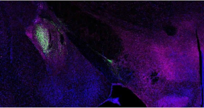
A CORNAL CUTTING OF A MACAQUE BRAIN, USING A NISSL STAIN. THE HIPPOCAMPUS IS CIRCLED
AND THE DENTATE GYRUS IS LABELED

Two studies using LSD found that the psychedelic enhanced the rate at which rabbits learned a new conditioned behavior, and that higher doses resulted in faster learning. The same researchers found that MDMA, MDA, and DOM all did as well. A more recent study using psilocybin found similar results, albeit only at low doses. It’s hard to draw any strong conclusions from a handful of studies like this―it’s a long way from simple associative learning in a rabbit or rat, to a complex human behavior (like playing the piano), but it’s a start. For researchers interested in treating debilitating psychological conditions like depression using psychedelic medicines, these are enormously promising results.

Why this doesn’t get talked about more in psychedelic circles is beyond me.

So what does this have to do with the idea of neurogenesis? Neurogensis is thought to be one of the mechanisms by which this kind of learning might occur. There have been studies that suggest that, for at least some kind of learning, neurogenesis in the hippocampus may be a key part of the acquisition of new behaviors and pattern recognition. In the interest of fairness, it is worth noting that not every study has validated this theory―there’s still quite a bit of science to be done, but the groundwork has been laid. The same team that was researching the effects of psilocybin on learning in rats found signs of new neural growths in the hippocampus in the rats that had been given the low dose psychedelic treatment and learned the new behavior faster. Unfortunately, as of now, this is the only study that has found a psychedelic triggered neurogenesis AND enhanced learning behavior.

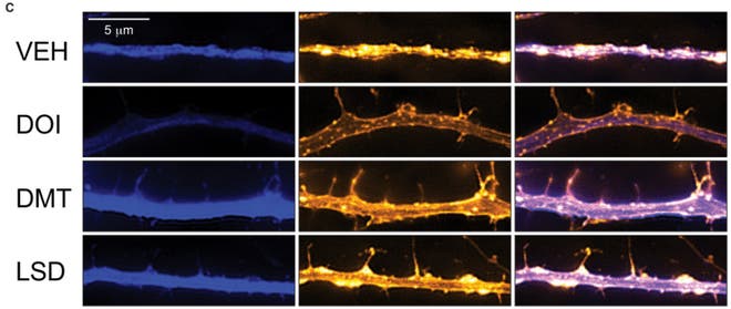
Don’t despair though, there is some circumstantial evidence that should be of interest to those banking on this theory of psychedelic neurogenesis. It has been known for quite a while that the receptor that psychedelic drugs target (the Serotonin 2A receptor) helps regulate the production of a molecule called Brain-Derived Neurotrophic Factor (BDNF, for short). Activate the receptor, and the brain secretes more BDNF. My own (unpublished) research found that that the psychedelic drug DPT (a close analogue of the more famous DMT) increased signs of BDNF in the brains of adult zebrafish, and studies using neurons in a dish and the drug DOI found similar results.

BDNF helps regulate neurogenesis and neuroplasticity: mice that have been artificially rendered unable to produce BDNF show severely distorted nervous systems, with behaviors thought to be related to psychiatric illnesses like eating disorders and OCD. Using genetic techniques to increase BDNF expression can enhance neurogenesis in certain brain regions as well. So far, no one has shown that BDNF causes increased learning capability directly, although participating in learning tasks causes a rapid increase in hippocampal BDNF expression in rats.

Gray739-emphasizing-hippocampus.png


My (tentative) hypothesis is that psychedelic drugs can enhance learning and memory capabilities by, at least partially, increasing the amount of BDNF (and related growth-factors) in the brain through activation of the Serotonin 2A receptor. The evidence for this idea is circumstantial right now―so far there hasn’t been a study that combines all of these different moving parts. At the very least, you would need to show that exposure to something like LSD increased how quickly an animal learned a new task, that levels of BDNF in the brain went up, and there was evidence of increased neurogenesis in the hippocampus. This is a fairly tall order, although not one that’s impossible, not by a long shot. All of the individual parts are well within the capabilities of modern science; it’s getting someone to throw time and money behind the question that’s the trick.

Interestingly, in 2016, the Beckley Foundation, working with scientists at the Sant Pau Institute for Biomedical Research in Spain announced findings that two of the key components in Ayahuasca, harmine and tetrahydroharmine stimulate the differentiation of stem cells into healthy neurons when they’re cultured in a dish. There’s still a lot of work to be done on this topic: researchers are moving ahead studying whether the same effect will be seen in living animals, and, if the findings are replicated, these findings would have big implications for the science of neurogenesis.

How these two molecules might stimulate neurogenesis is an open question: unlike drugs like psilocybin and LSD, which act directly at the 5-HT2A receptor, harmine and tetrahydroharmine act as inhibitors of the enzyme monoamine oxidase (MAO), which degrades natural neurotransmitters like serotonin and dopamine. It may be that, when MAO is inhibited, the increase in free-floating serotonin in the brain can trigger BDNF by binding to the 5-HT2A receptor, much like psilocybin would. It may also be an entirely new pathway that still needs to be discovered. For psychedelic scientists, this is the start of an exciting new world of possibilities.

Gray717-copy.png


So if all of this is true, what does it mean?

One of the front-line treatments for mental illnesses like anxiety, PTSD, and OCD is cognitive behavioral therapy (CBT), which is based on the same principles that the researchers are investigating with the classical conditioning studies. Anyone who’s participated in a session of CBT knows that the premise is actually very simple, and very reminiscent of the kind of mechanisms scientists use to train rats. Imagine you have PTSD with a specific trigger that sends you into panic attacks. A CBT approach to treatment might be exposure therapy: in a safe setting, guided by a therapist, you are gently exposed to your triggers, again and again. As time goes on and nothing terrible happens, your brain learns something new; the trigger isn’t dangerous, and slowly, your original response is made extinct. Similar techniques are used for patients with OCD (who might be unable to stop doing a particular ritual because they’re afraid something terrible will happen) and anxiety disorders. CBT is also used to treat depression, and while the mechanisms are a little different, the same basic principles apply.

It’s easy to see why, if it’s true, psychedelic neurogenesis might be useful. If we could use psychedelics to bolster and enhance our own innate capacity for learning, the applications for treatments and therapies would be tremendous. Humans are fundamentally pattern-making machines. Our most impressive technologies stem from our ability to recognize and make patterns, while some of our deepest illnesses, such as drug addiction, OCD, and depression are the result of getting caught in patterns we cannot control. If the theory laid out here is correct (and it may be entirely wrong), this could be another foundational piece on which to build psychedelic therapy. Beyond knowing just that it works, this might give us a robust, scientific understanding of why and how. It also may help us design new paradigms of psychedelic treatment: currently, almost all of the big studies being done are investigating the effects of single, medium-to-high doses of something like psilocybin, but, if psychedelic neurogenesis is real, there may be just as much therapeutic power in a series of repeated lower doses, in the right context. A medical microdose. The possibilities are endless.

Of course, such simplistic solutions should always be taken with a grain of salt: if someone tells you they have an easy-to-digest answer to a problem involving the brain, they’re probably trying to sell you something (keep that quote in mind next time someone tells you that depression is just a lack of serotonin!). Simple theories can ultimately be built into far more complete, and complex pictures, and even if this isn’t the whole picture (which it almost certainly isn’t), it seems like a pretty good place to start.

https://www.psymposia.com/magazine/do-psychedelics-trigger-neurogenesis-heres-what-we-know/
 
5U9JDXi.gif


Single dose of 5-MeO-DMT found to stimulate cell proliferation and neuronal survivability

Rafael Vitor Lima da Cruz, Thiago Moulin, Lyvia Lintzmaier Petiz, Richardson Leao

Psychoactive tryptamines are a class of molecules that act as neurotransmitter in the vertebrate brain. 5-MeO-DMT is found in a great variety of plants in South America, with an even greater diversity of chemical analogs. It is a serotonin agonist that acts in a non-selective manner in 5-HT2A >5-HT2C >5-HT1A receptors. However, 5-MeO-DMT also acts in many glutamate, dopamine and acetylcholine receptors. 5-MeO-DMT is one of the main active ingredient of Ayahuasca, a millenarian decoction used as sacrament by south American indigenous tribes, known to induce powerful hallucinogen states when administered together with monoamine oxidase inhibitors. At present, Ayahuasca is used by many syncretic churches ritualistically, as way to heal many physical and mental illness with or without scientific knowledge about the effects. Recent studies also suggest that Ayahuasca can potentially treat recurrent depression even in a placebo controlled frame.

Deficits in adult neurogenesis are associated with the physiopathology of depression and modulation of neurogenesis is behind the action of several antidepressants. Serotonin reuptake inhibitors, for example, rescue normal neurogenesis levels in animal models of depression. Adult neurogenesis is known to occur in two sites in the brain, the subgranular zone of the dentate gyrus and the subventricular zone of the lateral ventricle. There is some debate whether or not SVZ neurogenesis responds to mood disorders and psychoactive drugs, but the effect of mood disorders in SGZ Radial glial Like cell proliferation and neuronal survivor is prolifically described. Interestingly, alkaloids from one of the plants used in the Ayahuasca brew stimulate neurogenesis in vitro; however, it is not known whether in vivo adult neurogenesis is affected by psychoactive tryptamines.

In this study we tested if a single dose of 5-MeO-DMT affects neurogenesis in mice. We found that after a single intracerebroventricular injection of 5-MeO-DMT, cell proliferation in the DG was significantly larger in comparison to saline. Moreover, the number of DCX cells are also higher for experimental group, these same DG granule cells show more complex dendritic trees when compared to control animals. Finally, we found that after hyperpolarization, potential duration was shorter, and action potential threshold was higher in newborn neurons in mice treated with 5-MeO-DMT.

In this work we show that a single dose of 5-MeO-DMT increases proliferation of neural progenitors and accelerates the maturation of newborn GC. We first used BrdU staining to show that 5-MeO-DMT treatment increases proliferation in the DG Next, we used an inducible Cre recombinase line under the control of a marker of neurogenesis crossed with a fluorescent reporter to identify newborn neurons. We also show that the total number of DCX cells in the ventral hippocampus of adult mice are increased, and that those cells are indeed neurons. Dendritic trees of newborn neurons from 5-MeO-DMT-treated mice were significantly more complex as compared with saline-treated mice. AP threshold was lower and AHP potential was longer in newborn cells from 5-MeO-DMT-treated mice compared to controls.

The higher number of BrDU+ cells indicate that a larger number of cells are entering in the S-phase of cell-cycle, but cannot elucidate the type of progenitor cell that is being affected. Studies using antibodies against GFAP, nestin and Sox2, might confirm if those BrdU+ cells are indeed RGL cells, the neural stem progenitors cells from adult DG. Also, future experiments may confirm whether the increase in BrdU+ cells following 5-MeO-DMT injection is due to the lengthening of S-phase or a higher recruitment of RGL.

The choice of a single dose treatment, was made to address the gap between the molecular mechanisms, subjective and hormonal effects underlying Ayahuasca acute administration to depression diagnosed patients. The bulk of Ayahuasca tea, are composed of several psychoactive substances including DMT analogs and MAOi. The scope of present study is to unveil the effect of the main psychoactive compound in the Ayahuasca without adding any bias due to other psychoactive compounds also present in the concoction. To study the specific contribution of the 5-MeO-DMT to the adult neurogenic process, we needed to isolate the effect of the 5-MeO-DMT from another psychoactive components of the Ayahuasca. For example, the harmine and B-carbolines acting as MAOi, in such way, using oral or intraperitoneal administration may reduce the availability of 5-MeO-DMT to the central nervous system, since the monoamine oxidase will readily destroy any tryptamine, in the blood stream, gut and brain. Hence 5-MeO-DMT can be easily degraded, we choose to deliver the 5-MeO-DMT ICV to reduce the chemical inactivation prior to the arrival of the molecule to the brain. Additionally has been reported elsewhere that the harmine per se can increase neurogenesis at least in vitro cultured hippocampal cells.

Increased proliferation after 5-MeO-DMT injection does not indicate neuronal commitment. Thus, we performed histological analysis in mice injected with 5-MeO-DMT. Our results indicate a greater number of DCX cells in the ventral hippocampus of 5-MeO-DMT treated animals, showing that the total numbers of neuron that reach neuronal maturity are also increased, in addition to the initial increase in proliferation right after 5-MeO-DMT injection as evinced by our proliferation assay. Serotonin has been shown to increase granule cell proliferation in the adult DG. However, serotonin does not seem to affect specialization of newborn cells in the SGZ. Our results, on the other hand, suggest that 5-MeO-DMT not only has a positive effect on proliferation and survivability, but also on the maturation of GC. Hence, our results imply that the positive effect of 5-MeO-DMT in adult neurogenesis differs from that of serotonin alone.

Our current-clamp recordings indicate that young neurons from 5-MeO-DMT-treated mice show faster maturation than cells from control animals. Mature GC show a higher AP threshold and are able to fire in higher frequencies. These differences in maturation were also found in the morphology of dendritic trees. Dendritic complexity is a major indicative of cell maturation. Cells from animals submitted to a single 5-MeO-DMT injection showed dendrites with more branches and intersections. Interestingly, chronic antidepressant therapy also accelerates the maturation of dendrites. Future studies should address how tryptamine analogs affect the temporal expression of voltage-dependent currents. Our preliminary results indicate that the hyperpolarizing-activated current, Ih, is larger in novel GC in animals injected with 5-MeO-DMT when compared with saline Also, it will be interesting to examine changes in Cl− reversal potential as GC show a depolarized potential until adolescence.

Dorsal Raphe Nucleus profusely targets the SGZ but a previous work have shown that lowering serotonin levels in the brain can increase neurogenesis. Yet, serotonin agonists and serotonin uptake inhibitors seem to increase neurogenesis. Hence, specific 5HT receptors might be involved in neurogenesis modulation. 5-HT1A, 5-HT2A and 5-HT2C, 5-MeO-DMT targets, are all expressed in the DG. While 5-MeO-DMT is a strong 5-HT2A and 5-HT2C agonist, this compound acts in other receptors. Hence, we cannot affirm that the effect of 5-MeO-DMT in neurogenesis occurs through 5-HT2A and 5-HT2C receptors. Future studies using agonists and antagonists are necessary for dissecting the molecular mechanism of 5-MeO-DMT action in neurogenesis.

In conclusion, we show here that a single dose of 5-MeO-DMT can increase proliferation, survivability and accelerate maturation of newborn neurons in the DG. To our knowledge, this work was the first to demonstrate a direct effect of a naturally occurring psychoactive compound in adult neurogenesis. New lines of investigation have suggested that serotoninergic hallucinogens can significantly improve severe depression and anxiety. Thus, the effect of 5-MeO-DMT in modulating neurogenesis could throw light on the mechanism behind the beneficial effects of hallucinogenic compounds in mood disorders.

https://www.frontiersin.org/articles/10.3389/fnmol.2018.00312/full
 
sep6_2016_salkinst_epithelialinvivoregeneration1822142291-1.jpg


Psychedelic drugs reshape cells to "repair" neurons in our brains

by Peter Hess | INVERSE

Psychedelics could show the way to a new generation of medicine.

LSD, DMT, and psilocybin have shaken off a lot of their stigma and reputation as party drugs in the past few years, as scientists begin to investigate their significant healing potential for people with mental illness. Similarly, the drug ketamine, best known as a rave drug, has also shown promise in rapidly treating medication-resistant depression, and like psychedelics, its effects persist after treatment has ended.

New research shows that the way psychedelics repair the brain is very similar to ketamine’s action, which could pave the way for a future class of fast-acting drugs to treat conditions like depression, post-traumatic stress disorder, and substance use disorders. In a paper published Tuesday in the journal Cell Reports, a team of researchers showed evidence that psychedelic drugs can induce structural changes in nerve cells — a trait called neuroplasticity — that could, in turn, help repair brain dysfunction in people with mood and anxiety disorders.

“Psychedelics are some of the most powerful compounds known to impact brain function so I was very interested to know what their mechanisms of action are,” David Olson, Ph.D., an assistant professor of biochemistry and molecular medicine at UC Davis and the corresponding author on the study, tells Inverse. This paper adds to the on growing body of psychedelic neuroscience research by showing some of the changes induced by psychedelics.

Through experiments conducted on cultured rat neurons, as well as the actual brains of fruit flies and rats, Olson and his colleagues found that LSD, DMT, and DOI (2,5-dimethoxy-4-iodoamphetamine, a potent psychedelic amphetamine) increased the number of dendrites (branches) in nerve cells, increased the density of dendritic spines (protrusions on dendrites that help the neurons receive input from other cells), and increased number of synapses (functional connections between neurons). Altogether, these findings suggest that psychedelics induce structural changes to the brain, which Olson says can help treat mental illness.

“The structure of neurons affects their function, and in the case of a lot of neuropsychiatric diseases, particularly mood and anxiety disorders, these are characterized by an atrophy of neurons in the prefrontal cortex, a key brain region that regulates emotion, fear, and reward,” says Olson. “So if we can find compounds that promote growth of those neurons we might be able to repair the circuits are damaged in those diseases.”

Since prefrontal cortex helps control other areas of the brain involved in fear, anxiety, and reward, says Olson, it’s a critical region for the treatment of depression, PTSD, and substance use disorders.

But Olson and his co-authors aren’t just interested in using psychedelics to treat patients. They hope to use psychedelic compounds as tools to dig down into the biochemical signaling pathways that lead to the neuroplasticity observed in this study. By identifying the specific ways in which psychedelics act on the nervous system, Olson and his colleagues hope that they can develop a new generation of drugs that will replicate — or improve upon — the rapid, long-lasting healing effects of ketamine and psychedelics, but without the potential for abuse or challenging experiences.

“That’s the ultimate goal: to use psychedelics as inspiration for better medicine,” Olson says.

Of course, this is just one snapshot of the neuronal changes induced by psychedelics, so further research will be necessary to find out long-term effects on brain function.

“Plasticity is not universally a good thing. We were hoping to induce plasticity in prefrontal cortex, which can be potentially useful for treating mood and anxiety disorders, but promoting plasticity in other parts of the brain, like the amygdala can induce anxiety,” says Olson. “It’s very unclear what the risks are right now.”

https://www.inverse.com/article/53416-exercise-effects-on-circadian-rhythms
 
1*4aSCBwPIh0RM0-w19nmVgQ.jpeg


How psychedelics help neurons grow

by Alli Feduccia | Psychedelic.Support

A common question about the recent wave of psychedelic research is, “how do these substances work in the brain to produce a rapid reduction of symptoms and long-lasting improvements for a variety of mental health disorders?” Is it possible there is a common mechanism in the brain underlying the therapeutic effects of all psychedelics?

New findings published by Ly et al. in the prominent scientific journal Cell Reports are the first to show that classic psychedelics (DMT, LSD, psilocin), amphetamine analogs (MDMA, DOI), and ibogaine all converge at one target (mTor) in the brain to promote neuroplasticity. This is a notable finding because depression and stress-related disorders, e.g. PTSD, can cause a loss of synaptic connectivity – the major way that neurons and supporting cells communicate. The researchers showed that when rodent cortical neurons were put into a dish with each of the before mentioned substances, the number and complexity of dendritic branches and arbors greatly increased, meaning the neurons were changing their structure to make new connections. You can think of a tree being sprinkled with natural fertilizer that causes bolting of new branches and leaves to support optimal functioning of the entire system. Reversal of synaptic loss is also observed with ketamine and antidepressant drugs, and thought to the primary way that they reduce depression symptoms.

The research group at the University of California went on to show that the neuronal growth-enhancing properties of these substances occurred within a rodent brain, and not just in cell cultures. DMT infused into the prefrontal cortex of rats, a brain region that exhibits a lose in neurons in patients with neuropsychiatric illnesses, induced growth of dendritic spines comparable to ketamine. The elegant set of well-controlled experiments demonstrate that these drugs in fact do converge on a specific signaling pathway (BDNF – TrkB – mTOR) known to be involved in structural plasticity, and the effects are conserved across rodents and fruit flies. As previously documented, the substances increase brain derived neurotropic factor (BDNF) either through the serotonin system or by enhancing glutamate levels, and now this new evidence points to how the brain is structurally and functionally modified to produce fast-acting antidepressant effects.

Neuroplasticity, and the sprouting of new dendritic spines, is the basis for new learning.

Substances that can promote acquisition of new behaviors and ways of thinking are beneficial for treating mental health disorders, and may alleviate repetitive negative loops of thoughts, excessive rumination, and enable positive behavioral change. These experiments demonstrate neuroadaptations stimulated by many different psychedelics that follow a timeframe similar to the rapid onset of therapeutic effects with lasting gains even after the drug has left the body. The authors coined a new term to describe these related compounds, which could become in vogue if these underlying mechanisms prove correct in humans. “To classify the growing number of compounds capable of rapidly promoting induced plasticity, we introduce the term “psychoplastogen,” from the Greek roots psych- (mind), -plast (molded), and -gen (producing)”.

As exciting as these findings are, we must be cautious when extrapolating results from rodents and flies to humans. Little research has been done in humans with psychedelics and neuroimaging techniques. More is known about ketamine, which has been shown to reverse functional connectivity impairments in patients with major depressive disorder.

Could the same be true for the other psychedelics? The study published in Cell Reports also doesn’t address the added component of therapy that is used in human trials of psychedelic-assisted therapy. However, if neural networks are primed for change or new learning, then self-directed or therapist-directed processing of emotional memories could possibly guide the neuronal adaptations into a direction that supports positive behavioral change. The durable outcomes after MDMA-assisted psychotherapy, for example, suggests that brain circuits have been modified in some way.

This rigorous, well-designed study is particularly notable in present because many scientists and physicians are still skeptical about the large effects of psychedelics shown in recent clinical trials. By understanding neurobiological mechanisms, belief of the therapeutic potential of these substances will likely rapidly propagate amongst scientific communities, as seen with ketamine and cannabis. The expanding body of knowledge on mechanisms for therapeutic response will help fine-tune treatments and possibly aid in the discovery of new drugs for use in psychiatric medicine.

 
neurons.jpg


New study shows how psychedelic drugs repair neurons in the brain

by Alanna Ketler | Collective Evolution

In recent years, many psychedelic drugs, such as LSD, DMT, and psilocybin (magic mushrooms) have been able to shake off some of their stigma and bad reputation as scientists have discovered their promising results for the treatment of a variety of mental health disorders. Even ketamine, a common tranquilizer, often used as a party drug has been found to be able to treat cases of treatment-resistant depression, with the effects lasting long after the treatment has ended.

New research has been able to show that the way that psychedelics repair the brain is similar to how ketamine can repair the brain. This could signify the beginning of a class of fast-acting drugs to treat a wide array of mental health disorders from depression, post-traumatic stress disorder, and addiction. A paper which was recently published in the journal Cell Reports noted how a team of researchers showed evidence that psychedelic drugs can induce structural changes in nerve cells, this is also known as neuroplasticity, this could help repair brain dysfunction and aid those suffering from mood and anxiety disorders.

“Psychedelics are some of the most powerful compounds known to impact brain function so I was very interested to know what their mechanisms of action are,” David Olson, Ph.D., an assistant professor of biochemistry and molecular medicine at UC Davis and the corresponding author on the study, tells Inverse.

This paper merely adds to a large growing body of psychedelic neuroscience research by showing some of the direct changes in the brain while under the influence of psychedelic substances.

The Study

Experiments were conducted on cultured rat neurons as well as on the brains of fruit flies and rats. Olson and his colleagues were able to find that LSD, DMT, and DOI actually increased the number of dendrites (branches) in nerve cells, increased the density of dendritic spines and increased the number of synapses, which are the functional connection between neurons. These findings certainly suggest that psychedelics absolutely do induce structural changes to the brain, which is why Olson believes they are so effective at treating mental illness.

“The structure of neurons affects their function, and in the case of a lot of neuropsychiatric diseases, particularly mood and anxiety disorders, these are characterized by an atrophy of neurons in the prefrontal cortex, a key brain region that regulates emotion, fear, and reward,” says Olson. “So if we can find compounds that promote the growth of those neurons we might be able to repair the circuits are damaged in those diseases.”

The prefrontal cortex helps control the other areas of the brain that are involved in fear, anxiety, and reward. This is a critical region for the treatment of depression, PTSD and substance abuse disorders.

Olson and the co-authors of this study aren’t only interested in using psychedelics to treat patients. They want to be able to use psychedelic compounds as tools to dig deeper into the biochemical signalling pathways that lead to the neuroplasticity observed in this study. Being able to identify the specific ways in which psychedelic substances act on the nervous system, Olson and his colleagues hope to be able to develop a new generation of drugs that can emulate the same, long-lasting effects of ketamine and other psychedelic substances, but without the potential for abuse or other challenging experiences.

“That’s the ultimate goal: to use psychedelics as inspiration for better medicine,” Olson says.

https://www.collective-evolution.com...-in-the-brian/
 
Enhanced-MRI-scan-of-the-brain-by-Wellcome-Images-Creative-Commons.jpg


Psychedelics forge new connections between neurons

by Cici Zhang | Chemical & Engineering News

About one-third of people with depression do not find relief from their symptoms with current drugs for the disease. In recent years, psychiatrists have become interested in using low doses of the anesthetic ketamine as an antidepressant because it often works in patients who don’t respond to conventional drugs and it is fast acting—having an effect within hours instead of weeks or even months. But the drug has undesirable side effects. It can produce out-of-body feelings, hallucinations, and it has the potential for abuse. Scientists need to understand how ketamine works as an antidepressant so they can design new molecules that lack the negative aspects of the anesthetic.

Now, a study reports that other psychedelic compounds have similar effects on neurons as ketamine does: "They promote the growth of connections between neurons. The work suggests new chemical scaffolds that could mimic ketamine’s antidepressant properties," says lead author David Olson of the University of California, Davis.

Previous research had shown that ketamine can rapidly grow synapses—the connections between neurons—in brain areas that regulate emotion and mood, possibly accounting for the drug’s fast-acting therapeutic effects. In the current study, the UC Davis team found that DMT, DOI, and LSD increased the number of synaptic connections in the brains of rats and fruit flies, as well as in cultured neurons from the animals. As with ketamine, the effects were long lasting. When the researchers injected rats with DMT, they still observed synaptic changes 24 hours later, well after the animals had cleared the drug from their bodies, Olson says. The team previously had demonstrated that DMT produced antidepressant-like effects and stopped behaviors that resemble post traumatic stress disorder in rats.

In the new study, the scientists also determined that these other psychedelics promote synaptic growth through a similar signaling pathway involving the protein mTOR that ketamine does. Ronald Duman, a researcher at Yale University who studies ketamine, says "the findings are interesting and important because they could explain how these agents might treat mood disorders." While ketamine activates the mTOR pathway by blocking N-methyl-D-aspartate receptors on neurons, the psychedelic compounds do so via activating a different target, the 5-HT2A receptor, which according to Duman suggests a novel approach for treating depression.

Roland Griffiths, a neuroscientist at Johns Hopkins University School of Medicine agrees that the new study is important. “Almost nothing is known about the neural mechanisms underlying the ability of psychedelics to produce enduring change in moods, attitudes, and behavior,” he says.

But David Feifel, a professor emeritus of psychiatry at University of California, San Diego, who has used ketamine to treat patients with depression, says more work is needed before the reported mechanisms can be identified as those responsible for these drugs’ antidepressant properties. "The next step," he says, "would be to block the pathway outlined in this study and then see if that prevents the drugs from producing an antidepressant effect in animals."

https://webcache.googleusercontent.c...&ct=clnk&gl=ca
 
photo-1534803973914-062f44d06ff9


Everyone should understand the potential heart risk of psychedelics

The Psychedelic Scientist

An important part of building a healthy psychedelic community is being fully aware of the risks of psychedelics.

This includes both being educated about the risk of trauma and injury from challenging psychedelic experiences, but also the potential physiological harm they can do to us.

It appears that occasional large doses of psychedelics don’t do much harm to healthy individuals, as long as they are properly looked after to prevent really damaging traumatic experiences. But we don’t have any evidence yet that regular microdosing is safe.

There are reports of people microdosing for many months in succession, with no ill effects aside from tiredness. But there is always the chance that with longer term microdosing regimens, unwanted physiological side effects could start building up. Ingesting any substance over a long period, no matter how harmless they are in single doses, could cause significant changes in your body.

Here’s what we know so far about the potential risks of taking frequent doses of psychedelics…

MDMA and heart disease

Various studies have shown that there is a link between regular, high-dose MDMA use and heart defects. Although the conclusion of this research is that the occasional dose of MDMA will not harm you, it has potential implications for frequent, long-term psychedelic use – especially microdosing – and I’ll explain how.

MDMA’s harmful effects on the heart are due to its activation of the 5-HT2B receptor. This receptor is present all over the heart, and convincing evidence suggests that the long-term activation of this receptor leads to the formation of ‘valvular strands’, which can lead to Valvular Heart Disease (VHD) in extreme cases.

Classic psychedelics, including LSD and psilocybin, also activate this 5-HT2B receptor.

Again – cases of VHD are only found in people who use MDMA very frequently (several times a week) and at high doses. The question we want to answer is: do the classic psychedelics (LSD and psilocybin) activate the 5-HT2B receptors in our hearts as much as MDMA? And – is there a risk of VHD with long-term usage, like microdosing?

LSD, psilocybin, and the 5-HT2B receptor

LSD and psilocybin work by mimicking the effect of our natural neurotransmitter, serotonin. Therefore both these psychedelics activate a wide range of serotonin receptors, including the 5-HT2B receptor. The real question is, are these psychedelics activating the 5-HT2B receptor enough to cause damage to the heart?

Unfortunately, we don’t have a clear answer to that question yet. We know that LSD and psilocybin bind strongly to the 5-HT2B receptor, but we don’t know how comparable this is to the way that MDMA (and other cardiotoxic molecules) binds to 5-HT2B. So right now, there is no way of knowing for sure if there is any risk.

We can, however, make some educated speculation.

We can look at a previous study of a compound that definitely causes heart damage through the 5-HT2B receptor: fenfluramine. This was a weight-loss drug that was withdrawn in the 90s after a small percentage of people developed heart disease after using it.

Studies found that fenfluramine roughly doubled the risk of developing VHD after a 90-day treatment course, at a dose of around 30mg/day. Fenfluramine has an affinity (Ki) for the 5-HT2B receptor of around 30nM.

LSD has a similar affinity for the 5-HT2B receptor as fenfluramine, a Ki of around 30nM. However, when we take a dose of LSD, it is several hundred times less than a single dose of fenfluramine (100ug compared to 30mg). So it’s highly unlikely that a single dose of LSD, even if it’s a high dose, would have any immediate cardiotoxicity.

With microdosing, it’s a different story. A typical microdosing regimen involves taking the equivalent of around 3ug/day, several thousand times less than fenfluramine. However, the main reason that fenfluramine is cardiotoxic is because it is taken every day in a continuous regimen.

The comparison to fenfluramine isn’t great – it’s quite possible that a daily dose of fenfluramine affects the 5-HT2B receptor in a vastly more harmful way than intermittent microdoses.

Overall, it seems reasonable to assume that microdosing probably has nowhere near the heart risk associated with fenfluramine. At the same time, it’s also very possible that even very minor, but frequent activation of the 5-HT2B receptor could slightly increase our risk of heart disease.

Conclusions

It seems likely that single large dose psychedelic experiences, and short-term microdosing routines, are relatively safe for your body. Decades of anecdotal reports and epidemiological studies back this up.

What remains to be seen is whether long-term microdosing regimens (i.e. for many months or even years) have a potential to damage the heart. This is why it is sensible to microdose for no longer than 90 days, and spread out your microdosing regimens throughout the year. If you have a pre-existing heart condition, it is especially important to avoid extended periods of microdosing.

It’s important for the community to be aware of these potential risks. I often come under fire for “scaremongering” when I bring up this research. But the reality is that educating ourselves about the science, and showing that we have a firm understanding of psychedelic safety, gives us the best chance of defeating the authorities hell-bent on shutting down our movement.

 
Top