• Psychedelic Medicine

ALZHEIMER'S | +80 articles

extravirgino.jpg


Study shows extra virgin olive oil staves off multiple forms of dementia in mice

by Temple University | Nov 25 2019

Boosting brain function is key to staving off the effects of aging. And if there was one thing every person should consider doing right now to keep their brain young, it is to add extra virgin olive oil to their diet, according to research by scientists at the Lewis Katz School of Medicine at Temple University. EVOO is a superfood, rich in cell-protecting antioxidants and known for its multiple health benefits, including helping put the brakes on diseases linked to aging, most notably cardiovascular disease. Previous LKSOM research on mice also showed that EVOO preserves memory and protects the brain against Alzheimer's disease.

In a new study in mice published online in the journal Aging Cell, LKSOM scientists show that yet another group of aging-related diseases can be added to that list—tauopathies, which are characterized by the gradual buildup of an abnormal form of a protein called tau in the brain. This process leads to a decline in mental function, or dementia. The findings are the first to suggest that EVOO can defend against a specific type of mental decline linked to tauopathy known as frontotemporal dementia.

Alzheimer's disease is itself one form of dementia. It primarily affects the hippocampus—the memory storage center in the brain. Frontotemporal dementia affects the areas of the brain near the forehead and ears. Symptoms typically emerge between ages 40 and 65 and include changes in personality and behavior, difficulties with language and writing, and eventual deterioration of memory and ability to learn from prior experience.

Senior investigator Domenico Praticò, MD, Professor in the Departments of Pharmacology and Microbiology, and Director of the Alzheimer's Center at Temple at LKSOM, describes the new work as supplying another piece in the story about EVOO's ability to ward off cognitive decline and to protect the junctions where neurons come together to exchange information, which are known as synapses.

"EVOO has been a part of the human diet for a very long time and has many benefits for health, for reasons that we do not yet fully understand," he said. "The realization that EVOO can protect the brain against different forms of dementia gives us an opportunity to learn more about the mechanisms through which it acts to support brain health."

In previous work using a mouse model in which animals were destined to develop Alzheimer's disease, Dr. Praticò's team showed that EVOO supplied in the diet protected young mice from memory and learning impairment as they aged. Most notably, when the researchers looked at brain tissue from mice fed EVOO, they did not see features typical of cognitive decline, particularly amyloid plaques—sticky proteins that gum up communication pathways between neurons in the brain. Rather, the animals' brains looked normal.

The team's new study shows that the same is true in the case of mice engineered to develop tauopathy. In these mice, normal tau protein turns defective and accumulates in the brain, forming harmful tau deposits, also called tangles. Tau deposits, similar to amyloid plaques in Alzheimer's disease, block neuron communication and thereby impair thinking and memory, resulting in frontotemporal dementia.

Tau mice were put on a diet supplemented with EVOO at a young age, comparable to about age 30 or 40 in humans. Six months later, when mice were the equivalent of age 60 in humans, tauopathy-prone animals experienced a 60 percent reduction in damaging tau deposits, compared to littermates that were not fed EVOO. Animals on the EVOO diet also performed better on memory and learning tests than animals deprived of EVOO.

When Dr. Praticò and colleagues examined brain tissue from EVOO-fed mice, they found that improved brain function was likely facilitated by healthier synapse function, which in turn was associated with greater-than-normal levels of a protein known as complexin-1. Complexin-1 is known to play a critical role in maintaining healthy synapses.

Dr. Praticò and colleagues now plan to explore what happens when EVOO is fed to older animals that have begun to develop tau deposits and signs of cognitive decline, which more closely reflects the clinical scenario in humans. "We are particularly interested in knowing whether EVOO can reverse tau damage and ultimately treat tauopathy in older mice," Dr. Praticò added.

 
Last edited:
Panorama_of_Kings_Parade_in_Cambridge%2C_UK%2C_at_St._Mary%27s.jpg

Cambridge

Alzheimer’s trial finds favorable safety & tolerability of LSD in older volunteers*

by Barbara E. Bauer, MS | Psychedelic Science Review | 23 Jan 2020

There were no abnormalities or deviations in safety or cognitive outcome measures compared to the baseline data.

In December 2019, Eleusis Benefit Corporation announced the results of their Phase I clinical trials studying the use of LSD (lysergic acid diethylamide) in therapy for treating Alzheimer’s disease. The results of the trial were published in a paper in the journal Psychopharmacology.

The double-blind, placebo-controlled, and randomized trial used 48 volunteers with an average age of 63 years. They were randomly divided into four dose groups consisting of 5, 10, or 20 µg of LSD or a placebo. The volunteers were dosed six times over three weeks (every four days). Then, they had a follow-up visit after one month.​

Study results

The data showed that plasma levels of LSD were undetectable for anyone in the 5 µg dose group. Peak blood plasma levels for the 10 µg and 20 µg groups occurred 30 minutes after dosing.

There were no abnormalities or deviations in safety or cognitive outcome measures from the baseline readings. There were some mild to moderate headaches reported in the treatment groups. However, this is a known side-effect of LSD and was expected. The authors explained that because the headaches were mild to moderate in intensity, they likely would not interfere with or impede daily activities.

The results also indicated a dose-dependent increase in vigilance reduction, i.e., statements such as “feeling bad drug effects,” “feeling dizzy,” and “sleepy.” Possible explanations for this reduction include the results being highly influenced by the setting of the study. All the volunteers were sitting in their beds the whole day after dosing, which may have made them feel more fatigued. Also, being in bed all day can enhance the suggestion of feeling sleepy. The researchers said that both of these explanations are supported by historical and current LSD research.

Dr. Charles Nichols, a co-author of the study and professor of pharmacology at Louisiana State University, said,​
LSD’s unique polypharmacology may serve to enhance its capacity to simultaneously modulate multiple key pathological processes in the brain associated with Alzheimer’s disease, including neuroinflammation, that are implicated in its progression from mild cognitive impairment.

Dr. Robin Carhart-Harris, head of the Centre for Psychedelic Research at Imperial College London, commented on the impact of the study results:​
The study provides reassuring safety data and opens the door for larger scale clinical trials to evaluate the potential therapeutic effects of LSD.

Shlomi Raz, chairman and founder of Eleusis, said,​
We are committed to unlocking the therapeutic potential of psychedelics at subperceptual, non-psychoactive doses, to safely address the most urgent unmet needs in public health.

Founded in 2013, Eleusis is a private life sciences company that studies and develops psychedelic drugs for therapeutic use. They accomplish this by understanding how to mitigate and manage their psychoactivity.

*From the article here :
 
Last edited:
Sausage-French-Fries-Beer.jpg

The combination of foods you eat together may raise dementia risk.

How you combine foods may raise dementia risk

AAN | Neuroscience News | 23 April 2020

In the case of dementia, it might not be what you eat, but more of what combination of foods you eat, which increases your risk of developing the neurodegenerative disorder. Researchers found those whose diets consisted mostly of highly-processed meats, starchy foods, and sugary snacks were more likely to be diagnosed with dementia later in life that those who consumed healthier foods.

It’s no secret that a healthy diet may benefit the brain. However, it may not only be what foods you eat, but what foods you eat together that may be associated with your risk of dementia, according to a new study published in the April 22, 2020, online issue of Neurology. The study looked at “food networks” and found that people whose diets consisted mostly of highly processed meats, starchy foods like potatoes, and snacks like cookies and cakes, were more likely to have dementia years later than people who ate a wider variety of healthy foods.

“There is a complex inter-connectedness of foods in a person’s diet, and it is important to understand how these different connections, or food networks, may affect the brain because diet could be a promising way to prevent dementia,” said study author Cécilia Samieri, PhD, of the University of Bordeaux in France. “A number of studies have shown that eating a healthier diet, for example a diet rich in green leafy vegetables, berries, nuts, whole grains and fish, may lower a person’s risk of dementia. Many of those studies focused on quantity and frequency of foods. Our study went one step further to look at food networks and found important differences in the ways in which food items were co-consumed in people who went on to develop dementia and those who did not.”

The study involved 209 people with an average age of 78 who had dementia and 418 people, matched for age, sex and educational level, who did not have dementia.

Participants had completed a food questionnaire five years previously describing what types of food they ate over the year, and how frequently, from less than once a month to more than four times a day. They also had medical checkups every two to three years. Researchers used the data from the food questionnaire to compare what foods were often eaten together by the patients with and without dementia.

Researchers found while there were few differences in the amount of individual foods that people ate, overall food groups or networks differed substantially between people who had dementia and those who did not have dementia.

“Processed meats were a “hub” in the food networks of people with dementia,” said Samieri. “People who developed dementia were more likely to combine highly processed meats such as sausages, cured meats and patés with starchy foods like potatoes, alcohol, and snacks like cookies and cakes. This may suggest that frequency with which processed meat is combined with other unhealthy foods, rather than average quantity, may be important for dementia risk. For example, people with dementia were more likely, when they ate processed meat, to accompany it with potatoes and people without dementia were more likely to accompany meat with more diverse foods, including fruit and vegetables and seafood.”

Overall, people who did not have dementia were more likely to have a lot of diversity in their diet, demonstrated by many small food networks that usually included healthier foods, such as fruit and vegetables, seafood, poultry or meats.

“We found that more diversity in diet, and greater inclusion of a variety of healthy foods, is related to less dementia,” said Samieri. “In fact, we found differences in food networks that could be seen years before people with dementia were diagnosed. Our findings suggest that studying diet by looking at food networks may help untangle the complexity of diet and biology in health and disease.”

One limitation of the study was that participants completed a food questionnaire that relied on their ability to accurately recall diet rather than having researchers monitor their diets. Another limitation was that diets were only recorded once, years before the onset of dementia, so any changes in diet over time were unknown.

 
Last edited:
brown-mushroom-53494-753x500.jpg


Could psychedelics help treat Alzheimer’s?

by Lecia Bushak | Being Patient | 17 Mar 2020

“If you’re looking at cells like neurons and expose them to these drugs, they create this kind of synapto-genesis,” Dr. Albert Garcia-Romeu, assistant professor of psychiatry and behavioral sciences at Johns Hopkins, said. “There are new outgrowths off the neuron, so that’s one thing they’ve found in studies in people and cells derived from people.”

Then there’s the impact of psychedelic drugs on something called the brain’s default mode network — a network of brain regions that’s involved in a person’s sense of self and “autobiography,” as well as moral reasoning and processing the emotions of other people.

The default mode network is also the basis for what Garcia refers to as the state when the mind is wandering and not necessarily focused on anything. Psychedelics come in and shake those connections up — resulting in a rearranged way of thinking.

“The connectivity in the default mode network is drastically altered when the drug is on board, and you have parts of the brain communicating in different ways when they normally would not,” Garcia said. “It’s been correlated with a decrease in depression.”

There also appears to be a decrease in amygdala connectivity, which is associated with emotional responses, and what Garcia refers to as “psycho-plastogenic effects,” which changes brain structure by creating new outgrowth of neurons.

But scientists still don’t fully understand how psychedelics are beneficial to the brain — and it’s perhaps what’s so exciting about the emerging research.

“It’s still a mystery,” Garcia said. “We’re basically in the very early stages of studying the neurobiology of these drugs.”

Can psychedelics improve mood, memory and cognition in Alzheimer’s?

Currently, most of the solid scientific evidence behind psychedelics has focused on its potential to treat depression, addiction, anxiety disorders and other mental illnesses. But research is emerging to test the drugs’ ability to enhance mood, behavior or even memory in people with Alzheimer’s.

At Johns Hopkins, Garcia is currently working on a study focusing on a particular type of psychedelic known as psilocybin. Psilocybin is similar to LSD — both drugs work on what’s known as the serotonin 2A receptor in the brain — but it’s naturally-occurring, found in up to 200 different species of mushrooms. It’s known to cause a wide spectrum of visual and mental effects — from hallucinations to a changed sense of time, and heightened spiritual experiences.

The study at Johns Hopkins will look at whether moderate to high doses of psilocybin can work as a therapeutic agent in treating depression among people with mild cognitive impairment (MCI) or early onset Alzheimer’s.

“We’ve seen a lot of promising results using high doses of psilocybin,” Garcia said. “We found these can have lasting therapeutic benefits including helping treat depression, anxiety and addictions.”

The researchers also aim to understand whether quality of life in general could improve among people who take the drug. It’s currently recruiting participants.

“Often when people get mild cognitive impairment or dementia that can adversely affect their mood,” he added. “We know that treating cancer patients with high dose psilocybin leads to improvements in mood and anxiety. We’re targeting that as our primary outcome for people with Alzheimer’s.”

Though the evidence in humans is preliminary, there have been some promising animal studies that have shown improved learning and memory processes in rodents and monkeys given high doses of psilocybin. Whether that’s because psilocybin has been associated with anti-inflammatory effects (and inflammation has been linked to Alzheimer’s disease), or because influencing serotonin 2A receptors in the brain enhances cognition, is still being explored.

 
Last edited:
Compounds-in-cannabis-could-be-used-to-treat-Alzheimer-s-685214.jpg


Pharmaceutical THC may help treat Alzheimer's, study says

by Chris Moore | 26 Jun 2020

Austrian psychiatrists found that a patient was able to reduce her usage of several pharmaceutical medications for Alzheimer's after using dronabinol, a synthetic form of THC.

A new case report by Austrian psychiatrists suggests that dronabinol, a synthetic form of THC, may be a more effective treatment for Alzheimer's-induced dementia than traditional pharmaceutical medications.

The case study, recently published in the Frontiers in Psychiatry journal, focuses on a woman who was admitted to the Medical University of Innsbruck in 2008. The patient, who was 69-years-old at the time, was experiencing memory loss, spatial orientation problems, paranoia, and mild depression. Psychiatrists diagnosed the patient with Alzheimer's Dementia (AD), a degenerative disorder that affects around 17 percent of people aged 75-84.

Within a year after her initial hospital visit, the patient's condition worsened. She lost mobility, began having epileptic seizures, and largely stopped communicating with her caregivers. She also became increasingly disruptive and physically aggressive towards her nursing staff, a behavior that doctors suspect may have been triggered by anxiety and paranoid delusions.

Doctors prescribed the patient a number of standard pharmaceutical drugs to reduce the seizures and psychotic episodes, but concluded that the heavy side effects of these medications were seriously impacting her quality of life. In an attempt to find a safer treatment, doctors began prescribing a low dose of dronabinol, a synthetic form of THC initially created to help treat nausea and lack of appetite associated with chemotherapy or AIDS.

“The medical rationale behind this decision lay in the known positive anxiolytic, pain-relieving, and calming effect of cannabinoids,” the report's authors explain. The dronabinol treatment had a significantly positive effect, improving the patient's mental state and reducing her disruptive behavior, aggression, and sedation. Over the course of the treatment, doctors were able to cut her usage of six different pharmaceutical drugs down to just three.

Of course, this case report only details the experience of one individual patient, which makes it impossible to conclude that every AD patient would necessarily experience the same positive effects. The authors note that their report “underpins the need for randomized, controlled trials to explore the effect of cannabinoid receptor agonists on behavioral and psychological symptoms in patients in different stages of AD.”

“Cannabinoids have a distinct pharmacologic profile that may offer an alternative pharmacologic approach to antipsychotics and sedatives for treating NPs [neuropsychiatric symptoms] in patients with AD,”
the authors wrote. “In addition, the beneficial effect on appetite and pain may significantly improve quality of life of AD-patients and their caregivers. Further research is needed to investigate the effects of different doses and types of cannabinoids in more detail. Especially in patients with severe AD, controlled clinical trials comparing cannabinoids with atypical antipsychotics are urgently needed.”

Although this case report only studied one subject, the findings support prior research studies suggesting that cannabinoids could help treat Alzheimer's symptoms. In 2016, researchers found that THC and other cannabis compounds can help brain cells remove toxic proteins linked to AD. Other preliminary research has suggested that the “entourage effect” created by taking CBD, THC, and other cannabinoids together could effectively treat some symptoms of this disorder, as well.

So far, there has been little conclusive evidence confirming that cannabis can effectively treat AD, but several research teams have begun investigating the topic within the last six months. Last fall, Australian researchers began a 16-week study to determine if a THC/CBD blend could help treat Alzheimer's-related dementia, and Canadian researchers launched three separate studies on cannabis and neurodegenerative disorders in December. And in February, Denver awarded the city’s first cannabis research license to a company dedicated to studying whether cannabinoids can help treat AD.

 
Last edited:
Jul20_2020_GettyImages-651138712_Omegas-scaled.jpg


Larger doses of Omega-3s might be needed to fight Alzheimer’s

University of Southern California | Genetic Engineering & Biotechnology News | 20 Jul 2020

The results of numerous lab investigations and population studies support the preventive potential of omega-3 fatty acids, good fats found abundantly in fish, to fight Alzheimer’s Disease according to researchers at the University of Southern California (USC). However, to date the majority of studies evaluating omega-3s for averting or curtailing cognitive decline in human participants have failed to show benefits, they add.

Now, the team says a small clinical trial provides important clues about this discrepancy, in the first Alzheimer’s prevention study to compare levels of omega-3s in the blood with those in the central nervous system. The findings, “Brain delivery of supplemental docosahexaenoic acid (DHA): A randomized placebo-controlled clinical trial,” published in EBioMedicine, suggest that higher doses of omega-3 supplements may be needed to make a difference, because dramatic increases in blood levels of omega-3s are accompanied by far smaller increases within the brain.

“Trials have been built on the assumption that omega-3s get into the brain,” notes senior author Hussein Yassine, MD, associate professor of medicine and neurology at the Keck School of Medicine of USC. “Our study was specifically designed to address this question.”

The researchers recruited 33 participants who had risk factors for Alzheimer’s but were not cognitively impaired. All participants had a family history of the disease, a sedentary lifestyle, and a diet low in fatty fish. Fifteen carried a gene variant called APOE4, which is linked to inflammation in the brain and increases Alzheimer’s risk by a factor of four or more; the other 18 were noncarriers.

At random, participants were assigned to a treatment group or control group. Members of the treatment group were asked to take supplements containing more than 2 grams of an omega-3 called docosahexaenoic acid (DHA) daily for six months. Control group members took placebos each day over the same period. Participants in both groups also were asked to take daily B-complex vitamins, which help the body process omega-3s.

Yassine and his colleagues gathered samples of blood plasma and cerebrospinal fluid from participants at the outset, and again at the end of the study period. The scientists looked at levels of two omega-3 fatty acids: DHA and eicosapentaenoic acid (EPA), a potent anti-inflammatory that the body derives from a small portion of its DHA intake.

The researchers found that at the end of the six months, participants who took omega-3 supplements had 200 percent more DHA in their blood compared to those who took placebos. In contrast, the DHA found in cerebrospinal fluid was only 28 percent higher in the treatment group than the control group. This result hints that measuring omega-3 levels in the blood may not indicate how much is reaching the brain.

Yassine and his co-authors also report that, within the treatment group, those without the risk-inflating APOE4 mutation showed an increase of EPA (anti-inflammatory omega-3 fatty acid) in their cerebrospinal fluid three times greater than what was seen in carriers of the gene.

“E4 carriers, despite having the same dose, had less omega-3s in the brain,” he points out. “This finding suggests that EPA is either getting consumed, getting lost or not getting absorbed into the brain as efficiently with the E4 gene.”

The 2-gram dose of DHA in this study far exceeded what has been used in major clinical trials testing the preventive power of omega-3s, which typically administer 1 gram or less daily.

“If you use a lower dose, you can expect a less-than-10-percent increase in omega-3s in the brain, which may not be considered meaningful,” continues Yassine.

The preliminary data from the current study was intriguing enough that the scientists were able to attract funding for a larger trial for which recruitment is underway. Following 320 participants over two years, it will examine whether high doses of omega-3s can slow cognitive decline in carriers of the APOE4 gene.

Yassine believes that the progression from a small study to a bigger one is a good model for developing therapies and preventions targeting the brain.

“These pilot studies are so important as a step toward much larger, more complicated studies,” he explains. “The bottom line is, before you embark upon very expensive clinical trials, you need to show proof of concept, that your drug is getting into the brain and changing biomarkers of disease in the right direction.”

 
Last edited:
31301056-8568991-image-a-1_1595965612431.jpg


A simple blood test to diagnose Alzheimer's

by Sandee LaMotte and Andrea Kane | CNN | 29 Jul 2020

A simple blood test to diagnose Alzheimer's disease -- on the wish list of many doctors, researchers and patients -- may be closer to becoming a reality, according to the results of studies presented virtually this week during the Alzheimer's Association International Conference 2020.

In a study presented Tuesday at the conference and published in JAMA, a blood test to detect the tau protein, one of the hallmark signs of Alzheimer's, was as accurate as a spinal tap or a positron emission tomography (PET) scan, which are the current gold standards of diagnosis while a person is living.

The test could also differentiate between different types of cognitive dementia and even flag early signs of Alzheimer's. While more research is required and such a test is likely still years away from being available, experts say the results are encouraging.

"This research represents an exciting step towards developing a blood test that could help identify Alzheimer's disease by focusing on specific sub-types of tau, one of the key proteins that becomes abnormal as part of the Alzheimer's disease changes in the brain," said Clive Ballard, professor of age-related disease at the University of Exeter Medical School, in the UK, who was not involved in the study.

"A reliable blood test for Alzheimer's disease would be a huge boost for dementia research, allowing scientists to test treatments at a much earlier stage which in turn could lead to a breakthrough for those living with dementia," said Dr. Rosa Sancho, head of research at Alzheimer's Research UK, who was also not involved in the study.

Looking for a mutant protein

In a three-part study, researchers from the United States and Sweden measured an abnormal version of the tau protein called p-tau217 and found more of that modified tau in the blood of people with Alzheimer's disease than in healthy participants.

How does tau get into blood? It appears to cross the blood-brain barrier.

"Tau protein is modified and clumps abnormally in the brains of people with Alzheimer's disease and some frontotemporal dementias, and some of this tau protein leaks out of the brain into the blood," said Tara Spires-Jones, the deputy director of the Centre for Discovery Brain Sciences at the University of Edinburgh, who was not involved in the study.

21 ways to reduce your Alzheimer's risk, backed by research

The researchers report the blood test can distinguish Alzheimer's disease from other types of dementia and Parkinson's with a high degree of accuracy -- 89% to 98%.

Additionally, measuring p-tau217 levels may also be able to detect brain changes 20 years before dementia symptoms occur.

"This test, once verified and confirmed, opens the possibility of early diagnosis of Alzheimer's before the dementia stage, which is very important for clinical trials evaluating novel therapies that might stop or slow down the disease process," the researchers, led by Dr. Oskar Hansson from Lund University in Sweden, wrote.

Another study, published Tuesday in the Journal of Experimental Medicine, also verified that p-tau217 was more closely related to amyloid buildup in the brain than other chemicals that have been investigated.

"These two papers add to increasing evidence that modified tau proteins in the blood can accurately reflect Alzheimer's disease in processes occurring in the brain," said Amanda Heslegrave, a senior research fellow at the UK Dementia Research Institute at University College London, who was not involved in the study.

In your doctor's office

The formation of brain proteins, amyloid and tau, into clumps of amyloid plaques and tau tangles are defining physical features of Alzheimer's disease, but they are hard to detect without expensive PET scans or invasive spinal taps, neither of which is routinely covered by insurance.

Doctors are left with verbal and written tests of memory and cognitive impairment, combined with interviews with patients' family members and caregivers about their behavior. That approach makes it tough to accurately diagnosis Alzheimer's as distinct from other types of cognitive impairment.

Age-proof your brain by keeping your heart healthy, study says

In addition, brain scans and spinal taps can only identify beta amyloid plaques, not the tau protein. A test for tau is important, experts say -- beta amyloid alone isn't enough to diagnose Alzheimer's because some people with high levels don't develop the neurological disease.

The new blood test is able to detect both amyloid plaques and tau tangles and is very specific to Alzheimer's, experts said.

A third abstract presented at the Alzheimer's conference found the test could differentiate between less common frontotemporal dementia, which affects younger people and leads to changes in behavior and personality rather than memory loss, and Alzheimer's, illustrating the test's diagnostic abilities. These include changes in personality, behavior and difficulties with language.

"These studies show that blood based tau is a marker of amyloid pathology. Very interesting and not what would have been predicted 5 years ago," said John Hardy, the chair of Molecular Biology of Neurological Disease at University College London.

These advances mean a day may come in the next few years when your health care provider can do a blood test for Alzheimer's while you are at the doctor's office, experts say.

"It is very exciting, because we all know that blood tests are really important and they're necessary as a first pass in the clinic, in your general practitioner's office," said Maria Carrillo, chief medical officer of the Alzheimer's Association.

And unlike current detection methods, blood tests could more easily be scaled up to test many people at much lower cost.

Early detection and treatment before significant damage from Alzheimer's disease occurs to the brain would be a game changer for individuals, experts said. The test could also help to identify the right people for clinical drug trials.

"We know that brain changes in Alzheimer's disease can occur decades before symptoms start to show and the early stages of disease are likely to be the time when future drugs are most effective," Sancho said.

Carrillo and other experts caution that while promising, the blood test still needs to be tested among asymptomatic people and larger populations.

"Now we need longer and larger studies to validate these results and find out if this test could accelerate our ability to develop new treatments for Alzheimer's disease in the future." said Fiona Carragher, director of policy and research at Alzheimer's Society in the UK.

Ballard agrees: "Although this research looks extremely promising, further validation in people from more routine clinical settings are still needed, and a lot of work will be needed to achieve standardisation of the test across laboratories."

"So it could still be at least five years before we see an accurate blood biomarker test for dementia it in the clinic."


Last edited:
90


Microdosing LSD for Alzheimer’s proves safe in early human trial

by Rich Haridy | NEW ATLAS | December 18, 2019

New results have been published from one of the first placebo-controlled clinical trials investigating the effects of microdosing LSD. This Phase 1 trial is the first step in testing whether these kinds of psychedelic microdose methods could be useful as a therapeutic approach for treating Alzheimer’s disease, and while the early data doesn't identify significant cognitive benefits in microdosing, it certainly demonstrates the method is safe enough to proceed to larger efficacy trials.

The potential for psychedelic drugs such as LSD and psilocybin to confer potent anti-depressant effects has been demonstrated across a number of recent clinical trials. Psilocybin in particular has proved promising enough in this area for the FDA to give it Breakthrough Therapy status on two occasions over the past 12 months.

One of the ways these psychedelic drugs work is by stimulating serotonin 5-HT2A receptors in the brain. These particular brain receptors not only mediate cognitive function, but disruptions to these neural processes have been implicated in many early symptoms of Alzheimer’s disease, including psychiatric symptoms such as anxiety and depression.

“Our research with serotonin 5-HT2A receptor agonists, such as LSD, suggest that they may represent a new strategy to treat diseases associated with chronic inflammation,” explains Charles Nichols, co-author of the new study. “LSD’s unique polypharmacology may serve to enhance its capacity to simultaneously modulate multiple key pathological processes in the brain associated with Alzheimer’s disease, including neuroinflammation, that are implicated in its progression from mild cognitive impairment.”

Of course, the potent psychoactivity of LSD limits its ability to be administered broadly and consistently as a common medicine. One or two strong doses accompanied by structured psychotherapy sessions may be helpful in treating major depression or addiction, but these psychedelics are not realistically deployable in patients for sustained therapeutic treatments. At least not in high psychedelic doses …

So, a major unanswered question in the field of psychedelic science is whether sustained sub-perceptual microdoses of drugs such as LSD can effectively improve mood and cognition. And despite years of anecdotal reports enthusiastically supporting the broad benefits of psychedelic microdosing, until very recently there had been no clear placebo-controlled clinical trial data on the subject.

One of the first published studies reporting the physiological effects of LSD microdoses on human subjects was conducted by researchers from the University of Chicago. The trial did not examine the longer term effects of microdosing, but instead was simply investigating how single, very low LSD doses affect a person’s mood and cognition.

Perhaps the most interesting outcome from that particular trial was in effectively homing in on the most optimal microdose of LSD that doesn’t produce any subjective or physiological effects that would interfere with normal day-to-day functioning.

A general psychedelic LSD dose is around 100 to 200 micrograms (μg). Testing four different dosages – a placebo, 6.5, 13, or 26 μg – the researchers concluded 13 μg is the highest dose recommended for a microdose. At 26 μg, subjects began reporting "drug-like sensations."

This new study reports results from a trial conducted in the UK. This Phase 1 clinical trial set out primarily to explore the safety and tolerability of periodic LSD microdoses in healthy older adults. The trial is essentially a precursor to a larger Phase 2 study on the efficacy of LSD microdoses for Alzheimer’s disease.

The trial recruited a cohort of 48 healthy older adults with an average age of around 63 years. The subjects were randomly, and blindly, assigned to one of four dose groups – 5 , 10, 20 μg of LSD, or a placebo. Over three weeks the subjects received six doses in total. Doses were administered every four days, based on the most popularly accepted microdose regime proposed by anecdotal reports.

From a safety and tolerability standpoint the results suggest a promising path forward for future research. No adverse effects were detected in any of the dose groups, both across the three-week trial and in a follow-up examination one month later. No abnormalities were seen across blood pressure, heart rate, or ECG measurements.

“The study provides reassuring safety data and opens the door for larger scale clinical trials to evaluate the potential therapeutic effects of LSD,” says Robin Carhart-Harris, head of the Centre for Psychedelic Research at Imperial College London.

So, at the very least, this new research offers a useful safety profile for sustained LSD microdosing, allowing scientists to move forward with more focused efficacy studies. However, the early secondary data on the cognitive effects of this kind of microdosing is so far not pointing to the anecdotally popular technique as improving very much at all.

The University of Chicago trial from earlier this year concluded LSD microdoses did not affect the majority of mood, cognition and physiological measures that were examined. Of course, those conducting that particular trial made it clear they did not have the power to investigate whether microdosing over longer periods of time conferred cumulative benefits.

This new research offers a periodic dosing strategy that more resembles the experience of anecdotal microdoses, however, it also did not identify any particular cognitive or sensory alterations in its cohort. The most interesting cognitive outcome identified in this clinical trial was reported in a separate study, published late in 2018.

That article described the intriguing observation of LSD microdoses subtly influencing some participants' subjective perception of time. Despite the subjects in the trial reporting no perceivable effects from the microdose, a specific interval test designed to evaluate a subjective sense of time revealed interesting results.

When under the influence of a microdose the subjects consistently over estimated how long a couple of seconds lasted. While it is still unclear exactly what this finding means in terms of how a LSD microdose specifically influences a person’s subjective sense of time, what the study did make clear is that microdoses may confer some degree of cognitive impact in sub-perceptual doses, even when the subject is not perceiving any conscious effect from the drug.

The jury is certainly still out on whether psychedelic microdoses could be a clinically effective tool, or whether decades of anecdotal reports amount to not much more than a grand placebo effect akin to psychedelic homeopathy. This new study undoubtedly presents a promising pathway towards Phase 2 clinical trials that will hopefully begin to offer some solid clinical data elucidating exactly what psychedelic microdosing does in humans.

The new research was published in the journal Psychopharmacology.

 
Last edited:
gut.jpg


Fungi in the gut linked to higher Alzheimer’s risk can be reduced through ketogenic diet

by Hariom Yadav | Neuroscience News | 31 Aug 2020

Following the Mediterranean ketogenic diet can help modulate unique fungi found in the gut of those with mild cognitive impairment.

Specific fungi in the gut associated with a higher risk of Alzheimer’s disease and found in people with mild cognitive impairment (MCI) can be altered in a beneficial manner by eating a modified Mediterranean diet, researchers at Wake Forest School of Medicine have found.

The study is published in the current online edition of the journal EBioMedicine.

“Our study reveals that unique fungi co-living with bacteria in the gut of patients with MCI can be modulated through a Mediterranean ketogenic diet,” said principal investigator Hariom Yadav, assistant professor of molecular medicine at Wake Forest School of Medicine, part of Wake Forest Baptist Health.

In the single-center, randomized, double-blind crossover pilot study, Yadav’s team identified the organisms in the gut microbiome by sequencing the fungal rRNA ITS1 gene in 17 older adults (11 with diagnosed MCI and six with normal cognition) before and after a six-week intervention of a modified Mediterranean ketogenic diet or the American Heart Association Diet to determine its correlation with Alzheimer’s markers in cerebrospinal fluid and gut bacteria.

keto-diet-alzheiemrs-gut-neuroscineews.jpg


“Our study reveals that unique fungi co-living with bacteria in the gut of patients with MCI can be modulated through a Mediterranean ketogenic diet,” said principal investigator Hariom Yadav.

“Although we do not fully understand how these fungi contribute to Alzheimer’s disease, this is the first study of its kind to reveal their role in our mental health, which we hope will ignite thinking in the scientific community to develop better understanding of them in relation to Alzheimer’s disease,” Yadav said. “It also indicates that dietary habits such as eating a ketogenic diet can reduce harmful fungi in the gut which might help in reducing Alzheimer’s disease processes in the brain.”

Original Research: Open access
“Gut mycobiome and its interaction with diet, gut bacteria and alzheimer’s disease markers in subjects with mild cognitive impairment: A pilot study” by Hariom Yadav et al. EBioMedicine.

 
Last edited:
istock-114196916152-l.jpg


Poor sleep linked with amyloid-β accumulation years later*

by Abby Olena | The Scientist | 11 Sep 2020

Accumulation of the protein was more likely to be found in the brains of people who slept less well years earlier, according to a new study.

There’s evidence in people and animals that short-term sleep deprivation can change the levels of amyloid-β, a peptide that can accumulate in the aging brain and cause Alzheimer’s disease. Scientists now show long-term consequences may also result from sustained poor sleep. In a study published September 3 in Current Biology, researchers found that healthy individuals with lower-quality sleep were more likely to have amyloid-β accumulation in the brain years later. The study could not say whether poor sleep caused amyloid-β accumulation or vice versa, but the authors say that sleep could be an indicator of present and future amyloid-β levels.

“Traditionally, sleep disruptions have been accepted as a symptom of Alzheimer’s disease,” says Ksenia Kastanenka, a neuroscientist at Massachusetts General Hospital who was not involved in the work. Her group showed in 2017 that improving sleep in a mouse model of Alzheimer’s disease, in which the animals’ slow wave sleep is disrupted as it usually is in people with the disease, halted disease progression.

Collectively, the results from these studies and others raise the possibility that “sleep rhythm disruptions are not an artifact of disease progression, but actually are active contributors, if not a cause,” she says, hinting at the prospect of using these sleep measures as a biomarker for Alzheimer’s disease.

As a graduate student at the University of California, Berkeley, Joseph Winer, who is now a postdoc at Stanford University, and his colleagues were interested in whether or not sleep could predict how the brain changes over time. They collaborated with the team behind the Berkeley Aging Cohort Study, which includes a group of 32 cognitively healthy adults averaging about 75 years of age. They participated in a sleep study, then had periodic cognitive assessments and between two and five positron emission tomography (PET) scans to check for the presence of amyloid-β in their brains for an average of about four years after the sleep study.

The researchers found at their baseline PET scan, which happened within six months of their sleep study, that 20 of the 32 participants already had some amyloid-β accumulation, which was not unexpected based on their average age. They also showed that both slow wave sleep, an indicator of depth of sleep, and sleep efficiency, the amount of time sleeping compared to time in bed, were both predictive of the rate of amyloid change several years later. In other words, people with lower levels of slow wave sleep and sleep efficiency were more likely to have faster amyloid build up.

The subjects all remained cognitively healthy over the duration of the study, says Winer. “We do expect that they’re at higher risk for developing Alzheimer’s in their lifetime because of the amyloid plaque.”

The strengths of the study include the well-characterized participants with detailed sleep assessments, as well as cognitive testing and longitudinal amyloid PET imaging, says Brendan Lucey, a sleep neurologist at Washington University in St. Louis who did not participate in the work.

There are still open questions about the link between sleep and amyloid deposition over time. “Amyloid accumulation on PET increases at different rates in amyloid-negative and amyloid-positive individuals, and even within amyloid-positive individuals,” Lucey explains. “Without adjusting for participants’ starting amyloid [levels], we don’t know if some participants would have been more likely to have increased amyloid compared to others, independent of sleep.”

“It is very hard to untangle this question of baselines,”
acknowledges Winer. "Because the sleep measures the team identified in the study are related to amyloid levels, to actually tease apart the effect of sleep quality on amyloid deposition and vice versa, it’d be necessary to study people starting as early as their fifties, when they’re much less likely to have amyloid accumulation, he says.

"This study is a great start,” David Holtzman, a neurologist and collaborator of Lucey at Washington University in St. Louis tells The Scientist. In addition to controlling for the amount of amyloid deposition that is present in a subject’s brain at the beginning of the study, it would be important to see if the findings bear out in larger numbers of people and what role genetic factors play.

“The most important question down the road is to test the idea in some sort of a treatment paradigm,” Holtzman adds. “You can do something to improve the quality of sleep or increase slow wave sleep, and then determine if it actually slows down the onset of Alzheimer’s disease clinically.”

*From the article here :
 
Last edited:
microbes-alzheimers.jpg


Psychedelics as a treatment for Alzheimer’s Disease Dementia*

by Simon Andrew Vann Jones and Allison O’Kelly | FRONTIERS | 21 Aug 2020

Currently, there are no disease-modifying treatments for Alzheimer’s disease (AD) or any other dementia subtype. The renaissance in psychedelic research in recent years, in particular studies involving psilocybin and LSD, coupled with anecdotal reports of cognitive benefits from micro-dosing, suggests that they may have a therapeutic role in a range of psychiatric and neurological conditions due to their potential to stimulate neurogenesis, provoke neuroplastic changes and reduce neuroinflammation. This inevitably makes them interesting candidates for therapeutics in dementia. This mini-review will look at the basic science and current clinical evidence for the role of psychedelics in treating dementia, especially early AD, with a particular focus on micro-dosing of the classical psychedelics LSD and psilocybin.

Introduction

Globally an estimated 50 million people have a diagnosis of dementia and population prevalence continues to increase. Alzheimer’s disease (AD) accounts for approximately 50–70% of cases.

AD is a progressive neurological disorder characterized by extracellular amyloid protein deposition and intracellular tau protein aggregates (tangles) that, in accumulation, are associated with a variety of pathological processes including microtubular damage, axonal transport disruption and, ultimately, cell death. The hippocampus, a key structure in the ability to learn and retain information and a site for neurogenesis, is particularly vulnerable to AD pathology and one of the earliest parts of the brain to be affected by the disease.

Currently, there is a renaissance of research using psychedelics, potent 5HT2A receptor (5HT2A-R) agonists, in psychiatric and neurological disorders. The 5HT2A-R is found in high concentrations in regions of the brain vulnerable to dementia such as the prefrontal cortex and aforementioned hippocampus. Psychedelics induce brain plasticity and modify connectivity between brain regions and there is considerable anecdotal evidence of cognitive benefits from micro-dosing—a dose that does not cause perceptual change or impair functioning.

This mini-review will explore the role of classical psychedelics psilocybin and LSD in treating AD with a focus on sub-perceptual- or “micro”- dosing. Promoting neuroplasticity and neurogenesis via the 5HT2A-R in regions such as the hippocampus could theoretically help protect this and other brain structures and may, therefore, hold potential for treating AD.

the-key-questions-surrounding-the-discovery-that-alzheimers-can-be-transmitted-from-person-to-person-136400282180203901-150909222032.jpg


Cognitive effects

High dose psilocybin reduces attention to both clinical and electrophysiological parameters. However, this may be due to an increased awareness of sensory stimuli that are usually filtered out (cost of attention) rather than reduced attentional ability per se.

In younger adults, the only controlled studies of micro-dosing LSD to date, both using a within-subjects design, found no effects either positive or negative on the cognitive function of healthy volunteers at different sub-perceptual doses. Participants had all previously experienced psychedelics. The former study used a placebo, 6.5, 13, and 26 μg, and the latter study placebo, 5, 10, or 20 μg. In the latter study, subjects had objective increases in psychomotor vigilance coupled with a paradoxical reduction in concentration and reduced set-shifting ability at the highest microdose hours after ingestion. Participants also reported subjectively greater productivity at 10 micrograms and no discernible subjective or objective differences at five micrograms compared to placebo. Importantly, subjects were aware that they were on the active drug at the two higher doses and had the experience of recreational drug use.

A 2018 uncontrolled, open-label naturalistic trial found increased cognitive fluency, flexibility, and originality amongst the 33 participants at various micro-doses of psilocybin. However, results should be interpreted with a degree of caution due to the risk of selection bias, a lack of a placebo control arm, risk of practice effect bias, and no intention-to-treat analysis.

In older adults, a recent double-blind placebo-controlled study in older adults who had not taken LSD for at least the past 5 years found no difference in the number of adverse events (including cognitive impairment) between those taking placebo, 5, 10, or 20 μg doses every 5 days for 28 days. Headaches were reported more often in those taking LSD however the small number of participants and non-linear dose-response makes this difficult to interpret. In general, the medication was well tolerated with no serious adverse events or drop-outs.

Longer-term effects

In rat models, 5HT2A-R activation with mid-dose psilocybin enhances both prospective and retrospective learning with lesser effects at low-dose. Consecutive daily dosing diminished benefits, and older rodents learning was enhanced by an enriched environment.

An observational study of 89 recreational users micro-dosing psychedelics found self-reported improvement across multiple psychological domains, including creativity and attention, with a sustained improvement over 6 weeks. Studies of recreational micro-dosing, highly vulnerable to bias but arguably self-selecting for longer-term users, report improvement in cognitive focus and attention. However only one of these studies reported figures for the duration of use, with 61% of respondents using for 3 months of more.

There have been no properly controlled studies of micro-dosing in cognitively impaired humans or effects on cognition or mood beyond the acute phase. However, studies of high dose LSD and psilocybin have shown long-term benefits on mood. A study of 16 healthy subjects showed subjective benefits of a single dose of 200 μg LSD 12 months later, with 10 participants rating the experience as one of the top 10 most meaningful of their lives. In 10 patients with a life-threatening disease, LSD-assisted psychotherapy reduced anxiety significantly, an effect that persisted 12-months after therapy in 78%. Two-thirds of the respondents also reported that the experience has improved their quality of life.

Similar results have been observed following high dose psilocybin both in patients with anxiety related to life-threatening cancer, and cancer-related depression and anxiety, both cross-over design. Both studies reported that approximately 60–80% of participants had a clinically significant response that was sustained some 6 months later. The latter study also followed up 4.5 years later and found that these results were sustained, with 71–100% of participants reporting the experience being one of the most meaningful of their lives.

In treatment-resistant depression, 10 mg and 25 mg of psilocybin given 1 week apart (n = 20), led to clinical response or remission in 14 participants sustained at assessment 5 weeks later. This effect persisted at 6 months follow up despite no further treatment.

These encouraging results have led, in part, to approval for a trial of high-dose psilocybin specifically targeting depression in early AD.

psychedelic-therapy.jpg


Neurobiological effects

Specific 5HT2A-R polymorphisms impair verbal memory recall and object recognition and reduced 5HT2A-R density in areas of the brain responsible for key memory processes are associated with worse cognitive performance. Pre-task 5HT2A-R activation in mice enhances post-task hippocampal long-term potentiation and enables the re-consolidation of fear conditioning in the amygdala supporting a critical role in neuroplasticity. This effect can be reproduced in rats and rabbits by very low dose psychedelics but is abolished by higher doses.

In rats, 5HT2A-R activation stimulates neurogenesis and brain-derived neurotrophic factor (BDNF) expression in the neocortex but appears to consistently inhibit the same process in the hippocampus. This may be dose-dependent, with higher doses suppressing neurogenesis beyond a certain threshold. In cultured rat neurons, activation also stimulates dendritic spine proliferation and growth. In a mouse model of fear conditioning, both low and high dose psilocybin led to complete resolution of a cued fear response in animals that had been primed for a shock by an auditory tone. This process was more rapid at lower doses where hippocampal neurogenesis was unimpaired. In rat cortical neuron cultures and drosophila larvae, LSD promotes neurogenesis and synaptogenesis in a dose-dependent manner suggesting both an important cross-species evolutionary pathway for this effect and that there may be an optimal dose to which it may be therapeutic for this purpose.

There is also likely to be an optimal dose spacing. Repeated administration of LSD and/or psilocybin leads to rapid tolerance, or tachyphylaxis, of mental effects from 24 h which peaks after just four consecutive daily doses, cannot be overcome even with substantial dose increases or switching to the other substance (cross-tolerance) and is completely reversed by 5 days of abstinence. In rats, high doses of LSD given every 2 days for 90 days resulted in hyperactive and asocial symptoms. The aforementioned double-blind placebo-controlled safety study in older adults using a schedule of a dose every fourth day. This may be optimal as tachyphylaxis is unlikely at this infrequency and, importantly, side effects were minimal and not significantly different to those on placebo.

Neurophysiological effects

Human gamma frequency oscillations within neuronal networks are important for communication between brain regions, particularly those involving attention and memory. These networks become disrupted decades before the onset of symptoms in AD, possibly linked to dysfunctional inhibitory interneurons leading to the disruption of the gamma-mediated temporal structure for cortical processing which allows for the coherent packaging of sensory information.

Studies in mild cognitive impairment and AD show contradictory results on levels of gamma activity with some showing an increase and others a decrease in vulnerable brain regions and networks. However, a recent study found that gamma frequency response is slowed in response to stimulus in Alzheimer patients suggesting that the increase in gamma power seen in some studies with AD patients may the greater use of brain resources to maintain resting state. The same researchers found that long-distance gamma-related connectivity was heightened in AD patients compared to controls. It is possible that this increase in gamma activity is an initial response to brain failure but that this process is fatigable.

In recent studies, enhancing gamma frequency oscillations via external stimuli reduced amyloid burden, possibly via increased microglia activity, and improved cognitive function in rodents. 5HT2A-R agonists enhance the power of gamma-frequency recordings suggesting a role for the 5HT2A-R both in mediating long-range projections and reducing focal Alzheimer’s pathology.
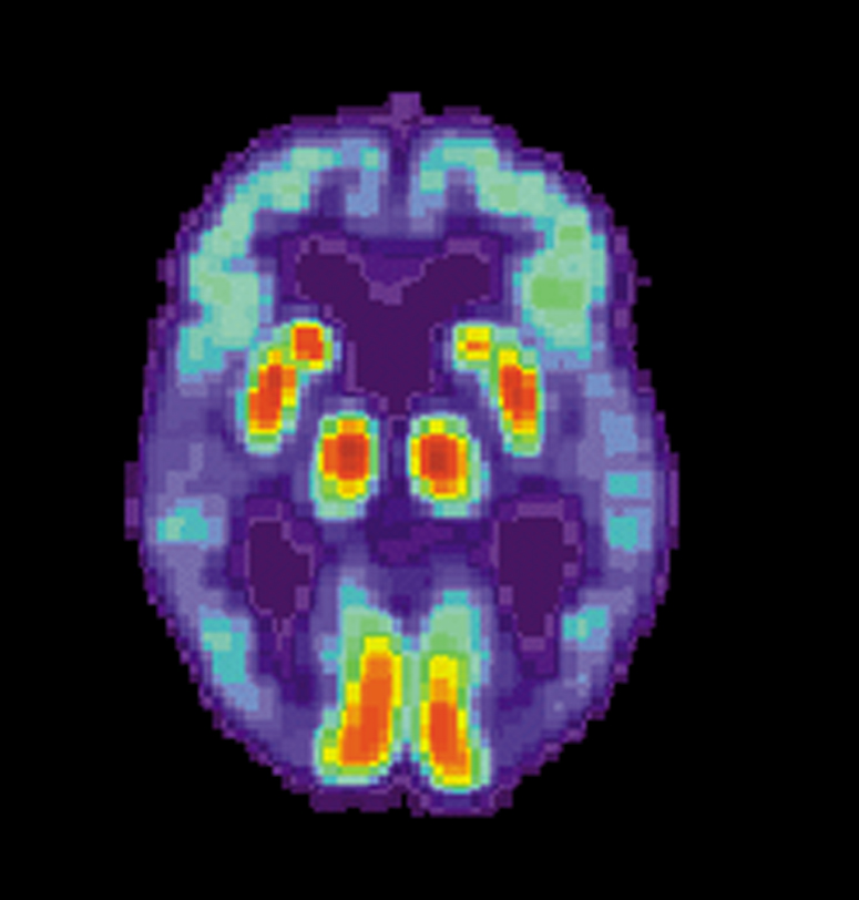
GettyImages-105693503-56e0a3aa3df78c5ba05670a3.jpg


Neuroimaging

In AD there is a reduction in global brain glucose metabolism which is marked in frontal and temporal-parietal areas. In the only psilocybin FDG-PET study to date, in healthy volunteers, acute ingestion of a 15 mg or 20 mg dose increased global brain glucose metabolism by approximately 25%, particularly in the frontal and medial temporal cortex.

A 2019 fMRI study showed lasting benefits 4 months after a single dose of 315 μg/kg psilocybin in a group of 38 meditators. Acute MRI changes—reduced connectivity between self-perceiving medial prefrontal and ventral cingulate areas—were associated with positive changes at 4 months. A 2020 fMRI study involving 16 patients with depression who took a single dose of 10 mg of psilocybin and 25 mg a week later, found increased functional connectivity between the ventromedial prefrontal cortex and the default mode network in responders the day following treatment completion, with changes sustained 5 weeks post-dosing. Functional connectivity was increased between regions with high 5HT2A-R density suggesting that reorganizing of dysfunctional neural circuitry is an important component of the neuroplastic effects of 5HT2A-R agonists.

Studies suggest that at least some of the antidepressant effect from psilocybin may be mediated via improved top-down control of the limbic system, which holds significant promise for impulse control and well-being in dementia where this control has diminished. In healthy adults, high-dose psilocybin has been shown to attenuate amygdala reactivity to emotional stimuli. In 19 adults with treatment-resistant depression, 10 mg and 25 mg psilocybin given a week apart, improved functional connectivity between the cortex and amygdala a day after the higher dose. In another study, also in healthy adults, sub-perceptual doses of LSD was shown to significantly influence functional connectivity between the amygdala and other key regions within the limbic system suggesting 5HT2A-R mediated reorganization of more primitive networks is possible without profound acute perceptual changes, although whether these changes were lasting is unclear.

These imaging studies reveal the potent reorganization of dysfunctional brain networks in affective and anxiety states. Such changes may also yield improvements in cognition, mood, and behavior by ameliorating dysfunctional circuits in cognitive impairment and dementia.

Anti-inflammatory mechanisms

All known genetic and environmental risk factors for AD are associated with increased inflammation, suggesting that reducing inflammation could be a target for preventing AD. Psychedelics have been shown to have potent anti-inflammatory properties and, given their affinity for the 5HT2A-R, may represent a unique anti-inflammatory overwhelmingly targeted to brain tissue.

In a rodent model of AD induced by chronic intracerebral inoculation of streptozotocin, 5HT1A- and 5HT2A-R agonists had a significant independent and synergistic neuroprotective effect in hippocampal neurons at 35 days via anti-apoptotic pathways. This neuroprotection infers activation of anti-inflammatory pathways, the corollary to this being that activation of 5HT2A-R in rodent neurons protects against reactive oxygen species (ROS) via the upregulation of neuroprotective Sirtuin 1 expression. This pathway simultaneously stimulates mitochondrial biogenesis leading to greater availability of adenosine triphosphate and suggesting the potential for psychedelics to address impaired energy metabolism, another key pathological pathway to cognitive dysfunction in AD.

two-examples-observational-studies.jpg


Discussion

After decades of repeated failure of treatments for dementia, there is an urgent need to develop new treatments for AD. The potential for psychedelic compounds to influence and enhance functional neuronal connectivity, stimulate neurogenesis, restore brain plasticity, reduce inflammation and enhance cognition provides a new therapeutic target and compelling argument for further investigation of the potential for psychedelics as a disease-modifying compound in conditions where currently none exists.

Animal models testing the neurobiological effects of psychedelic compounds have demonstrated hippocampal neurogenesis at lower doses and suppression at higher doses and potent neuroprotective properties. Studies in people suffering from depression and anxiety disorders have demonstrated lasting neuroplastic changes following just one or two large doses. This suggests a potential role for both sub-perceptual “micro”- and psychedelic-doses as a strategy for neuroprotection and cognitive enhancement in prodromal AD. For cognitive enhancement, the ideal dose and frequency have yet to be determined however the rapid desensitization of 5HT2A receptors by both psilocybin and LSD suggests that daily dosing is unlikely to be the optimal strategy.

Despite anecdotal evidence of widespread recreational use of micro-dosing for cognitive enhancement, robust scientific studies of the cognitive effects of micro-dosing in humans have so far been limited to acute changes in very small studies in cognitively normal individuals with no reports of persistent cognitive changes, either positive or negative, at psychedelic doses. Studies looking at both micro-dosing and psychedelic doses, longer-term, in cognitively impaired individuals are lacking and urgently needed.

*From the article here :
 
Last edited:
alzheimersdi.jpg

PET scan of a human brain with Alzheimer's disease.

New vaccine could help halt Alzheimer's progression*

by Anne Delotto Baier | University of South Florida | 20 Oct 2020

Our immune system's capacity to mount a well-regulated defense against foreign substances, including toxins, weakens with age and makes vaccines less effective in people over age 65. At the same time, research has shown that immunotherapy targeting neurotoxic forms of the peptide amyloid beta (oligomeric Aβ) may halt the progression of Alzheimer's disease, the most common age-related neurodegenerative disease.

A team led by Chuanhai Cao, Ph.D., of the University of South Florida Health (USF Health), has focused on overcoming, in those with impaired immunity, excess inflammation and other complications that interfere with development of a therapeutic Alzheimer's vaccine.

Now, a preclinical study by Dr. Cao and colleagues indicates that an antigen-presenting dendritic vaccine with a specific antibody response to oligomeric Aβ may be safer and offer clinical benefit in treating Alzheimer's disease. The vaccine, called E22W42 DC, uses immune cells known as dendritic cells (DC) loaded with a modified Aβ peptide as the antigen.

The Alzheimer's mouse model study of this new investigational vaccine was published early online Oct. 13 in the Journal of Alzheimer's Disease.

One of the two hallmark pathologies of Alzheimer's disease is hardened deposits of Aβ that clump together between nerve cells (amyloid protein plaques) in the brain; the other is neurofibrillary tangles of tau protein inside brain cells. Both lead to damaged neurological cell signaling, ultimately causing the onset of Alzheimer's disease and symptoms.

"This therapeutic vaccine uses the body's own immune cells to target the toxic Aβ molecules that accumulate harmfully in the brain," said principal investigator Dr. Cao, a neuroscientist at the USF Health Taneja College of Pharmacy, USF Health Morsani College of Medicine and the university's Byrd Alzheimer's Center. "And, importantly, it provides strong immunomodulatory effects without inducing an unwanted, vaccine-associated autoimmune reaction in the aging mice."

Unfortunately, clinical trials of all anti-amyloid treatments for Alzheimer's disease so far have failed—including the initial vaccine trial targeting Aβ (AN-1792), which was suspended in 2002 after several immunized patients developed central nervous system inflammation. "Inflammation is a primary symptom of Alzheimer's disease, so any possible treatment with neural inflammation as a side effect essentially pours gas on the fire," Dr. Cao said.

A next-generation anti-amyloid vaccine for Alzheimer's would ideally produce long-lasting, moderate antibody levels needed to prevent Aβ oligomers from further aggregating into destructive Alzheimer's plaques, without over-stimulating the immune systems of elderly people, Dr. Cao added.

In this study, the researchers tested the vaccine they formulated using modified Aβ-sensitized dendritic cells derived from mouse bone marrow. Dendritic cells interact with other immune cells (T-cells and B-cells) to help regulate immunity, including suppressing harmful responses against healthy tissues.

"Because we use dendritic cells to generate antibodies, this vaccine can coordinate both innate and acquired immunity to potentially overcome age-related impairments of the immune system," Dr. Cao said.

The study included three groups of transgenic (APP/PS1) mice genetically engineered to develop high levels of Aβ and behavioral/cognitive abnormalities that mimic human Alzheimer's disease. One group was vaccinated with the investigational E22W42 DC vaccine, another received an endogenous amyloid beta peptide to stimulate dendritic cells (wild-type vaccine group), and the third was injected with dendritic cells only, containing no Aβ peptide (DC control group). A fourth group was comprised of untreated healthy, older mice (nontransgenic control group).

"Though the E22W42-sensitized DC vaccine is being developed for patients with Alzheimer's disease, it can potentially help strengthen the immune system of elderly patients (with other age-related disorders) as well," the study authors concluded.

*From the article here:
 
Last edited:
alzheimers-microbiome-neuroscinews.jpg

Proteins produced by certain intestinal bacteria, could indeed modify the interaction
between the immune and the nervous systems and trigger the disease.


Link between Alzheimer’s and gut microbiota confirmed

University of Geneva | Neuroscience News | 13 Nov 2020

Study confirms an association between an imbalance in gut bacteria and the development of amyloid plaques in the brains of humans.

Alzheimer’s disease is the most common cause of dementia. Still incurable, it directly affects nearly one million people in Europe, and indirectly millions of family members as well as society as a whole. In recent years, the scientific community has suspected that the gut microbiota plays a role in the development of the disease.

A team from the University of Geneva (UNIGE) and the University Hospitals of Geneva (HUG) in Switzerland, together with Italian colleagues from the National Research and Care Center for Alzheimer’s and Psychiatric Diseases Fatebenefratelli in Brescia, University of Naples and the IRCCS SDN Research Center in Naples, confirm the correlation, in humans, between an imbalance in the gut microbiota and the development of amyloid plaques in the brain, which are at the origin of the neurodegenerative disorders characteristic of Alzheimer’s disease.

Proteins produced by certain intestinal bacteria, identified in the blood of patients, could indeed modify the interaction between the immune and the nervous systems and trigger the disease.

These results, to be discovered in the Journal of Alzheimer’s Disease, make it possible to envisage new preventive strategies based on the modulation of the microbiota of people at risk.

The research laboratory of neurologist Giovanni Frisoni, director of the HUG Memory Centre and professor at the Department of Rehabilitation and Geriatrics of the UNIGE Faculty of Medicine, has been working for several years now on the potential influence of the gut microbiota on the brain, and more particularly on neurodegenerative diseases. “We have already shown that the gut microbiota composition in patients with Alzheimer’s disease was altered, compared to people who do not suffer from such disorders,” he explains.

“Their microbiota has indeed a reduced microbial diversity, with an over-representation of certain bacteria and a strong decrease in other microbes. Furthermore, we have also discovered an association between an inflammatory phenomenon detected in the blood, certain intestinal bacteria, and Alzheimer’s disease; hence the hypothesis that we wanted to test here: could inflammation in the blood be a mediator between the microbiota and the brain?”

The brain under influence

Intestinal bacteria can influence the functioning of the brain and promote neurodegeneration through several pathways: they can indeed influence the regulation of the immune system and, consequently, can modify the interaction between the immune system and the nervous system. Lipopolysaccharides, a protein located on the membrane of bacteria with pro-inflammatory properties, have been found in amyloid plaques and around vessels in the brains of people with Alzheimer’s disease. In addition, the intestinal microbiota produces metabolites – in particular some short-chain fatty acids – which, having neuroprotective and anti-inflammatory properties, directly or indirectly affect brain function.

“To determine whether inflammation mediators and bacterial metabolites constitute a link between the gut microbiota and amyloid pathology in Alzheimer’s disease, we studied a cohort of 89 people between 65 and 85 years of age. Some suffered from Alzheimer’s disease or other neurodegenerative diseases causing similar memory problems, while others did not have any memory problems,” reports Moira Marizzoni, a researcher at the Fatebenefratelli Center in Brescia and first author of this work.

“Using PET imaging, we measured their amyloid deposition and then quantified the presence in their blood of various inflammation markers and proteins produced by intestinal bacteria, such as lipopolysaccharides and short-chain fatty acids.”

A very clear correlation
“Our results are indisputable: certain bacterial products of the intestinal microbiota are correlated with the quantity of amyloid plaques in the brain,” explains Moira Marizzoni.
“Indeed, high blood levels of lipopolysaccharides and certain short-chain fatty acids (acetate and valerate) were associated with both large amyloid deposits in the brain. Conversely, high levels of another short-chain fatty acid, butyrate, were associated with less amyloid pathology.”

This work thus provides proof of an association between certain proteins of the gut microbiota and cerebral amyloidosis through a blood inflammatory phenomenon. Scientists will now work to identify specific bacteria, or a group of bacteria, involved in this phenomenon.

A strategy based on prevention

This discovery paves the way for potentially highly innovative protective strategies – through the administration of a bacterial cocktail, for example, or of pre-biotics to feed the ‘good’ bacteria in our intestine. “However, we shouldn’t be too quick to rejoice,” says Frisoni.

“Indeed, we must first identify the strains of the cocktail. Then, a neuroprotective effect could only be effective at a very early stage of the disease, with a view to prevention rather than therapy. However, early diagnosis is still one of the main challenges in the management of neurodegenerative diseases, as protocols must be developed to identify high-risk individuals and treat them well before the appearance of detectable symptoms.”

 
Last edited:
blood-draw-banner-l.jpg


First Alzheimer’s blood test rolled out for clinical use in the US

by Shawna Williams | The Scientist | 2 Nov 2020

The test will be a cheaper and more accessible alternative to currently available diagnostic tools, researchers say.

The first blood test designed to assist physicians in determining whether a patient has Alzheimer’s disease is now available in most US states, the company C2N Diagnostics announced October 29. The test measures biomarkers that frequently reflect the presence of amyloid plaques in the brain—a hallmark of Alzheimer’s—as well as the presence of a gene variant that increases the risk of the disease.

“I’m very excited about it,” says Suzanne Schindler, a neurologist at the Washington University School of Medicine in St. Louis who was involved in testing an earlier version of the assay but is not connected to C2N. While there are two other tests for Alzheimer’s-associated brain changes, she notes, both have logistical and financial challenges: one that collects biomarkers in the cerebrospinal fluid (CSF) requires a spinal tap, while the other, a scan called amyloid PET, involves injecting a radioactive tracer, costs thousands of dollars, and is only performed at specialized centers. “I think patients really like the idea of a blood test,” she says. “And I think that it really has the potential to allow us to do a lot more testing than we have done in the past.”

The price of the test is $1,250, says C2N CEO Joel Braunstein, but patients who qualify for financial assistance will be charged between $25 and $400. Health insurance companies don’t currently pay for the test, he adds, but qualifying for this reimbursement “is a very high priority” for the company.

“If you asked me [five or ten] years ago if there would ever be a blood test for Alzheimer’s, I would have been very skeptical,” says Howard Fillit, the executive director and chief science officer of the Alzheimer’s Drug Discovery Foundation, which invested in C2N’s development of the test. “So the fact that this is on the market now is just amazing.”

Fillit wasn’t alone in thinking that, due to the blood-brain barrier, biomarkers from the brain wouldn’t be found in peripheral blood in sufficient quantities to deliver a diagnosis. But analytical techniques have advanced in sensitivity in recent years, allowing small amounts of biomarkers in the blood to be detected.

The C2N test relies on the ratio of two isoforms of the amyloid-β protein, Aβ42 and Aβ40, that aggregate to form amyloid plaques in the brain, combined with the presence of isoforms of apolipoprotein E (ApoE) that reflect whether the patient caries a genetic variant associated with Alzheimer’s risk. The results are combined into a score that indicates the probability that a patient would be found to have amyloid plaques if they were to undergo an amyloid PET scan. Doctors can then consider the test results along with other information about the patient to arrive at a diagnosis. According to data posted on the company’s website, a study in 686 patients with cognitive impairment found that those with scores above a certain cutoff point had a positive amyloid PET scan 92 percent of the time, while those with scores below a certain cutoff had a 77 percent chance of having a negative result on the PET scan.

While no drugs have yet been approved to treat Alzheimer’s, being able to distinguish it from other possible causes of cognitive impairment is nonetheless valuable, Fillit and Schindler say. As a clinician, Schindler says, “I want to know what my patients have. That can be complicated because in many cases, they don’t just have memory impairment, they’re taking multiple medications, they have all sorts of health issues,” she says. “And sometimes it’s really hard to know whether the symptoms they’re experiencing are due to something like Alzheimer’s or something else.”

Symptom-based diagnosis of Alzheimer’s disease is only about 70 percent accurate, notes Colin Masters of the Florey Institute of Neuroscience and Mental Health in Australia who was part of the team that first characterized the amyloid plaques in the disease. "If a clinician suspects the disease but the blood test returns a negative result, this will force the clinicians to go and look harder for other causes of dementia,” says Masters, who collaborates with one of the test’s developers but has no ties to C2N. He adds that once an amyloid-targeting Alzheimer’s drug is approved, the test will be useful in determining who should receive it. Such a treatment may not be far off: the Biogen drug candidate aducanumab is set for review by a US Food and Drug Administration panel later this week.

The blood test is also likely to be a boon for clinical trials to develop other drugs, as it will make it easier to screen patients for recruitment, Fillit says. “Screening and enrollment represents up to fifty percent of the cost of clinical trials in Alzheimer’s disease, and one Phase 3 trial costs three to four hundred million dollars,” he says, calling the blood test "a huge advance in clinical trials.”

 
Last edited:
3-cells.jpg


Drug reverses age-related cognitive decline within days

by UCSF | Medical Xpress | 2 Dec 2020

Just a few doses of an experimental drug can reverse age-related declines in memory and mental flexibility in mice, according to a new study by UC San Francisco scientists. The drug, called ISRIB, has already been shown in laboratory studies to restore memory function months after traumatic brain injury (TBI), reverse cognitive impairments in Down Syndrome, prevent noise-related hearing loss, fight certain types of prostate cancer , and even enhance cognition in healthy animals.

In the new study, published December 1, 2020 in the open-access journal eLife , researchers showed rapid restoration of youthful cognitive abilities in aged mice, accompanied by a rejuvenation of brain and immune cells that could help explain improvements in brain function.

"ISRIB's extremely rapid effects show for the first time that a significant component of age-related cognitive losses may be caused by a kind of reversible physiological "blockage" rather than more permanent degradation," said Susanna Rosi, Ph.D., Lewis and Ruth Cozen Chair II and professor in the departments of Neurological Surgery and of Physical Therapy and Rehabilitation Science.

"The data suggest that the aged brain has not permanently lost essential cognitive capacities, as was commonly assumed, but rather that these cognitive resources are still there but have been somehow blocked, trapped by a vicious cycle of cellular stress," added Peter Walter , Ph.D., a professor in the UCSF Department of Biochemistry and Biophysics and a Howard Hughes Medical Institute investigator. "Our work with ISRIB demonstrates a way to break that cycle and restore cognitive abilities that had become walled off over time."

Could rebooting cellular protein production hold the key to aging and other diseases?

Walter has won numerous scientific awards, including the Breakthrough , Lasker and Shaw prizes, for his decades-long studies of cellular stress responses. ISRIB, discovered in 2013 in Walter's lab, works by rebooting cells' protein production machinery after it gets throttled by one of these stress responses—a cellular quality control mechanism called the integrated stress response (ISR; ISRIB stands for ISR InhiBitor).

The ISR normally detects problems with protein production in a cell—a potential sign of viral infection or cancer-promoting gene mutations—and responds by putting the brakes on cell's protein-synthesis machinery. This safety mechanism is critical for weeding out misbehaving cells, but if stuck in the on position in a tissue like the brain, it can lead to serious problems, as cells lose the ability to perform their normal activities, Walter and colleagues have found.

In particular, recent animal studies by Walter and Rosi, made possible by early philanthropic support from The Rogers Family Foundation, have implicated chronic ISR activation in the persistent cognitive and behavioral deficits seen in patients after TBI, by showing that, in mice, brief ISRIB treatment can reboot the ISR and restore normal brain function almost overnight.

The cognitive deficits in TBI patients are often likened to premature aging, which led Rosi and Walter to wonder if the ISR could also underlie purely age-related cognitive decline. Aging is well known to compromise cellular protein production across the body, as life's many insults pile up and stressors like chronic inflammation wear away at cells, potentially leading to widespread activation of the ISR.

"We've seen how ISRIB restores cognition in animals with traumatic brain injury, which in many ways is like a sped-up version of age-related cognitive decline," said Rosi, who is director of neurocognitive research in the UCSF Brain and Spinal Injury Center and a member of the UCSF Weill Institute for Neurosciences. "It may seem like a crazy idea, but asking whether the drug could reverse symptoms of aging itself was just a logical next step."

ISRIB improves cognition, boosts neuron and immune cell function

In the new study, researchers led by Rosi lab postdoc Karen Krukowski , Ph.D., trained aged animals to escape from a watery maze by finding a hidden platform, a task that is typically hard for older animals to learn. But animals who received small daily doses of ISRIB during the three-day training process were able to accomplish the task as well as youthful mice, much better than animals of the same age who didn't receive the drug.

The researchers then tested how long this cognitive rejuvenation lasted and whether it could generalize to other cognitive skills. Several weeks after the initial ISRIB treatment, they trained the same mice to find their way out of a maze whose exit changed daily—a test of mental flexibility for aged mice who, like humans, tend to get increasingly stuck in their ways. The mice who had received brief ISRIB treatment three weeks before still performed at youthful levels, while untreated mice continued to struggle.

To understand how ISRIB might be improving brain function, the researchers studied the activity and anatomy of cells in the hippocampus, a brain region with a key role in learning and memory, just one day after giving animals a single dose of ISRIB. They found that common signatures of neuronal aging disappeared literally overnight: neurons' electrical activity became more sprightly and responsive to stimulation, and cells showed more robust connectivity with cells around them while also showing an ability to form stable connections with one another usually only seen in younger mice.

The researchers are continuing to study exactly how the ISR disrupts cognition in aging and other conditions and to understand how long ISRIB's cognitive benefits may last. Among other puzzles raised by the new findings is the discovery that ISRIB also alters the function of the immune system's T cells, which also are prone to age-related dysfunction. The findings suggest another path by which the drug could be improving cognition in aged animals, and could have implications for diseases from Alzheimer's to diabetes that have been linked to heightened inflammation caused by an aging immune system.

"This was very exciting to me because we know that aging has a profound and persistent effect on T cells and that these changes can affect brain function in the hippocampus," said Rosi. "At the moment, this is just an interesting observation, but it gives us a very exciting set of biological puzzles to solve."

 
Last edited:
the-key-questions-surrounding-the-discovery-that-alzheimers-can-be-transmitted-from-person-to-person-136400282180203901-150909222032.jpg


Regulating copper in the brain stops memory loss associated with Alzheimer’s

CNRS | Neuroscience News | 17 Dec 2020

Researchers have developed a molecule that regulates the circulation of copper in the brain. The molecule extracts copper trapped in amyloid plaques in mouse models of Alzheimer’s disease. Administered orally, the molecule inhibits memory loss associated with Alzheimer’s.

Alzheimer’s disease is characterized by the presence of amyloid plaques in the patient’s brain. These plaques sequester copper, and contain approximately five times as much as a healthy brain.

Two CNRS scientists from the Coordination Chemistry Laboratory recently developed, with their colleagues from the Guangdong University of Technology and Shenzhen University in China, a molecule that regulates the circulation of copper in the brain.

This patented molecule extracts the copper trapped in amyloid plaques, and reintroduces it in the brain’s normal enzymatic circuit (which needs copper to function).

Administered orally to “Alzheimer” mice, this molecule inhibits memory loss among sick mice.

This shows a swirly head

These plaques sequester copper, and contain approximately five times as much as a healthy brain.

These results, which were published in ACS Chemical Neuroscience, open a new therapeutic avenue that could prove effective in early stages of Alzheimer’s disease among humans.

The scientists are now seeking a pharmaceutical partner to develop preclinical trials for this drug candidate.

 
Last edited:
Darby_Functional-Brain-Networks_1200x700.jpg

Stanford researchers suggest that if their findings are transferable to humans
they could lead to new therapies that battle age-related cognitive decline.


Breakthrough study finds age-related cognitive decline may be reversible

by Rich Haridy | NEW ATLAS | 20 Jan 2021

A breakthrough study from a team of neurologists at Stanford University claims to have discovered one way immune cells become dysfunctional as we age, leading to the inflammatory hyperdrive that plays a role in most age-related disease from cancer to cognitive decline. Preliminary study suggests this immune dysfunction can be reversed, pointing to compelling future anti-aging therapies.

As we age, our immune system slowly grows increasingly dysfunctional. For some that means the immune system can be slower to respond to infection, and for others it means defective immune cells begin to consistently attack healthy cells, causing chronic low-grade inflammation. Some researchers hypothesize this chronic inflammation plays a major role in many age-related diseases, particularly in the brain.

So this new research started out with some general questions. What could be causing this age-related immune dysfunction? Does this chronic inflammatory activity play a role in age-related cognitive decline? And, perhaps most importantly, can this mechanism be either slowed, or reversed?

The specific focus of the new study, published in the journal Nature, was a hormone called prostaglandin E2 (PGE2). Levels of this particular hormone have previously been found to rise with aging. PGE2 is also known to promote inflammatory activity in immune cells.

The first part of the research unpacked exactly how PGE2 initiates inflammatory activity in macrophages, a fundamental immune white blood cell. Across animal experiments and in vitro human cell tests the researchers uncovered a comprehensive chain of events showing how PGE2 directly triggers dysfunctional inflammatory activity in macrophages.

The researchers then confirmed, again in both mouse and human cells, that older macrophages produce significantly more PGE2 than their younger counterparts. Alongside that, these aging macrophages present with greater surface numbers of EP2 receptors, the receptors that bind PGE2.

Katrin Andreasson, senior author on the new study, calls this age-induced inflammatory mechanism, “a double-whammy – a positive feedback loop.”

The final part of the research explored what happens when you inhibit this PGE2-EP2 mechanism. Initial in vitro experiments revealed old macrophage cells transforming when this mechanism was disrupted. Inflammatory characteristics disappeared and the old cells were effectively rejuvenated.

Inhibiting this mechanism in aged mouse models of cognitive decline resulted in even more impressive results. The old mice, receiving an experimental drug that blocks PGE2-EP2 binding, displayed a reversal of cognitive decline, ultimately performing as well as young mice across a series of cognitive tests.

Andreasson suggests this mechanism of modulating the immune system can essentially “de-age the brain.” Even when PGE2-EP2 binding was isolated to cells outside of the brain, using a compound that cannot cross the blood-brain barrier, cognitive improvements and reductions in neural inflammation were still detected.

Researchers not involved with the new study have variously described the novel research as “well-conducted”, “intriguing”, and “important.” But they also suggest it is a very preliminary finding and needs validation in humans before we can even consider being close to an anti-aging or anti-dementia therapy.

“This study is important because it shows that in mice, age-related memory decline can be reversed,” explains Tara Spires-Jones from the University of Edinburgh, “however, it is important to note that this research was in small groups of mice and more work will need to be done to determine whether the same effects will be possible in people.”

Andreasson is clear in noting the long research path that lies ahead. The experimental drugs used in the study to block PGE2-EP2 binding, for example, are not at all ready for human clinical use. In fact, they may not even be safe for humans, Andreasson suggests.

Instead, the study offers researchers new directions for developing novel compounds that can disrupt this dysfunctional age-related inflammatory activity. It may be a long time before a tangible anti-aging clinical therapy arises from these findings but, if validated, these foundational discoveries offer an exciting road map for producing therapies that could battle a broad array of age-related diseases.

“Our study suggests that cognitive aging is not a static or irrevocable condition but can be reversed by reprogramming myeloid glucose metabolism to restore youthful immune functions,” the researchers conclude in the new study.

The new study was published in the journal Nature.

 
Last edited:
inflammation-reversed-dementia-neurosciencenews.jpg

UC Berkeley scientists propose radical new theory that AD memory loss and cognitive
dysfunction are due to a leaky barrier between the blood stream and the brain.


Drugs that quell brain inflammation found to reverse dementia

UC Berkeley | Neuroscience News | Dec 29 2019

Older mice given anti-inflammatory medication were better able to learn new tasks and became almost as adept at learning as mice half their age.

Drugs that tamp down inflammation in the brain could slow or even reverse the cognitive decline that comes with age. In a publication appearing today in the journal Science Translational Medicine, University of California, Berkeley, and Ben-Gurion University scientists report that senile mice given one such drug had fewer signs of brain inflammation and were better able to learn new tasks, becoming almost as adept as mice half their age.

“We tend to think about the aged brain in the same way we think about neurodegeneration: Age involves loss of function and dead cells. But our new data tell a different story about why the aged brain is not functioning well: It is because of this “fog” of inflammatory load,” said Daniela Kaufer, a UC Berkeley professor of integrative biology and a senior author, along with Alon Friedman of Ben-Gurion University of the Negev in Israel and Dalhousie University in Canada. “But when you remove that inflammatory fog, within days the aged brain acts like a young brain. It is a really, really optimistic finding, in terms of the capacity for plasticity that exists in the brain. We can reverse brain aging.”

The successful treatment in mice supports a radical new view of what causes the confusion and dementia that often accompany aging. More and more research shows that, with age, the filtration system that prevents molecules or infectious organisms in the blood from leaking into the brain — the so-called blood-brain barrier — becomes leaky, letting in chemicals that cause inflammation and a cascade of cell death. After age 70, nearly 60% of adults have leaky blood- brain barriers, according to Friedman’s magnetic resonance imaging (MRI) studies.

An accompanying paper by the two researchers and Dan Milikovsky of Ben-Gurion University shows that the inflammatory fog induced by a leaky blood-brain barrier alters the mouse brain’s normal rhythms, causing microseizure-like events — momentary lapses in the normal rhythm within the hippocampus — that could produce some of the symptoms seen in degenerative brain diseases like Alzheimer’s disease. Electroencephalograms (EEGs) revealed similar brain wave disruption, or paroxysmal slow wave events, in humans with epilepsy and with cognitive dysfunction, including Alzheimer’s and mild cognitive impairment (MCI).

Together, the papers give doctors two biomarkers — leaky barriers detectable by MRI and abnormal brain rhythms detectable by EEG — that can be used to flag people with blood-brain barrier problems, as well as a potential drug to slow or reverse the consequences.

“We now have two biomarkers that tell you exactly where the blood-brain barrier is leaking, so you can select patients for treatment and make decisions about how long you give the drug,” said Kaufer, a member of UC Berkeley’s Helen Wills Neuroscience Institute. “You can follow them, and when the blood-brain barrier is healed, you no longer need the drug.”

Diagram-of-blood-brain-barrier-BBB-and-other-components-of-a-brain.png


Blood-brain barrier

Scientists have long suspected that a leaky blood-brain barrier causes at least some of the tissue damage after brain injury and some of the mental decline that comes with age. But no one knew how.

In 2007, however, Friedman and Kaufer linked these problems to a blood protein, albumin. In 2009, they showed that when albumin leaks into the brain after trauma, it binds to the TGF-β (TGF-beta) receptor in brain cells called astrocytes. This triggers a cascade of inflammatory responses that damage other brain cells and neural circuits, leading to decreased inhibition and increased excitation of neurons and a propensity toward seizures.

They also showed in mice that blocking the receptor with an antihypertension drug, losartan, prevented the development of epilepsy after brain trauma. Epilepsy is a frequent consequence of concussions like those sustained by soldiers from roadside bombs.

Subsequent studies revealed leakiness in the barrier after stroke, traumatic brain injury and football concussions, solidly linking albumin and an overexcited TGF-β receptor to the damage resulting from these traumas.

In their new studies, Kaufer and Friedman showed that introducing albumin into the brain can, within a week, make the brains of young mice look like those of old mice, in terms of hyperexcitability and their susceptibility to seizures. These albumin-treated mice also navigated a maze as poorly as aged mice.

“When we infused albumin into the brains of young mice, we recapitulated aging of the brain: the gene expression, the inflammatory response, resilience to induced seizures and mortality after seizures, performance in a maze. And when we recorded their brain activity, we found these paroxysmal slow wave events,” Kaufer said. “And all were specific to the site we infused. So, doing this is sufficient to get an aged phenotype of this very young brain.”

When they genetically engineered mice so that they could knock out the TGF-β receptor in astrocytes after they’d reached old age, the senile mouse brains looked young again. The mice were as resistant to induced seizures as a young mouse, and they learned a maze like a young mouse.

Serendipitously, a Palo Alto, California, medicinal chemist, Barry Hart, offered to synthesize a small-molecule drug that blocks the TGF-β receptor in astrocytes only, and that could traverse the blood-brain barrier. When they gave the drug, called IPW, to mice in doses that lowered the receptor activity level to that found in young mice, the brains of the aged mice looked younger, too. They showed young brain-like gene expression, reduced inflammation and improved rhythms — that is, reduced paroxysmal slow wave events — as well as reduced seizure susceptibility. They also navigated a maze or learned a spatial task like a young mouse.

In analyzing brain tissue from humans, Kaufer found evidence of albumin in aged brains and increased neuroinflammation and TGF-β production with age. Friedman developed a special type of MRI imaging — dynamic contrast-enhanced (DCE) imaging — to detect leakage in the blood-brain barrier and found more leakage in people with greater cognitive dysfunction.

"Altogether, the evidence points to a dysfunction in the brain’s blood filtration system as one of the earliest triggers of neurological aging," Kaufer said.

Kaufer, Friedman and Hart have started a company to develop a drug to heal the blood-brain barrier for clinical treatment and hope that the drug will help reduce brain inflammation — and, thus, permanent damage — after stroke, concussion or traumatic brain injury, and eventually help older adults with dementia or Alzheimer’s disease who have demonstrated leakage of the blood-brain barrier.

“We got to this through the back door — we started with questions about plasticity having to do with the blood-brain barrier, traumatic brain injury and how epilepsy develops,” Kaufer said. “But after we’d learned a lot about the mechanisms, we started thinking that maybe in aging it is the same story. This is new biology, a completely new angle on why neurological function deteriorates as the brain ages.”

 
tingri-everest-base-camp-trek.jpg


The latest on psychedelics for treating dementia

by Nate Seltenrich | Psychedelic Science Review | 26 Jan 2021

“…this stands up to scientific rigor and is worthy of further exploration.”

Among the many medical and therapeutic applications of psychedelics being explored in labs and boardrooms today, Alzheimer’s disease has maintained a relatively low profile. But that may be changing, thanks in part to a review article published last summer in Frontiers in Synaptic Neuroscience.

“We have been stunned by the popularity of our article on the Frontiers platform,” lead author Simon Vann Jones, an old-age psychiatrist, and researcher with the UK-based Cornwall Partnership NHS Foundation Trust, told Psychedelic Science Review in an email earlier this month. “I think this speaks to both the interest and excitement around psychedelic research but also the desire for something that might make a difference to Alzheimer’s disease and other dementias.”

The paper is joined by an ongoing study at Johns Hopkins University, currently recruiting volunteers, that is designed to investigate whether high-dose psilocybin can ameliorate depression — or, as secondary outcomes, improve memory and cognition — in patients with early Alzheimer’s.

And in December 2019, a Phase 1 trial published in the journal Psychopharmacology by the psychedelic medicine company Eleusis — with noteworthy contributors Robin Carhart-Harris of Imperial College London, Charles Nichols of Louisiana State University, and Eleusis CEO Shlomi Raz — showed that low doses of LSD were safe and well-tolerated in 48 healthy older volunteers. The paper is part of a larger study intended to evaluate the drug as a therapeutic for neuroinflammation associated with neurodegenerative diseases like Alzheimer’s.

Beyond these preliminary efforts, there’s not much to report yet — especially compared with the attention given to addiction, depression, anxiety, and post-traumatic stress disorder as targets of psychedelic drugs in recent years.

The prospect that psilocybin and LSD could be used to treat dementia has a mixed yet promising foundation in the scientific literature, write Vann Jones and coauthor Allison O’Kelly, also a memory and dementia expert with Cornwall Partnership NHS Foundation Trust.

Literature review gives perspective

The August 2020 article by Vann Jones and O’Kelly — for which Nichols, one of the world’s leading experts on the neurological effects of psychedelics, served as a reviewer — ranks among the most popular articles published over the last 12 months in all 109 Frontiers journals. It reviews the evidence linking psychedelics to dementia in a methodological fashion, pulling together various threads to ultimately conclude that existing research...​
…suggests a potential role for both sub-perceptual ‘micro’- and psychedelic-doses as a strategy for neuroprotection and cognitive enhancement in prodromal Alzheimer’s disease.
The paper begins on the topic of acute cognitive effects, citing the Eleusis Phase 1 trial and two other controlled studies from 20193 and 20204 that found no effects on cognition associated with sub-perceptual doses of LSD in healthy volunteers.

A 2018 uncontrolled, naturalistic study organized by the Dutch Psychedelic Society found increased cognitive “fluency, flexibility, and originality” among the 33 participants at various micro-doses of psilocybin, but Vann Jones and O’Kelly stress these results should be interpreted with caution.​

The authors go on to review recent evidence of longer-term psychological effects of psychedelics, citing some of the “encouraging results” on anxiety and depression that have informed the current Johns Hopkins study on depression in Alzheimer’s patients.

Then comes a section on neurobiological effects (which would have included a reference to a subsequently published August 2020 study reporting an acute elevation in circulating brain-derived neurotrophic factor levels in healthy volunteers following a microdose of LSD, Vann Jones said).

Finally, the authors address anti-inflammatory mechanisms — a section that could have come first, given, on one hand, the link between Alzheimer’s and inflammation, and, on the other, psychedelics’ known anti-inflammatory properties.

Review article authors are optimistic

With nearly 21,000 views, and, according to Vann Jones, interest from journalists and researchers around the world, this short review article is poised to point the way toward more targeted clinical research and investment in the field.

“It is very exciting that there are a number of research groups and private companies currently investigating the role of psychedelics in dementia. This confirms our belief (and findings) that this stands up to scientific rigor and is worthy of further exploration,” Vann Jones told Psychedelic Science Review. “We are optimistic that we are on the path to something that may make an enormous difference to those suffering with dementia.”

 
Last edited:
tingri-everest-base-camp-trek.jpg


Is inflammation the driver of Alzheimer’s?*

Science News | May 2 2018

A new research study by scientists in Australia and the US provides an explanation for why clinical trials of drugs reducing proteins in the brain that were thought to cause dementia and Alzheimer's have failed. The study has opened the way for potential new treatments with existing drugs.

Published online in the journal Human Molecular Genetics, the researchers assembled evidence from a wide range of human studies and animal models of dementia-related diseases to show that inflammation is a major cause, not just a consequence.

They show that many genes linked with dementia regulate our susceptibility and response to inflammatory damage.

"For decades, scientists have thought that dementia and Alzheimer's Disease are caused by protein aggregates forming in the brain. But recent clinical trials of drugs that reduce the aggregates have failed," says project leader Professor Robert Richards, from the University of Adelaide's School of Biological Sciences. He is working in collaboration with the University's Adelaide Medical School and the National Institutes of Health, in the US.

Inflammation has long been known to increase as dementia-related diseases progress, but only now is it identified as the cause. Previously it was thought to act simply to clean up tissue damage caused by the protein aggregates.

"We know that inflammation has different phases -- early on it can be protective against a threat by actively degrading it, but if the threat is not removed, then persistent inflammation actually causes cell death," says Professor Richards.

The new work turns previous thinking around. The genetic linkages imply that the inflammation comes first -- and the tissue damage second.

"Many genes linked with dementia operate at the level of controlling cellular inflammation. Both internal and external triggers interact with these genes to play a part. Inflammation is the point through which many triggers converge," says Professor Richards.

He likens the brain inflammation to a virus infection. "Inflammation is a very effective defence against foreign agents like viruses. But as we get older and accumulate mutations, our cells can make proteins and DNA products that mimic viruses, and these build up in the system," he says.

"Normally, our cells bar-code their own products to tell them apart from foreign agents. When these bar-codes aren't in place, our cells can't properly distinguish 'self' and 'non-self' trigger molecules. The result is inflammation that escalates and spreads -- hence the term autoinflammatory disease."

Certain types of gene mutation cause these systems to fail earlier or more often, and can increase as we age -- possibly accounting for age-related increased risk of developing dementia.

The good news is that by reducing some elements of inflammation, it may be possible to reduce dementia symptoms.

"With this new understanding of the disease, we now need to test existing anti-inflammatory drugs for their effectiveness in treating dementia," he says.

*From the article here :
 
Top