• Psychedelic Medicine

ALS | +50 articles

als-pathway-neurosciencew-publiv.jpg

The research team found that the LSM12-EPAC1 pathway is an important suppressor
of the NCT-related pathologies in C9-ALS/FTD.

Genetic pathway that suppresses ALS identified

UNIST | Neuroscience News | 19 Mar 2021

Professor Chunghun Lim and his research team in the Department of Biological Sciences unveiled a neuroprotective pathway that suppresses Lou Gehrig’s Disease (ALS).

Nucleocytoplasmic transport (NCT) defects have been implicated in neurodegenerative diseases, such as C9ORF72-associated Amyotrophic Lateral Sclerosis and frontotemporal dementia (C9-ALS/FTD).

In this study, the research team has identified a neuroprotective pathway of like-Sm protein 12 (LSM12) and exchange protein directly activated by cyclic AMP 1 (EPAC1) that sustains the nucleocytoplasmic RAN gradient and thereby suppresses NCT dysfunction by the C9ORF72-derived poly (glycine-arginine) protein.

The research team found that the LSM12-EPAC1 pathway is an important suppressor of the NCT-related pathologies in C9-ALS/FTD. The EPAC1 protein, which was expressed in this genetic pathway, normalizes the abnormal RAN gradient that determines the transport direction between the cell nucleus and the cytoplasm, thereby restoring its cellular function.

In general, the RAN proteins are more abundant in the cell nucleus, but in patients with Lou Gehrig’s disease, they begin to leak out into the cytoplasm, thereby resulting in abnormal concentration differences. Their findings revealed that the EPAC1 protein, which was expressed through the LSM12-EPAC1 pathway, indeed helped EPAC1 return to the cell nucleus, thereby restoring the RAN gradient.

The research team also identified that the EPAC1 protein, which was expressed through the LSM12-EPAC1 pathway, regulates the distribution of RAN proteins. The EPAC1 protein increases the binding force between the nuclear pore complex (NPC) and the RAN proteins. Because of this, RAN proteins lost within the cytoplasm are, then, captured by the NPC and returned to the nucleus.

“Although the distribution of RAN proteins is critical for the nucleocytoplasmic transport (NTC), its molecular biological mechanism has relatively been unknown,” says Professor Lim. “In this study, we have identified that LSM12-EPAC1 defines a neuroprotective pathway that sustains the nucleocytoplasmic RAN gradient.”

He adds, “Our findings are expected to contribute greatly to the prediction and treatment of neurodegenerative diseases, such as frontotemporal dementia and Lou Gehrig’s disease, as well as for the understanding of the molecular mechanisms underlying aging.”


3313250919_cca08d653b_b-1023x341.jpg

University of Copenhagen

ALS development could be triggered by loss of network connections in the spinal cord

University of Copenhagen | Neuroscience News | 1 Jun 2021

Summary: Inhibitory neurons in the spinal cord lose their connections to motor neurons in mouse models of ALS. While no connection between this deterioration to the development of ALS has been made, researchers say the loss of inhibitory signals could explain why motor neurons die in those with ALS.

ALS is a very severe neurodegenerative disease in which nerve cells in the spinal cord controlling muscles and movement slowly die. There is no effective treatment and the average life expectancy after being diagnosed with ALS is usually short. Because of this, new knowledge about the disease is urgently needed.

Now, researchers from the University of Copenhagen have gained new insights about ALS, by investigating the early development of the disease in a mouse model.

“We have found that networks of nerve cells in the spinal cord called inhibitory interneurons lose connection to motor neurons, the nerve cells that directly control muscle contraction. We do not yet know if these changes cause the disease. But the loss of the inhibitory signal could explain why the motor neurons end up dying in ALS patients,” says first and co-corresponding author to the new study Ilary Allodi, Assistant Professor at the Department of Neuroscience.

A lot of ALS research have focused on the motor neurons themselves, but the research group at the University of Copenhagen had a different approach.

“It is only natural that motor neurons have received major attention. They control our muscles, which is the challenge for ALS patients. Here, we wanted to investigate the circuit of interneurons in the spinal cord because they determine the activity of motor neurons. Since we found that there is a loss of connections between inhibitory interneurons and motor neurons that happens before the motor neuron death, we think that this loss could be a possible explanation for why the motor neurons ends up dying in ALS patients,” says Ole Kiehn, senior, co-corresponding author and Professor at the Department of Neuroscience.

Fast-twitch first

In ALS patients, the degeneration typically starts with what is called the fast-twitch motor neurons and then goes on to other motor neurons. This means that certain muscles and bodily functions are affected before others. Normally, patients lose coordination and speed in movement before more basic functions such as breathing. This is mirrored in the new findings, according to the researchers.

spinal-cord-als-neurosceincces.jpg

The spinal cord of a mouse with ALS. The green cells are inhibitory interneurons.

“In our mouse model, we show that the loss of connection happens to fast motor neurons first and then slow motor neurons later on involve a particular type of inhibitory neurons, the so called V1 interneurons,” says Roser Montañana-Rosell, who is PhD student and shared first author on the study.

“The V1 interneuron connectivity loss is paralleled by the development of a specific locomotor deficit in the pre-symptomatic phase with lower speed and changes in limb coordination in the ALS mice that is dependent on V1 interneuron connections to motor neuron,” says Ole Kiehn.

Expanding the window of opportunity

The researchers underline that the mechanisms should be investigated in human patients as well. However, they do not have any reason to believe that the same or similar biological mechanisms are not at play in humans.

Given the new understanding of the disease, Ilary Allodi hopes further research into the signaling process could reveal how to repair the nerve cell connection loss in ALS.

“We definitely hope that our findings can contribute with a new way of thinking about ALS development. With a distinct focus on interneurons, we might be able, in future experiments, to increase the signaling processes from the interneurons to the motor neurons and prevent or delay the motor neuron degeneration from an early stage,” ends Ilary Allodi.

 
Last edited:
brian-wallach-sandra-abrevaya-photo-julie-dietz.jpg

Brian Wallach and Sandra Abrevaya

Race to a cure for ALS

by Deirdre Cohen | CBS | 11 Jul 2021

You can be forgiven if you're a little jealous of Brian Wallach, at first; he's good-looking, smart, a track and field athlete at Yale, a graduate of Georgetown law. All in all, a pretty good catch, as they say.

Correspondent Lee Cowan asked Wallach's wife, Sandra, "What was it that struck you about Brian?"

"I mean, he's very cute,"
she replied. "I was trying to impress him since the moment I met him."

That moment was in New Hampshire, when Sandra Abrevaya was working for the 2008 Obama presidential campaign. Wallach was the political director for the state – it's a job he took instead of taking an offer from a big powerhouse law firm.

"It was literally our job to work together," Abrevaya said.

Eventually, they both became staffers in the Obama White House. They married in 2013, and moved to Chicago and started a family.

But a few years later, their charmed life took a turn.

While working for the Assistant U.S. Attorney in Chicago, Wallach went to the doctor about a simple cough. But it was much more than that…

He was eventually diagnosed with ALS – amyotrophic lateral sclerosis. It's already nearly robbed him of his ability to walk and his ability to speak.

"So, there was more going on than just the cough?" Cowan asked.

"Yes."

"But you hadn't even told her?"

"No."


Abrevaya said, "I had no idea."

There were other things bothering Wallach that he had kept to himself, thinking they were probably nothing to worry about: "My muscles were twitching," he said. "I also had weakness in my left hand."

That's how ALS works; it kills the nerves that move muscles. The average lifespan is only two to five years.

Wallach didn't know much about ALS when he was diagnosed. But he did know the legend of Lou Gehrig, the New York Yankee who bid goodbye to his fans after he, too, was diagnosed with ALS.

On July 4, 1939, Gehrig told the crowd at Yankee Stadium, "I consider myself the luckiest man on the face of the Earth." And he was dead less than two years later.

Time became Wallach's most pressing question for his doctor, who told him, "I think you have about six months or so."

Cowan asked, "And how do you even process something like that?"

"I didn't,"
he replied.

Wallach was only 36. He and Abrevaya had a two-year old, and a newborn just home from the hospital.

"We started crying on the floor of our living room," she recalled. "And then Brian said, 'We have to go to the Verizon store.' And I was like, 'What are you talking about?' 'We need to get more memory on my iPhone, because if I die, I don't want the girls to forget who I am.'"


Wallach did beat the odds – he's almost four years into his diagnosis – but its cruelty is so plainly evident. Despite the decline, though, he and Abrevaya are determined to fight the disease the same way they fought a winning political campaign.

For the last two-and-a-half years they have been crisscrossing the halls of the Congress. In 2019 Wallach testified, "I'm here to ask you to see us, to hear us."

And in May, he testified to Congress via Zoom: "The question now is how quickly we can end ALS."

The vehicle they're using to bring about change is their organization, I Am ALS. They are tireless – working every day, usually from the "ALS war room" upstairs in their home. Their goal is to give patients the power to make change, to give them a voice in their own care, and help them gain access to therapies that they might not otherwise get.

Abrevaya said, "When you are a patient, and you are driving the advocacy agenda, and you're running out of time, you push hard and you do work differently."

Since it was started, I Am ALS says it helped increase federal funding for research by $83 million. The infusion of money has helped launch dozens of clinical trials for promising new therapies. So far, there have only been two drugs that have been approved by the Food and Drug Administration (FDA) to treat ALS. But science is perhaps closer today than ever before to a possible breakthrough.

"For the first time ever, there are a host of drugs in the pipeline that are showing really promising results," Abrevaya said. "And yet, we can't get access."

Cowan asked, "So, what's the holdup?"

"FDA is afraid of getting it wrong,"
Wallach replied.

The FDA's job, in part, is to make sure therapies are safe and effective. But letting patients accept the risks for themselves is the kind of flexible regulation that Wallach and Abrevaya are begging the FDA to grant to the ALS community. "We understand they may not work," he said. "But we are facing a disease that is 100% fatal. So, we are willing to take those risks."

One experimental therapy shows particular promise for ALS patients: AMX0035, made by Amylyx Pharmaceuticals.

"That therapy showed, in clinical trial studies, that it extended life by six-and-a-half months," said Abrevaya, "which, when you have a prognosis of two years to five years ..."

"Or, like in Brian's case, six months ..."

"That's living twice as long."


Canadian and European regulatory agencies are edging ever closer to approving that drug, but in this country, the FDA wants more time to study it.

"The extra study is going to take four years to five years," Abrevaya said. "By then, pretty much every ALS patient alive today will be dead. How can you look people in the face and tell them that that's acceptable?"

There is legislation pending that could help, allowing people to get potentially lifesaving drugs before they're fully approved by the FDA. But the wheels of government sometimes need a push.

"The HIV community faced the same battles when they needed accelerated therapies," said Abrevaya. "It took people who didn't have HIV caring, and letting government know that it is unconscionable to deny therapies to dying people."

The most promising experimental ALS treatment so far is something called Tofersen, from Biogen. It appears to slow the progression for a very rare form of ALS.

Chris Snow got access to Tofersen through a clinical trial. It's given via a spinal injection every four weeks. "I don't necessarily feel like myself, or look like myself, but I act like myself," Snow said. "I coach sports. I play sports. I work every day."

Tofersen only targets a particularly aggressive form of ALS, but it's just that that also took the lives of Snow's father, two uncles, and his 28-year-old cousin.

At the time he started on the new drug, he was given less than a year to live. That was almost two years ago.

"He should be dead; that's the simplest way to put it," said his wife, Kelsie. "He looks pretty good for a dead person!"

Kelsie watched Chris get back on the ice and play hockey (he's the assistant general manager for the Calgary Flames), and he's still able to keep up with their two young children.

"The quality of life that this has given us is really a miracle," she said.

Tofersen hasn't completely halted ALS. Snow has still seen changes in his facial muscles; he can't really smile anymore, and he has trouble speaking and trouble swallowing ... but many of his other symptoms have indeed slowed.

"I don't think I'm dying," he told Cowan. "My fear is, can I play hockey with my son next winter? It's not, will my son have a dad next winter?"

Last month he teed off for a round of golf – and played all 18 holes. He posts those kinds of achievements regularly; Kelsie has a blog of her own about living with ALS.

"I'm offering up our family," she said. "And so, if you can see and care about my family and that makes you care about this cause, that's what I'm going for."

For all its promise, though, Tofersen won't help Brian Wallach's form of ALS, which in some ways makes his efforts all the more remarkable. His ability to still keep his humor while fighting for what at times seems like an insurmountable battle is really courage of a different sort.

Snow said, "You don't see this – people with this disease disappear into their homes, and there's actually not enough of them for the world to see."

Wallach has inked his commitment to curing ALS right on his arm, with a tattoo: "ALS: YG." [The YG means "You Gone."]

Cowan said, "This takes a lot of effort, and it takes an awful lotta time – and you don't have a lot of either, really."

"I don't know what tomorrow will bring,"
said Wallach, "but I'm sure that if it is the last day for me, I want to look back on my life and say, 'I tried, and I hopefully made a difference.'"

Brian Wallach has already beaten the odds. Four years ago, he was diagnosed with ALS – amyotrophic lateral sclerosis – and given just six months to live. He's used that time to lobby for more research funds for dozens of clinical trials of promising drugs to combat the disease. Wallach and his wife, Sandra Abrevaya, talk about their organization, I Am ALS, and their unceasing efforts to expand treatments for ALS patients; and with Chris Snow, whose use of a promising experimental ALS treatment has already more than doubled his life expectancy.


Last edited:
09-ancient-ayahuasca_4x3.jpg


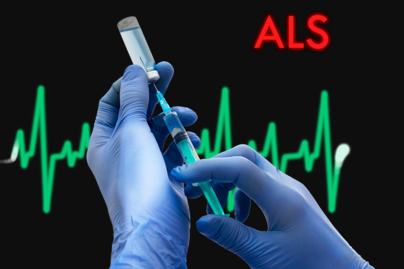
Ayahuasca and the potential treatment of ALS*

by Daniel Gustafsson

This article is the culmination of six years work, having studied ethnobotanical natural medicine and the field of motor neuron disease, making connections between the two in search for viable treatment options for ALS – amyotrophic lateral sclerosis, and similar neurodegenerative conditions.

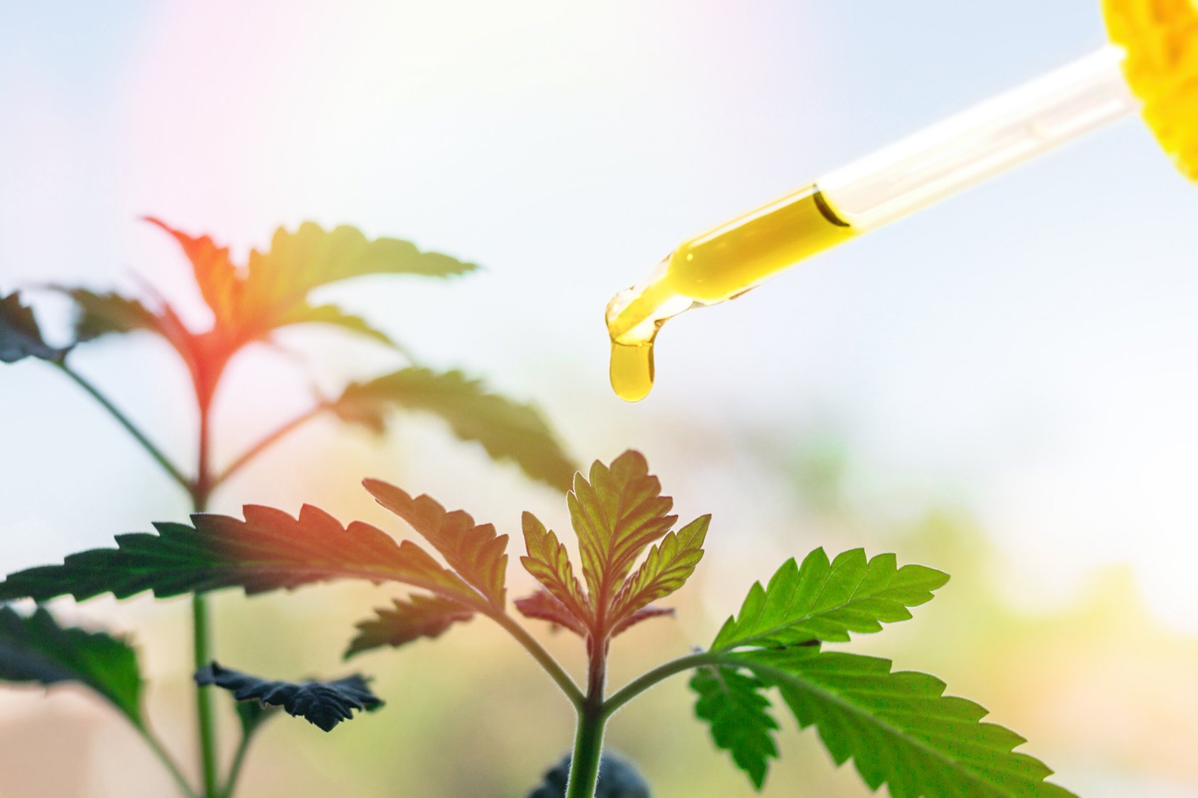
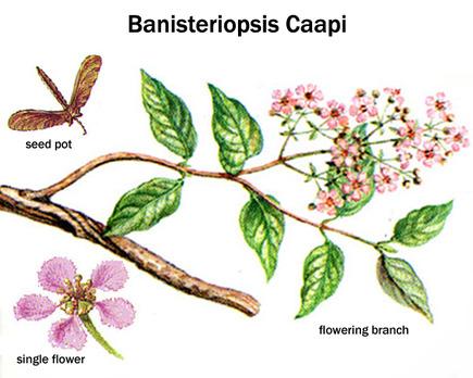
In south and central america, native people from tribes living within the Amazon rainforest have a long historical tradition of preparing and consuming a natural medicinal tea called ayahuasca. It is harvested and made mainly from a wild growing vine, its latin name being Banisteriopsis Caapi. Often, but not always, leaves from trees named Chacruna and Chaliponga (Psychotria Viridis and Diplopterys Cabrerana) are added to the tea, or in some regions Jurema tree bark (Mimosa Tenuiflora).

The rainforests of the earth are known to be enormously resourceful and vital to the ecosystem of the entire planet. An estimated abundance of undiscovered medicinally valuable plants remain to be explored within these forests, and many current conventional pharmaceuticals originate from substances found in rainforest plants. Ethnopharmacologists are long since aware of widespread support for the medicinal value of ayahuasca and its use in treatment of a wide variety of diseases, but until recently evidence has been limited. This however has changed in the 21st century, as interest and studies in ayahuasca have increased.

Natural substances extracted from ayahuasca plants have been found to possess unique restorative and targeted antioxidative properties on specific nerve cells in the brain and central nervous system, controlling neurotransmission, muscle and motor activity, memory and coordination. This gives probable cause to the theory that ayahuasca could be an effective treatment for neurodegenerative diseases such as ALS, Alzheimer’s, and Parkinson’s disease. Promising results have also been obtained from studying the substance psilocybin, remarkably close in molecular structure to substances found in ayahuasca, naturally occuring in certain species of medicinal mushrooms consumed by the indigenous people where ayahuasca is also used.

According to Dr. Juan Ramos, head of the neurological disease department at the South Florida university, USA, initial studies show that mushroom tryptamine substances semi-identical to the ones contained in ayahuasca stimulate the development of new cells in areas of the brain controlling above mentioned functions. If this could lead to an eventual cure through complete restoration of damaged and lost nerve cells remains to be seen, but initial results indicate this could potentially be the case. Other studies led by Dr. Jordi Riba at the spanish university of Sant Pau, Barcelona, show connections between ayahuasca and neural pathway redevelopment in the neocortex.

Cancer researchers have also shown interest in B. Caapi, as its alkaloids have shown to be effective against the growth of cancer cells, and are believed to be able to stabilize and balance mitochondrial function. This relates also to ALS research in that mitochondrial dysfunction is nominated one of the main causes of cell damage in ALS, and that the normalization of mitochondrial metabolism through modulation of calcium influx from beneficial alkaloids contained in ayahuasca could prevent motor neuron damage and increase nerve cell survival rate. Mitochondrial function is directly related to neuronal survival, and unregulated levels of intracellular calcium are thought to initiate motor neuron dysfunction, or amplify other mechanisms prone to injure motor neurons.

Eduardo E. Schenberg, Federal University of Sao Paulo:

“There are enough available evidence that the active substances in ayahuasca, especially dimethyltryptamine and harmine, have the positive effect of preventing cancer cells in cultures used for cancer research, and that these substances affect the biochemical processes that are crucial to the treatment of cancer in-vitro as well as in-vivo. The reports available about people with experience from ayahuasca in the treatment of cancer should be taken seriously. The hypothesis is that the combination of (beta-carboline) alkaloids and dimethyltryptamine present in ayahuasca blocks the transportation of nutrients to tumours, lessens the dividing process of cancer cells, and changes the unbalanced mutation-causing metabolism in cancer cells.”

ayacan.jpg


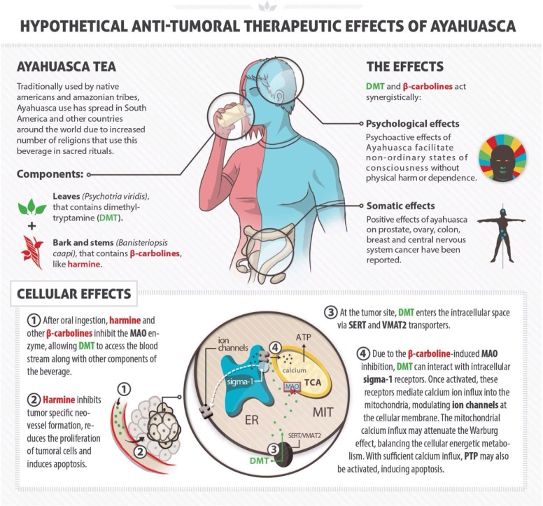
A recent study by Icahn School of Medicine, New York, singled out harmine (from the ayahuasca Caapi plant) among 100.000 substances, as the only one able to cause beta cells in the pancreas (the internal organ that produces insulin) to regenerate, a discovery of great interest to diabetes researchers. Other evidence suggest that ayahuasca may have the potential to regenerate several different types of cells, in many places in the body where needed, the specifics of which calls for medical research in many areas – especially neurodegenerative diseases without a known cure so far. There is also a growing interest in exploring the cell regenerative properties of these plants in spinal cord injury research. Harmine in ayahuasca has also been found to regulate glutamate pump expression in the central nervous system, thereby reducing glutamate toxicity – one of the causes believed to trigger and aggravate ALS through excitotoxic reactions occuring through excessive receptor stimulation by neurotransmitters.

Previously controversial about ayahuasca, was the fact that the plants included used to be thought of simply as “hallucinogenenic” by western science. In other words, ayahuasca and similar plants were neglected by the scientific community and disregarded as if they were nothing more than natural drugs. An objective and more accurate term for this category of plants, in respect to the indigenous culture in which ayahuasca is a part, is the term “entheogenic” – meaning plant that are used in a context sacred to native people, inducing spiritually oriented experiences (according to their own cultural perspective and worldview). In several countries, such as Peru, ayahuasca is fully legal and accepted as a complement to conventional medicine, and during recent decade, western countries have increasingly become more accepting towards entheogens such as ayahuasca, as more studies of entheogenic plants have been completed, changing a formerly unfounded an negative attitude.

Along several other similar harmala-alkaloids found in B. Caapi, harmaline is a monoamine oxidase inhibitor. Monoamine oxidase (MAO) is an enzyme in the body that breaks down signal substances (such as serotonin). The inhibition of MAO allows the signal substance to remain in the synapse for a longer period of time. Many anti-depressants work in a similar manner, as they stimulate receptors in a targeted area. However, the alkaloids present in ayahuasca should not be compared to antidepressants, as they are not exactly comparable though they both have the ability to affect the same receptors. A comparison is that Caapi alkaloids and antidepressants have the same type of delivery system, but different contents. The biochemical properties of plants used in ayahuasca, and the effects they cause on a multitude of bodily functions remain unique to these plants alone. Various types of harmala alkaloids exert suppression of neurotoxic metabolites, such as quinolinic acid and kynurenine – metabolites correlating with ALS, Alzheimer’s disease, Parkinson’s disease and Huntington’s disease, all in which elevated levels of given metabolites are found and thought to contribute to onset of disease through interaction with spinal motor neurons.

harmaline_3d_mid.jpg

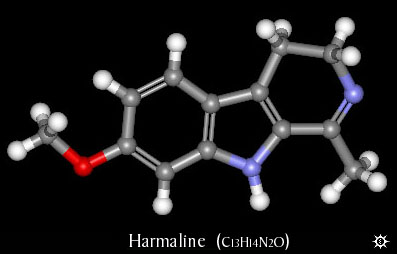
Harmaline together with harmine and tetrahydroharmine,
all forms of mao-inhibitors, are the main components in
the medicinal plant Banisteriopsis Caapi

Ayahuasca in itself is proven unharmful, its compounds being non-toxic, although temporary side effects such as nausea and vertigo are common when used in amounts above medicinal purposes. However, combining certain medical drugs with MAO-inhibitors (such as the ones found in ayahuasca) can be dangerous, even lethal in some cases. This means that in order to safely consume ayahuasca, it must not be combined with any counterindicated pharmaceutical drugs, and those suffering from certain health conditions such as epilepsy or high blood pressure are adviced to either refrain from use or to proceed only with caution and supervision. Uncomfortable side effects from ayahuasca are greatly dose dependent, and a smaller amount consumed for medicinal purpose can thus easily equal few if any side effects experienced.

When searching for information about ayahuasca, a few negative articles can be found, emotionally angled (understandably so), since they tell stories of unfortunate tourists who on their own, or having been duped into doing so, drink something entirely else than ayahuasca – for instance the toxic plant datura, or liquid made from tobacco plant – with serious outcome to their health (including death in a few known cases, from apparent nicotine poisoning). This leads to fear and misinformation, and is not only tragic for the diseased and their families, but also for the natural medicine community trying to promote safe and responsible use of natural medicine for health benefits, and treatment of diseases that regular medical care have failed providing options for. Sensationalistic headlines making unfounded claims, written by people without any knowledge about ethnobotanical medicine, will definitely not help neither ALS patients or people searching for information, and only makes medicinal plant research more difficult. In several countries, including Peru, Brazil and Costa Rica, established retreats offer ayahuasca treatment where the correct plants are harvested (and sometimes organically grown on the property) and prepared by experienced botanists.

ayaflora1.jpg


One of the earliest studies on B. Caapi were done in the 1920’s, and involved patients with Parkinsons’s disease. The patients experienced great symptom relief in early trials, but unfortunately the research was prematurely discontinued due to lack of profit potential – as substances already present in natural plants could not be applicable for patents useful to pharmaceutical companies.

Ayahuasca therapy is likely to gain further attention in coming years, but is already well established in some places. Should discoveries eventually lead to production of a therapeutic pharmaceutical drug derived from these plants, it would take many years until a final widespread and established product reaches and helps actual patients. The process from studies, through trials, to eventual launch of an approved pharmaceutical made for use in the medical care system, is slow due to obvious reasons. The real and interesting fact is that ayahuasca in its natural form is something already available to people with diagnoses lacking other treatment options. For those who can and want to use ayahuasca, there is, while not in any way guaranteed, a possibility for improvement that is still real. As in many other cases, individual results vary, and there should be an emphasis on not overly promoting this medicine (and thereby people’s hope) while questions and work remain. There is also the importance of emphasizing and thereby minimizing the risks involved concerning contraindications with other medications and conditions. Awaiting further studies, this information should still be worth the attention of those suffering from a debilitating progressive disease such as ALS, and researchers in medical science.

My personal connection to this topic and project, was the passing of a close friends’ mother in ALS which occured in 2012. The course of her disease was rapid and aggressive, and unfortunately several of the now available studies referred to in this article, were not yet published at the time. This led me into investigating biochemical aspects of entheogenic plant medicine in search for treatment of neurological disease, and making information on the topic publicly available, while connecting it to a study currently in progress.

B. Caapi is legally obtainable in most countries and states much in the same way as other known herbal remedies, such as Echinacea and Ginseng. However, as with any other potent natural supplement, it is up to the consumer to use and combine supplements and herbal remedies in an informed and responsible way. In the same way as conventional pharmaceuticals, natural medicines should always be treated with proper respect. One commonly known species that actually contains small amounts of harmala alkaloids is passion flower, or passion fruit tree, although the concentrations in its leaf foliage and flowers are far too low (and the fruit contains none) to be used effectively for monoamine oxidase inhibition and ayahuasca purposes. Also, while similar to some degree, the alkaloid profile in terms of proportions and molecular structural deviations between distinct alkaloids does not match up exactly with that of B. Caapi, making the two species related from a certain aspect, but far from equal regarding their medicinal properties. Extracts from various passiflora species are however produced and sold worldwide as mild herbal relaxants and sleeping aids, and as an antispasmodic for treating Parkinson’s.

The substance known as dimethyltryptamine, found in plants traditionally added to ayahuasca, is regulated by law in a number of countries classified as a scheduled substance. (Questionably so, due to medicinal value in multiple areas). This particular substance contained in the additional aforementioned plants induces the altered state of consciousness, a many times misunderstood and stigmatized phenomenon. A description of this altered state is that it is dreamlike, that it stimulates memory and the ability to think abstract, and has self-therapeutic qualities. Even though dimethyltryptamine is naturally occuring in the human body, thought to be produced in small amounts by the pineal gland in the brain during sleep, it remains an illegal substance in a number of western countries since the 1960’s and 70’s, when lawmakers prematurely criminalized almost any substances suspected of having any effect on the mind, including natural ones, due to the widespread moral panic at the time – regardless of the fact that many of them, including dimethyltryptamine, were never proven unhealthy, having in fact been used by indigenous people, in the form extracted from plants, to successfully treat diseases and health problems for centuries. Leaf juice from Chacruna has been used traditionally as a remedy for migraines and ant bites, and Jurema bark for treating burn wounds – significantly quickening regeneration of skin and scar tissue. Dimethyltryptamine also targets chaperone sigma 1, a receptor subtype expressed in both neurons and glia of multiple regions within the central nervous system, with capacity to modulate biological mechanisms implicated with neurodegeneration. Sigma 1-receptors present compelling targets for pharmacologically treating neurodegenerative disorders, and dimethyltryptamine acts as an endogenous Sigma-1 receptor regulator, but interactions between the two in association with motor neuron disease is not understood.

molecules.jpg

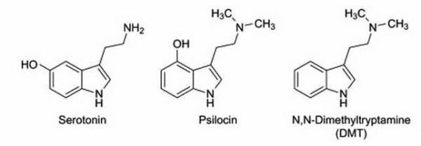
Comparison between Dimethyltryptamine, Psilocin- and Serotonin-molecules
(Psilocybin being the precursor to Psilocin)

Although, several european countries have redefined their policy regarding many formerly frowned upon medicinal plants in recent years, much due to an increasing awareness and access to new and unbiased information and up-to-date research regarding these plants. In Scandinavia, Sami natives from Sapmi, Sweden, were recently aquitted from all charges in the court of law, for having brought Peruvian medicinal cactus into the country. The court established that natural plant material alone cannot be defined as a scheduled substance, and that the therapeutic work involved people were doing, which included Echinopsis Pachanoi cactus, was indeed not a criminal act, but served the purpose to help and heal people. Another similar case with the same outcome involved ayahuasca additive plants. Cacti from the Echinopsis and Lophophora species are known for their restorative and cleansing effects on the body, and are used as such in ethnobotanical medicine.

To be precise, the definition of Ayahuasca is any tea made from either the plant Banisteriopsis Caapi alone, or from B. Caapi and additional plants containing dimethyltryptamine. A tea made from B. Caapi alone does not have what is sometimes referred to as “visionary” qualities, as only the addition of dimethyltryptamine from additive plants, or actually the combination of mao-inhibiting alkaloids in B. Caapi together with dimethyltryptamine content induces a state of mind formerly mislabeled “hallucinogenic”. It needs to be clarified that this word brings up negative associations in many people, and is thus feared and misunderstood. Unlike what some people tend to think, one does not hallucinate things appearing out of thin air after having consumed ayahuasca, but rather there are sequences of inner dreamlike visions taking place while resting, while still awake and fully conscious, provided a significant amount of tea has been consumed. It is actually quite undramatic, aside from the unpleasant vomiting which does affect some people (which is entirely intentional in traditional in cleansing rituals).

And herein lies the essence many times misunderstood: It does not take a great amount of ayahuasca to experience strictly its medicinal effects – without abstractions and visionary effects, or nausea/vomiting which naturally people want to avoid. Several medicinal health benefits can be obtained even by using B. Caapi alone – without additive plants, thereby ensuring no visual effects experienced at all, should this be desired. It should be noted though, that the synergistic effect between the two plants used simultaneously will bring the best medicinal and bodily response. Exaggerations regarding ayahuasca made these medicinal plants overlooked for many years in the west, but the reputation has been revised as more people with experience in a medicinal context have made statements, claiming the medicinal value of ayahuasca relevant to medical conditions of different types – the field of neurological disease being the latest. Ayahuasca has already been effectively used for symptom relief from Multiple Sclerosis and rheumatoid arthritis, by a growing number of people in Europe since at least 2006. ALS, Multiple Sclerosis, Alzheimer’s and Parkinson’s disease all share a lot of common ground, being that they all involve varieties of nerve cell degeneration. Thus it is likely that any type of natural broad spectrum medicine able to affect the process of nerve cell regeneration, and that also has substantially antioxidative and cell protective properties, could prevent and slow the progression of neurological disease in general.

Whatever wild or strange stories about ayahuasca that may occasionally be found circulating, they stem mostly from people who went to live with native tribes during the late 80’s and early 90’s, taking part in traditional ceremonial use of ayahuasca – consuming exceptionally generous or concentrated amounts of the medicine, enfolding themselves in deep cleansing experiences not necessarily easily endured. This medicine, like all others, should definitely be well respected, but not subject to exaggeration or downright misrepresentation – causing people to dismiss what they are simply uneducated about, which in turn may lead to people never getting treatment that could slow the progression of their terminal health condition, or in some cases even reverse it. The vivid and fascinating visions induced by strong tea often seem to have a theme rooted in nature, perhaps tied into the underlying psychological expectations associated with the revered history of ayahuasca itself, as depicted quite beautifully in colorful paintings by Peruvian artist Pablo Amaringo (1938-2009). The visions arise from the simple fact that the alkaloids and tryptamines dissolved in the tea, combine to affect receptors that in turn stimulate the processing of memory relating to images and words – noticeably of relevance to Alzheimer’s research.

Ayahuasca is proven non-addictive, and is even used to aid people in breaking their drug dependencies, as ayahuasca has a detoxifying and documented effect of ridding the user of drug related abstinence issues.

MAO-inhibition makes uptake of dimethyltryptamine in the body possible, unable to occur otherwise as dimethyltryptamine without MAO-inhibition is rapidly broken down by enzymes in the stomach, unable to cause effect. Dimethyltryptamine is almost molecularly identical to the above mentioned psilocybin from Dr. Ramos research. Researchers think that psilocybin and dimethyltryptamine binds to brain receptors that stimulate growth and healing, acting on the hippocampus, a part of the brain that is essential to learning and forming memories, recieving sensory impulses and that has target cells and receptors for important signal substances. Hippocampus atrophy occurs when nerve cells die, or when abnormal levels of stress hormones prevent neurogenesis, and is a known sign in Parkinson’s disease. It is theorized that the unique combination of a variety of harmala-alkaloids from B. Caapi, and dimethyltryptamine from additional plant sources used in ayahuasca, work on cellular level to repair and restore nerve cells, stimulate and enhance motor neuron transmission, and to protect remaining nerve cells from degeneration. This is without doubt valuable from a neuromedical perspective.

21food_1305567977015.jpg

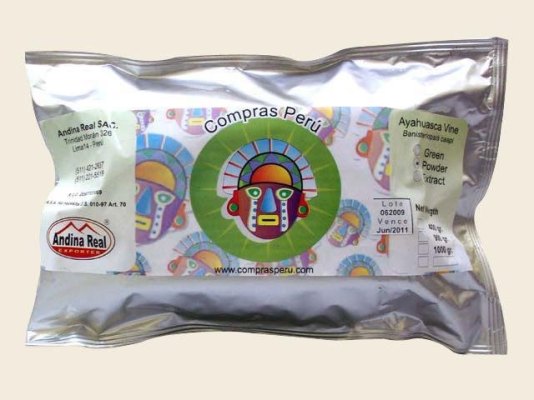
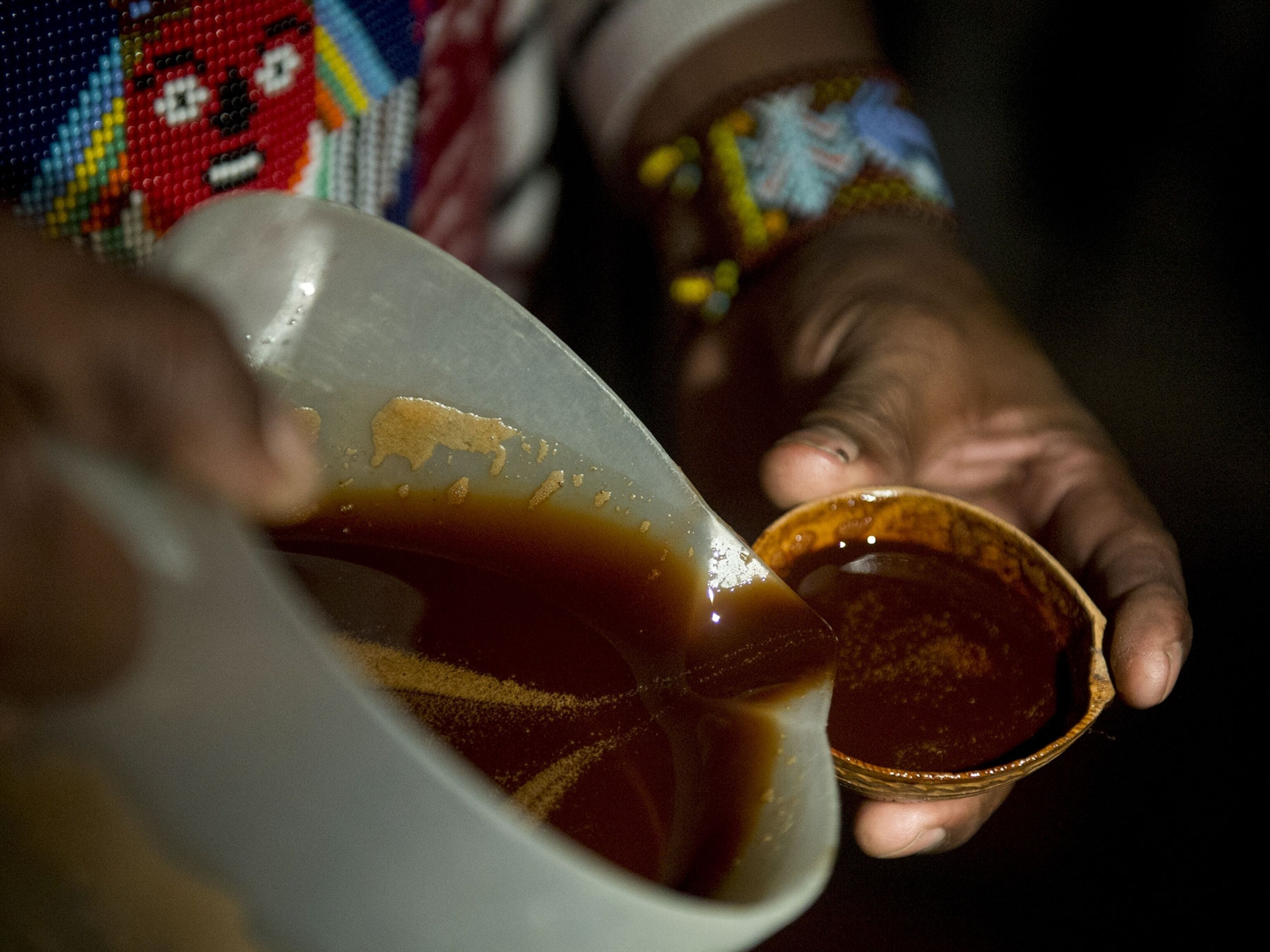
Ayahuasca plants as packaged and sold in health food stores in, Peru.

As the non regulated B. Caapi alone has proven positive abilities, potentially effective against neurological and cancer diseases, it is thus something real and available that may be a valid treatment option. For anyone who experience positive results to any degree, but does not live in a state or country where the use of plants containing dimethyltryptamine is permitted, there is the possibility to travel to one of the many countries (or states) which by law allows the use of added secondary plants with their combined medicinal properties for evalution of full-spectrum ayahuasca treatment. In Europe, Spain is one of several countries where ayahuasca is becoming an established therapy, and Spain is also the chosen location for an international conference 2014, where ethnopharmacologists, psychologists and researchers from all over the world gather around the topics of ayahuasca and medicinal entheogens.

Among others, Ede Frecska, M.D., Ph.D, University of Debrecen, lectures on the possibilities of recreating braincells and regulating the immune defense system through this plant-based medicine and others. This event is held by ICEERS – International Center for Ethnobotanical Education Research and Service, and can be followed at:

caapi_examine.png

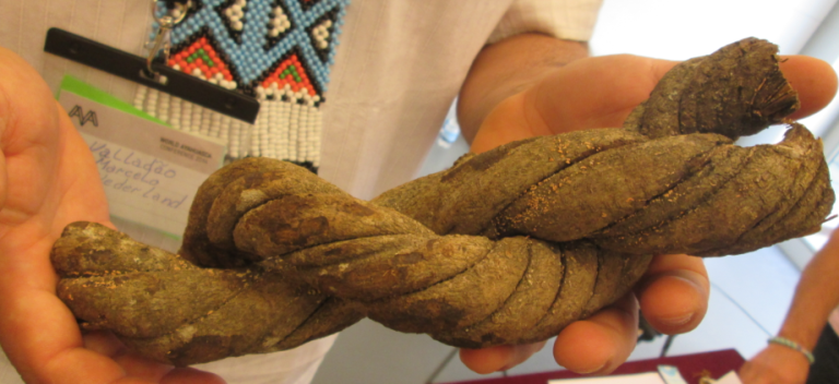
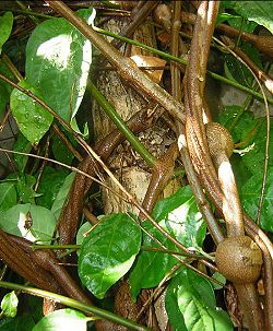
Examination of Caapi medicinal vine specimen.

Furthermore, besides ability to aid and enhance the process of nerve cellular repair and protection against cell oxidation, several entheogenic plants (and fungi), including ayahuasca, do possess psychotherapeutical qualities as well. Coping with degenerative illness is obviously stressful to patients, and a great deal of emotional relief, personal insight, and ability to better cope with personal situation is achievable through the single or repeated experience of entheogenic medicinal plants/mushrooms in a comfortable and supportive environment, according to renowned Johns Hopkins medical university.

The fact that many of these medicinal plants are becoming revived as they recieve scientific approval, is great news in several ways. Sustainability and environmental issues comes to mind, and so far the outlook remains positive. Many organic farms have developed in south and central america, cultivating ayahuasca plants for both local use and for export, providing work and income for people in rural areas otherwise stricken with poverty. This also serves as a way for numerous local people to reconnect with their cultural past, as ayahuasca is declared a national heritage in Peru among other places.

dmckenna.jpg


Dennis Mckenna, PH.D, one of the world’s most renowned ethnopharmacology researchers speaks on the importance of sustainability, regarding cultivation of ayahuasca and the preservation of rainforests. This should concern all people who benefit from treatment using ayahuasca medicinal plants. Dr. Mckenna is co-founder and director of ethnopharmachology at Heffter Research Institute, New Mexico. He is also a faculty member of the Academic Health Center at the University of Minnesota, and was key organizer in the Hoasca Project, an international biomedical study of ayahuasca, funded by the Stanley Medical Research Institute. While ayahuasca plants take years to mature for harvest, they can actually be grown at home in gardens anywhere in the world where the climate allows, or indoors or in heated greenhouses elsewhere, and the seeds are both cheap and abundant – available from numerous online ethnobotanical vendors worldwide, ensuring the survival of these medicinal plant species and their sustained availability for future generations.

Formerly unknown to science, we are beginning to understand the potential and medicinal aspects of this particular plant, replacing neglect of entheogens with knowledge as scientific groundwork in this matter becomes firm.

People should not be led into thinking this is some kind of natural miracle cure, but used the right way it could provide longterm aid in the restorement of body and mind function in certain neurological conditions. Together we can all inform people in an unbiased, ethical and safe way about any and all viable treatment options, and about the medicinal and therapeutical value of entheogens in general.

Common sense should be applied to determine dosage in order to ensure a sufficient degree of medicinal activity, while not being harmful or overwhelming. Specific diet is essential in conjunction with ayahuasca, in order to minimize side effects and to maximize utilization of medicinal compounds, and basic knowledge on the process of preparation of the medicine is helpful in order for the plants to synergize correctly. The help and information needed for these purposes, along with protocols for detoxification are individually provided to participants in the below pilot study.

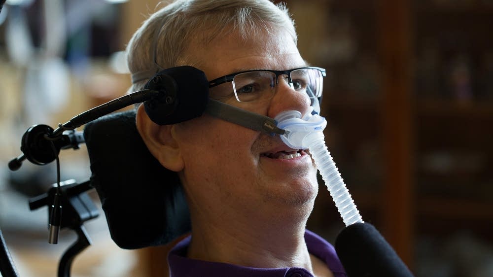
Ayahuasca has been used for a very long time historically, and only recently for treatment of the conditions brought up in this article. Any substantial health improvement from this natural medicine in cases of motor neuron disease would be likely to reveal itself long-term at first. Initial updates from people participating in the ayahuasca ALS treatment pilot study report a few things in common; wider range of movement, weight gain in muscle mass, muscle tension relief, reduced spasticity and improved strength in affected limbs, though it should be noted that none of the participants had lost all muscle control prior to treatment, and that whether or not this effect turns out to be persistent or permanent is not known at the moment.

Summary of key points:

Ayahuasca could effectively be used in treatment of ALS and other motor neuron diseases based on the fact that studies suggest uniquely antioxidative effects that seem to protect brain/nerve cells, targeting motor neurons through a unique biochemical transport system, and that it and other moleculary similar substances, also naturally occuring, stimulate neurogenesis – the development of new brain/nerve cells, and improved neural connectivity. In studies it has been found to reduce symptoms in Parkinsons’s patients – all neurodegenerative diseases share common ground, making it likely that something that improves one major neurological condition could also be beneficial to other closely related conditions. Also based on credible personal accounts from people having used ayahuasca for symptom relief from their multiple sclerosis (once again – the common ground of neurodegenerative diseases), documented in books about ayahuasca, and descriptions of early stage minor improvement by Ayahuasca ALS Treatment study participants, already having used the medicine for a period of time. Studies also indicate ability to normalize metabolism in mitochondria crucial to motor neuron survival, and to regulate and decrease levels of excitotoxicity in the central nervous system.

Ayahuasca and other entheogens can and will gain the credibility and amends they truly deserve, and bring new possibilities to many people living with diseases lacking clinical healthcare treatment options. Until then, these medicinal plants remain available for personal evaluation by individuals who choose to explore the option. In relation to the medical conditions brought up in this article, these plants may hold future keys in becoming a powerful tool for reversal of ALS progression and related conditions. Share this important information if you find it valuable.

*From the article here :
 
Last edited:
shutterstock_506591185-1400x480.jpg


Research clears up how 'brain cleaners’ fail in ALS

Mayo Clinic | Neuroscience | 9 Jan 2022

Findings point to microglial TREM2 as a potential target for the treatment of ALS.

In preclinical studies, Mayo Clinic scientists and collaborators have identified the molecular mechanism used by the brain’s “cleaners” as they remove a problematic protein in the brain.

This work, published in Nature Neuroscience, demonstrates that the cleaners—resident immune cells in the brain called microglia—play a protective role in a mouse model of Lou Gehrig’s disease, also known as amyotrophic lateral sclerosis (ALS). The study may provide a potential therapeutic target for that disease.

The loss of neurons characterizes the neurodegenerative diseases of Alzheimer’s and ALS. In Alzheimer’s, the lost neurons handle memory retrieval, and in ALS, it is the neurons that manage movement that are damaged. In the brain of people diagnosed with either of these diseases, pathologists can see a buildup of specific proteins: beta-amyloid and tau in those with Alzheimer’s, and a protein called TAR-DNA binding protein 43 kDa (TDP-43) in people with ALS.

Microglia are the cells tasked with cleaning up debris in the brain, and they have a unique receptor called TREM2. When this receptor is mutated, evidence suggests risks increase for developing Alzheimer’s disease, the theory being that microglia cannot clean up the brain efficiently. But the evidence for TREM2 and ALS was more tenuous until now.

“The aggregation of TDP-43 in the brain is a hallmark for most ALS patients,” explains Long-Jun Wu, Ph.D., a Mayo Clinic neuroimmunologist and senior author of the paper. “Our study shows for the first time that TDP-43 is a potential ligand for microglial TREM2. Further, we found that this interaction mediates microglial TREM2 sensing and clearance of pathological TDP-43 protein."

microglia-als-neurosicences.jpg

Using samples donated to Mayo Clinic’s Brain Bank for Neurodegenerative Disorders, as well as mouse models,
biochemistry, confocal microscopy, and computational simulations, researchers showed that a receptor on
microglial cells, called TREM2, can clear a protein that builds up in the brains of patients with ALS.

Image created with BioRender. Credit: Mayo Clinic

As a ligand, TDP-43 binds to the TREM2 receptor, which is important for microglia cells to clear up the protein. Using biochemistry, computational simulations, confocal microscopy, mouse models, and samples from the Mayo Clinic Brain Bank for Neurodegenerative Disorders, the researchers were able to decipher the interaction between TREM2 and TDP-43, thereby potentially revealing a target for therapy for ALS.

“The mechanisms underlying ALS initiation and progression are poorly understood,” says Dr. Wu. “Microglia comprise a unique subset of glial cells and are the principal immune cells in the central nervous system. Our current findings point out microglial TREM2 as a potential therapeutic target for ameliorating TDP-43-related neurodegeneration, including ALS.”

The Neuroimmune Interaction in Heath and Disease Laboratory led by Dr. Wu plans to delve into the exact binding sites of human TDP-43 and the TREM2 receptor. They also want to investigate a specific population of microglia that seem to be supercharged to remove TDP-43. And the eventual goal is to explore if TREM2 activators might be a candidate for treatment in mouse models of ALS, which is a first step toward potentially treating human disease.

 
Panorama_of_Kings_Parade_in_Cambridge%2C_UK%2C_at_St._Mary%27s.jpg

Cambridge

Ibogaine and Neurodegenerative Diseases*

Genesis Ibogaine Centre

This is a summary on existing research into the influence of Ibogaine on glial cell line-derived neurotrophic factor (GDNF) levels in the brain, and the beneficial impact that an increase in this protein can have. While existing studies have examined these areas, few have identified a possible link between Ibogaine, GDNF expression and neurodegenerative diseases.

Both Parkinson's Disease and ALS are chronic disorders with no known cure, and require management with drugs that can have considerable side effects, causing a very poor quality of life for terminal stage sufferers of these diseases. By contrast, a low dose regime of Ibogaine or Iboga alkaloid extract would be of low toxicity and free of serious side effects.

GDNF has been shown to have potent neurotrophic factor in both rodent and primate models of Parkinson's Disease. Direct brain infusion of GDNF into the brains of five Parkinson sufferers resulted in a 39% improvement in the off-medication motor sub-score of the Unite Parkinsons Disease Rating Scale and a 61% improvement in the activities of daily living sub score. Positron emission tomography (PET) scans of dopamine uptake showed a significant 28% increase in putamen dopamine storage after 18 months, indicating a direct effect of GDNF on dopamine function. Furthermore, after one year, no serious clinical side effects were observed. The use of Iboga alkaloid extract or Ibogaine would provide a longer term and much less invasive method of GDNF administration than direct brain infusion. Thus, further research on Ibogaine and GDNF is certainly warranted.

Regarding motor neuron disease (ALS), the little research that has occurred in this area, such as gene transfer of neurotrophic factors, suggests potential in the treatment of motor neuron disease. Again, Ibogaine therapy may offer a straightforward, non-invasive, cheap, low-toxicity method of treatment for sufferers of this disease.

*From the article here :
 
Last edited:
als-blood-test-neurosinces-public.jpg


Simple and reliable ALS diagnosis with blood tests

University of Gothenburg | Neuroscience | 28 Jan 2022

Measuring the level of neurofilaments in the blood may be a reliable biomarker for the early diagnosis of ALS.
Blood tests may enable more accurate diagnosis of ALS at an earlier stage of the disease. As described in a study by researchers at University of Gothenburg and Umeå University, it involves measuring the blood level of a substance that, as they have also shown, varies in concentration depending on which variant of ALS the patient has.

The study, published in Scientific Reports, include Fani Pujol-Calderón, postdoctoral fellow at Sahlgrenska Academy, University of Gothenburg, and Arvin Behzadi, doctoral student at Umeå University and medical intern at Örnsköldsvik Hospital, as shared first authors.

Currently, it is difficult to diagnose amyotrophic lateral sclerosis (ALS), the most common form of motor neuron disease, early in the course of the disease. Even after a prolonged investigation, there is a risk of misdiagnosis due to other diseases that may resemble ALS in early stages. Much would be gained from earlier correct diagnosis and, according to the researchers, the current findings look promising.

Neurofilaments—proteins with a special role in the cells and fibers of nerves—are the substances of interest. When the nervous system is damaged, neurofilaments leak into the cerebrospinal fluid (CSF) and in lower concetrations in blood compared to CSF.

In their study, scientists at Umeå University and the University Hospital of Umeå, as well as at the University of Gothenburg and Sahlgrenska University Hospital in Gothenburg, demonstrated that CSF and blood levels of neurofilaments can differentiate ALS from other diseases that may resemble early ALS.

More sensitive methods of analysis

Compared with several other neurological diseases, previous studies have shown higher concentrations of neurofilaments in CSF in ALS. Measuring neurofilament levels in the blood has previously been difficult since they occur at much lower concentrations compared to CSF. In recent years, however, new and more sensitive analytical methods have generated new scope for doing so.

The current study shows a strong association, in patients with ALS, between the quantity of neurofilaments in the blood and in CSF. The study is based on blood and CSF samples collected from 287 patients who had been referred to the Department of Neurology at the University Hospital of Umeå for investigation of possible motor neuron disease. After extensive investigation, 234 of these patients were diagnosed with ALS. These had significantly higher levels of neurofilaments in CSF and blood compared to patients who were not diagnosed with ALS.

Higher concentrations

Differences among various subgroups of ALS were also investigated and detected. Patients whose pathological symptoms started in the head and neck region had higher neurofilament concentrations in the blood and worse survival than patients whose disease onset began in an arm or a leg. The study has also succeeded in quantifying differences in blood levels of neurofilaments and survival for the two most common mutations associated to ALS.

“Finding suspected cases of ALS through a blood test opens up completely new opportunities for screening and measuring neurofilaments in blood collected longitudinally enables easier quantification of treatment effects in clinical drug trials compared to longitudinal collection of CSF. Finding ALS early in the disease course may facilitate earlier administration of pharmaceutical treatment, before the muscles have atrophied,” Arvin Behzadi says.

ALS is a neurodegenerative syndrome that leads to loss of nerve cells in both the brain and the spinal cord, resulting in muscle weakness and atrophy. Most of these patients die within two to four years after the symptom onset, but roughly one in ten survive more than ten years after the symptoms first appeared.

Several genetic mutations have been associated to ALS. At present, there is no curative treatment. Nevertheless, the current drug available has been shown to prolong the survival in some ALS patients if it is administered in time.

Original Research: Open access.
Neurofilaments can differentiate ALS subgroups and ALS from common diagnostic mimics” by Arvin Behzadi et al. Scientific Reports

 
Last edited:
Fotolia_233087215_Subscription_Monthly_M.jpg


CBD may prolong life for those suffering with ALS

by Lisa Rough | Leafly | 19 June 2015

Based on some exciting new anecdotal evidence, the use of cannabis oil to treat ALS may actually prolong life in addition to relieving many of the diseases devastating day-to-day symptoms.

ALS or Lou Gehrig's disease, named after the baseball legend, is a debilitating disorder that affects an estimated 30,000 people in the United States at any given time. You might remember ALS from that craze last summer when everyone decided to dump ice on their heads to raise awareness for the disease and to raise money for ALS research. Even Leafly hopped on the bandwagon for a good cause, and overall, the ALS Foundation received over $100 million in donations by the time the summer of the Ice Bucket Challenge was over.

ALS is a degenerative neurological disorder that causes muscle weakness, wasting, and paralysis of the limbs, as well as those that control vital functions such as speech, swallowing, and breathing. It is often the deterioration of these crucial muscles that leads to respiratory failure; even with breathing aids or a tracheotomy, the risk of respiratory failure persists.

Being diagnosed with ALS is devastating, and for many it can feel like a death sentence. The average life expectancy of a person with ALS is two to five years. However, more than half of all people with ALS live more than three years from diagnosis and there are definitely outliers, such as Stephen Hawking, one of the most iconic figures with ALS. Hawking was diagnosed in 1963, and now, more than 30 years later, he is among only 5% of those diagnosed with ALS who live more than 20 years after diagnosis.

Cannabis has long been known as a viable treatment option to relieve symptoms of ALS, which was outlined in this handy chart created by the American Journal of Hospice and Palliative Medicine, but the most astonishing results have come from several ALS sufferers who have managed to slow the progression of the disease through regular, controlled doses of cannabis oil.

Bob Strider began experiencing the symptoms of ALS in 1998, specifically the loss of function in his right arm and problems swallowing. An avid cannabis enthusiast, he had used cannabis heavily for decades, which he believes has kept the progression of his disease slow but steady. In 2012, Strider began manufacturing his own cannabis oils, dosing himself with approximately a gram a day for 60 days. Within 10 days of his regimen, he regained control of his right arm and was able to stop using opiates to manage his pain.

Another remarkable case comes from Cathy Jordan, who was diagnosed with ALS in 1986 and given less than five years to live. In the winter of 1989, Jordan spent the holiday in Florida, preparing for the end of her life, when she made a crucial discovery. While walking on the beach one night, she smoked a joint of Myakka Gold and felt her symptoms cease, essentially experiencing the neuroprotective effects of cannabis before they'd been proven.

Jordan never set out to be a cannabis activist, preferring instead to quietly continue treating her disease. She tried to tell her neurologist in 1989 that cannabis had helped her, and he tried to convince her husband to have her committed to a mental facility. In 1994, Jordan met a new doctor who was astonished by her progress. When he asked what shed been doing to stay alive, she informed him, and he advised her, Smoke all the cannabis you can and never tell a soul, because they will never believe you.

Unfortunately, Donnie Clark, the grower of Myakka Gold, was arrested and sent to prison for 12 years until his sentence was commuted by former President Clinton. The strain itself, which Jordan credits with stopping the progression of her ALS, has since been eradicated by the DEA.

Cathy Jordan, however, ended up becoming the inspiration for Amendment 2, the medical marijuana initiative in Florida that was edged out last November. Ironically, on February 25, 2013, the same day that the Cathy Jordan Medical Cannabis Amendment was announced, Jordan's home was raided and 23 plants were seized by local authorities, although charges were dropped when it became evident that she was using cannabis for medicinal purposes.

Now, finally, years later, there has been more and more conclusive research indicating that these patients self-medicating regimen was beneficial, after all. Pre-clinical data shows that cannabis has powerful anti-oxidative, anti-inflammatory, and neuroprotective effects, and with regular applications, it may actually slow the progress of the disease and prolong the lives of the individuals affected by ALS.

Amyotrophic Lateral Sclerosis is a qualifying condition for medical marijuana programs in Delaware, Washington, D.C., Georgia, Illinois, Maine, Massachusetts, Michigan, New Jersey, New Mexico, and New York. However, with more promising research emerging, other states medical marijuana programs need to follow suit and allow suffering patients the Right to Try and treat this horrible disease with cannabis if it can provide them with even a little bit of relief.

https://www.leafly.com/news/science-...gression-of-al
 
maxresdefault-e1410995215392-1020x423.jpg


Treating Neurodegenerative Disorders with Cannabinoids*

by Cornerstone Wellness

Neuroinflammation is known to play a significant role in essentially all neurodegenerative processes. Diseases such as Alzheimer’s, Multiple Sclerosis (MS), Huntington’s Disease, and Parkinson’s Disease all involve hyperactive microglia, which are the live-in macrophages of the brain, spinal cord, and central nervous system. Macrophages are immune cells that capture and dissolve foreign substances, germs, and cancer cells within the body. The microglia in the brain and spinal cord form the first line of immune defense in the central nervous system. Unfortunately, in the case of aforementioned diseases, these cells have become overactive causing them to secrete excess substances, such as cytokines (cell signals that regulate cell group growth and response), glutamate, and harmful free radicals. This excessive production of chemicals causes inflammation, which leads to further cell death.

Cannabis and the family of chemicals it produces are known to act on two major cell receptor types named CB1 and CB2 respectively. The CB1 receptor is most commonly found in neurons throughout the brain. The psychedelic effects of cannabis come from this receptor’s function, which re-wires the way neurons signal each other. The CB2 receptor on the other hand, is found throughout the body, especially within the immune system cells. The effects of activating the CB2 receptor are more myriad, but within the immune system specifically four groups of effects have been identified:

1. Induction of apoptosis or forced cell death

2. Suppression of cell proliferation

3. Induction of regulatory T cells

4. Inhibition of pro-inflammatory cytokine/chemokine production and increase in anti-inflammatory cytokines

The last of these effects is the basis upon exploring using cannabinoids to halt the progress of neurodegenerative disorders. The idea is that if cannabinoids can prevent excess production of cytokine, inflammation will decrease, and the resultant cell death around that inflammation will not occur. This would go a long measure toward slowing progression of neuro-inflammatory diseases. However, it is important to note that tempering inflammation would still not allow the brain to recover to its pre-disease state and slow neural damage would inevitably continue to occur. Likewise, modern medicine currently utilizes a variety of treatments in these diseases as more or less palliative care.

Based on these observations, research groups worldwide have been testing specific cannabinoids and other CB2 agonists with various models of neuro-inflammatory disease, generally in rodents.

The following is a general review of effects noticed, grouped by disease:

Alzheimer’s Disease – In Alzheimer’s cannabidiol has been shown to “reduce the transcription and expression of pro-inflammatory molecules in the hippocampus of an in-vivo model of induced neuroinflammation”. The hippocampus is the part of the brain that controls conversion of memory from short to long-term and controls spatial navigation. In Alzheimer’s, it’s one of the first areas of the brain to suffer damage and why patients have memory problems. Another agonist, which has the name SR141716A, also prevents amnesia induced by certain peptides, so these both promise a future in treating the disease.

Parkinson’s Disease – In this disease, the agonist WIN55,212-2 has been shown to protect mouse neurons from the neurotoxin MPTP, which is the chemical which leads to the death of dopaminergic neurons and causes Parkinson’s Disease.

Multiple Sclerosis (MS) – Although the cause of MS is unclear, researchers have determined that both genetic susceptibility and environmental trigger play a role. Some patients develop their symptoms of MS after contracting a virus. Likewise, testing rodents injected with a virus that intentionally lead to animal models of MS provided ground to investigate the effects that CB2 agonists might have on MS in humans. As is, cannabis concentrate is already prescribed under the name Sativex to alleviate neuropathic pain, spasticity, and overactive bladder symptoms associated with MS. Although some agonists did increase symptoms, several, including THC, delayed onset and reduced severity of symptoms. Three agonists, WIN55,212-2, ACEA, and JWH-015 were shown to improve motor function by attenuating microglia and immune cell infiltration into the spinal cord.

Exactly how these effects are achieved is still unknown. Although it is presumed that the effects of the CB2 agonists (any molecules that can activate CB2 receptors, including cannabinoids) stem from CB2 receptor activation, other theories have been proposed. One research group at the University of Bari has explored the extra-cannabinoid receptor binding activity of cannabidiol (CBD). Researchers there found that CBD can surprisingly communicate with the nucleus of the cell directly through interaction with nuclear hormone receptors. There are several possible chemical pathways through which cannabinoids can achieve their effects, beyond CB2 receptors. Further research will illuminate these pathways.

*From the article (including references) here :
https://cornerstonecollective.com/ho...der-treatment/
 
Last edited:
cfedc1-20140916-090414brucecathy03.jpg


Harmine and ALS

Yun Lia, Rita Sattlera, Eun Ju Yang, Alice Nunes, Yoko Ayukawa, Sadia Akhtar, Grace Ji, Ping-Wu Zhang, Jeffrey Rothstein

Harmine is a beta-carboline and a harmala alkaloid. It occurs in a number of psychedelic plants, most notably the Syrian rue and Banisteriopsis caapi.

Recently, the excitatory amino acid neurotransmitter glutamate was implicated in the pathogenesis of various chronic degenerative neurological diseases in humans and animals. This report describes abnormalities in excitatory amino acids in the central nervous system of 18 patients with amyotrophic lateral sclerosis (ALS). We conclude that excitatory amino acid metabolism is altered in patients with ALS. Based on neurodegenerative disease models, these changes may play a role in motor neuron loss in ALS.(1)

Glutamate is the predominant excitatory amino acid neurotransmitter in the mammalian central nervous system (CNS). Glutamate transporter EAAT2/GLT-1 is the dominant astroglial protein that inactivates synaptic glutamate. Previous studies have shown that EAAT2 dysfunction leads to excessive extracellular glutamate and may contribute to various neurological disorders including amyotrophic lateral sclerosis (ALS).

Few compounds have been shown to activate increased glutamate transporter-EAAT2/GLT1 protein expression and function in vivo. Our new cellular screen has provided insight into a class of compounds that could serve as a starting point for new GLT1 activators. Given harmine’s CNS side effects and its multi-target activities, harmine itself is not a suitable candidate for immediate clinical applications. However, it may eventually be possible to separate its effects on GLT-1 upregulation and on CNS pharmacology through optimization of its chemical structure. Medicinal chemistry efforts to do so are ongoing, which will hopefully lead to a more potent and selective drug candidate that can be moved through the drug development process reaching from preclinical studies to clinical trials. These trials may include clinical applications such as ALS, and other neurodegenerative disorders.(2)

(1) https://onlinelibrary.wiley.com/doi/abs/10.1002/ana.410280106
(2) https://www.ncbi.nlm.nih.gov/pmc/articles/PMC3220934/
 
stephen-hawking-on-october-10-1979-in-princeton-new-jersey-photo-by-santi-visalligetty-images.jpg

Prof. Stephen Hawking

Ayahuasca and ALS

Male, 65, UK, diagnosed with ALS (not Prof. Hawking) :

What can you tell us in general about your medical condition, and to what degree has it affected you? (Prior to treatment using ayahuasca)

My prognosis as of March 2013 was that of a one-year life expectancy, I did get a specific MND diagnosis confirmed by a consultant neurologist, on the back of which an insurance company paid up on my critical illness cover, there being a clear understanding that I would probably not last the year. I was very weak and trembly, had lost over 20 pounds in muscle mass, my knees were prone to swelling and I was racked by cramps and muscle twitching. I could barely manage stairs, and couldn't get out of the bath unassisted. I remember being a little challenged by a four inch step across the end of the room, choosing my best leg to step down.

Describe a typical experience with ayahuasca. What is it like during the immediate time the medicine is active in the body? What has been experienced afterwards?

Initially a fair amount of retching, although I didn't actually vomit much. Then some spastic activity, after which a warmth spread throughout my chest cavity, and sensations from the connection between muscle tissue and skeletal bone. It left me exhausted, but it mellowed out? after a while into a contemplation state of mind where the concept of fear had a major part. Stressful emotions such as fear and anxiety seems to be a major vexation (in relation to neurological diseases), and the ayahuasca seemed to soothe that too. I didn't feel that much different immediately afterwards, I suppose I felt a little more relaxed, but the following weeks (after sessions) my massage therapist said the muscle twitching was much improved, and I continued to recover steadily.

Has this treatment relieved or improved your condition in any way so far?

I have made essentially a full recovery, slow but persistent, and NHS consultants involved are mystified. Ayahuasca seems to me to have been instrumental in my recovery, which is fully documented. I am now fully healthy, I cycle to work. Now, after today doing a nine mile coastal walk for the pleasure it, I feel pretty much up to snuff, for a man of my age. A year after my diagnosis I held a party to express my gratitude to all the people who had helped me through, including T.H., the consultant neurologist (he didn't actually come). I was by that time much better, not up to full strength, but could party on and play the saxophone. I told my doctors about ayahuasca in August of 2013 I think, it was for a six monthly reassessment of my condition. T.H was frankly astonished, and when challenged said that the diagnosis was absolutely sound adding that the nerve conductivity tests were particularly unequivocal. It was then that I told him about the ayahuasca, and rather sheepishly, and mistakenly, he asked me if I had had a good trip? I replied that it had been unpleasant and that it was not a therapy for the faint-hearted. Since then T.H. has presented my case to some regional peer group, and a professor K.T. Their response was that perhaps 'I had a mimi', which seems to be exactly the same as the disease except for the outcome; or that the condition 'might have been brought upon me by the cocktail of therapies' I was taking at the time. I have yet to make a measured response to this, but it was not until I had serious symptoms that were initially interpreted as Lyme disease or an atypical sero-negative rheumatoid arthritic condition, that I embarked on any kind of medical therapy at all. The history presents a persuasive argument. My family, friends and some of my patients are aware of my ayahuasca treatment and are generally supportive. My wife plays tennis with a number of retired GPs, and their general opinion is that whatever I did cant be too bad. Hell, I am still walking about and they were initially supporting my wife and family on the clear expectation of my imminent demise.

Will you continue this treatment? If so, will you make any changes to your current regimen?

I don't see any necessity to carry on being treated. I might do the ayahuasca again but that would really be just out of curiosity.

Have you been following any particular diet prior to, or during your treatment?

Lots of organic fruit and vegetables, and I have a small holding so we have organic lamb and chicken too. I haven't eaten dairy for the past two years, and I seldom drink alcohol though I had a glass of cider the other day.

What made you try this alternative treatment?

It was an inspired patient who in doing an art degree in Plymouth came across the (ayahuasca) visionary art, and drew my attention to its source. Curiously some members of the church group with whom I sit proved an unexpected source of interest and loaned me various books on the topic, including those of the late Alexander Shulgin.

-----

Amyotrophic Lateral Sclerosis, or Lou Gehrig’s Disease, is a progressive, degenerative neuromuscular disease that affects the nerve cells in the brain and spinal cord. These motor neurons carry messages from the brain to the spinal cord and, ultimately, to the muscles that are necessary for voluntary and involuntary movement and function.

In people living with ALS, motor neurons progressively die and the brain can no longer communicate with the muscles through the spinal cord. As muscles are used less and less frequently, they can atrophy, causing people with ALS to lose the ability to perform everyday activities, such as walking, speaking, and eating. ALS also affects the diaphragm, an essential muscle responsible for breathing, so people with ALS eventually lose their ability to breathe on their own. The average life expectancy for ALS patients is three to five years after diagnosis and death is generally caused by respiratory failure. There is no known cause or cure for ALS.

Cytokinetics is driven to improve the lives of people fighting ALS by applying our understanding of the mechanics of muscle function and contractility to the discovery and development of novel potential treatments that may directly improve muscle function, potentially delaying disease progression and preserving independence.

 
Last edited:
iStock-1156668929-696x392.jpg


Scientists identify compound to reverse neuron damage from ALS*

Northwestern University | Neuroscience News

U-9, a novel, non-toxic compound, targets upper motor neurons and reverses damage associated with ALS within 60 days of treatment.

Northwestern University scientists have identified the first compound that eliminates the ongoing degeneration of upper motor neurons that become diseased and are a key contributor to ALS (amyotrophic lateral sclerosis), a swift and fatal neurodegenerative disease that paralyzes its victims.

In addition to ALS, upper motor neuron degeneration also results in other motor neuron diseases, such as hereditary spastic paraplegia (HSP) and primary lateral sclerosis (PLS).

In ALS, movement-initiating nerve cells in the brain (upper motor neurons) and muscle-controlling nerve cells in the spinal cord (lower motor neurons) die. The disease results in rapidly progressing paralysis and death.

So far, there has been no drug or treatment for the brain component of ALS, and no drug for HSP and PLS patients.

“Even though the upper motor neurons are responsible for the initiation and modulation of movement, and their degeneration is an early event in ALS, so far there has been no treatment option to improve their health,” said senior author Hande Ozdinler, associate professor of neurology at Northwestern University Feinberg School of Medicine. “We have identified the first compound that improves the health of upper motor neurons that become diseased.”

The study will be published in Clinical and Translational Medicine on February 23.

Ozdinler collaborated on the research with study author Richard B. Silverman, the Patrick G. Ryan/Aon Professor of Chemistry at Northwestern.

The study was initiated after Silverman identified a compound, NU-9, developed in his lab for its ability to reduce protein misfolding in critical cell lines. The compound is not toxic and crosses the blood brain barrier.

The NU-9 compound addresses two of the important factors that cause upper motor neurons to become diseased in ALS: protein misfolding and protein clumping inside the cell. Proteins fold in a unique way to function; when they misfold they become toxic to the neuron. Sometimes proteins aggregate inside the cell and cause pathology as in the TDP-43 protein pathology. This happens in about 90% of all ALS patient brains and is one of the most common problems in neurodegeneration.

The research team began to investigate whether NU-9 would be able to help repair upper motor neurons that become diseased due to increased protein misfolding in ALS. The results in mice were positive. Scientists next performed experiments to reveal how and why the diseased upper motor neurons regained their health.

Neuron-Illustration-777x583.jpg


New compound restores neurons to robust health

After administering NU-9, both the mitochondria (the cell’s energy producer) and the endoplasmic reticulum (the cell’s protein producer) began to regain their health and integrity resulting in improved neuron health. The upper motor neurons were more intact, their cell bodies were larger and the dendrites were not riddled with holes. They stopped degenerating so much that the diseased neurons became similar to healthy control neurons after 60 days of NU-9 treatment.
“Improving the health of brain neurons is important for ALS and other motor neuron diseases,” Ozdinler said.

Upper motor neurons are the brain’s commanders-in-chief of movement. They carry the brain’s input to spinal cord targets to initiate voluntary movement. The degeneration of these neurons impairs the connection from the brain to the spinal cord and leads to paralysis in patients.

Lower motor neurons have direct connections with the muscle, contracting muscle to execute movement. Thus, the lower motor neuron activity is in part controlled by the upper motor neurons.

Ozdinler and colleagues will now complete more detailed toxicology and pharmacokinetic studies prior to initiating a Phase 1 clinical trial.

*From the article here :
 
ketamine_treatment_als_psychedelic_spotlight-800x533.jpg


Ketamine for Amyotrophic Lateral Sclerosis*

PHARMATHER | 15 Apr 2021

PharmaTher Holdings, a specialty psychedelic pharmaceutical company, is pleased to provide an update on its psychedelic product programs as it relates to the clinical and commercial development of ketamine and the research and development of proprietary microneedle patch delivery systems for psychedelics such as ketamine, psilocybin, DMT, MDMA and LSD.

“Over the last 12 months, we have been focused on building our intellectual property portfolio, which currently consists of 17 granted patent and patent applications, advancing the clinical development of our ketamine product pipeline targeting Parkinson’s disease, depression and Lou Gehrig’s disease, and developing novel microneedle patch delivery systems to unlock the clinical value of psychedelics to treat neuropsychiatric, neurodegenerative and pain disorders,” said Fabio Chianelli, CEO of PharmaTher.

Currently, there is no known cure for ALS. ALS is a progressive neuromuscular disease with a life expectancy of only 2 to 6 years after diagnosis. ALS affects approximately 50,000 people in the U.S. and Europe, with over 5,000 new cases diagnosed annually. As ALS advances, upper and lower motor neurons die, causing the brain to lose its ability to control muscle movement. Peak sales for a new drug to treat ALS can achieve over USD $1 billion.

PharmaTher entered into an exclusive license agreement with The University of Kansas to develop and commercialize the intellectual property of ketamine to treat ALS. Inventors Dr. Richard J. Barohn, M.D., John A. Stanford, Ph.D., and Dr. Matthew Macaluso, D.O., have made the promising discovery that ketamine can be administered as an effective treatment for ALS. Their preclinical research has shown that the administration of ketamine preserves muscle function in advancing ALS and increases life expectancy when given in the early stages of muscle decline.

PharmaTher plans to apply for orphan drug designation with the FDA in Q2-2021 and file its IND with the FDA in Q3-2021 to support the clinical development of ketamine in ALS.

PharmaTher is advancing the clinical development of ketamine for Amyotrophic lateral sclerosis (“ALS”), also known as Lou Gehrig’s disease under the FDA 505(b)(2) regulatory pathway. The 505(b)(2) regulatory pathway provides for FDA approval based on data (i.e., preclinical and clinical data) previously reviewed by the FDA for the approval of a separate drug application. PharmaTher seeks to leverage this regulatory pathway to potentially accelerate the development and lower the clinical and regulatory risk of its ketamine-focused product pipeline. The Company plans to have investigational new drug applications with the FDA this year to evaluate ketamine in Phase 2 clinical trials for ALS.

*From the article here :
 
Last edited:
ion-channels-at-heart-of-upper-motor-neuron-degeneration-in-als-335245-1280x720.jpg


Psychedelics as prospective therapeutics for neurodegenerative disorders*

Urszula Kozlowska, Charles Nichols, Kalina Wiatr, Maciej Figiel | https://doi.org/10.1111/jnc.15509 | 13 September 2021

The studies of psychedelics, especially psychedelic tryptamines like psilocybin, are rapidly gaining interest in neuroscience research. Much of this interest stems from recent clinical studies demonstrating that they have a unique ability to improve the debilitating symptoms of major depressive disorder (MDD) long-term after only a single treatment. Indeed, the Food and Drug Administration (FDA) has recently designated two Phase III clinical trials studying the ability of psilocybin to treat forms of MDD with "Breakthrough Therapy" status. If successful, the use of psychedelics to treat psychiatric diseases like depression would be revolutionary. As more evidence appears in the scientific literature to support their use in psychiatry to treat MDD on and substance use disorders (SUD), recent studies with rodents revealed that their therapeutic effects might extend beyond treating MDD and SUD. For example, psychedelics may have efficacy in the treatment and prevention of brain injury and neurodegenerative diseases such as Alzheimer's Disease. Preclinical work has highlighted psychedelics’ ability to induce neuroplasticity and synaptogenesis, and neural progenitor cell proliferation. Psychedelics may also act as immunomodulators by reducing levels of proinflammatory biomarkers, including IL-1β, IL-6, and tumor necrosis factor-α (TNF-α). Their exact molecular mechanisms, and induction of cellular interactions, especially between neural and glial cells, leading to therapeutic efficacy, remain to be determined. In this review, we discuss recent findings and information on how psychedelics may act therapeutically on cells within the central nervous system (CNS) during brain injuries and neurodegenerative diseases.
------------------------------------------------------------------------------​

INTRODUCTION

We are in the midst of a renaissance of research into a class of drugs named psychedelics. This class of drugs was made illegal to use or possess worldwide in the late 1960s, but is now making a comeback as a possible clinical therapy for treating psychiatric conditions such as treatment-resistant depression (TRD), post-traumatic stress disorder (PTSD), and other neuropsychiatric diseases. There is no doubt that psychedelics influence essential functions of the Central Nervous System (CNS). Therefore, they are increasingly recognized and being studied as therapeutic agents for psychiatric disorders. In modern pharmacology, the term "psychedelic" refers to a class of CNS active drugs that primarily produce their effects through serotonin 5-HT2A receptor activation. Classic psychedelics are the natural products: psilocybin, DMT, 5-MeO-DMT, mescaline, and LSD. Non-classic psychedelics are newer derivatives of these classic compounds and also include DOx and 2C compounds such as (R)-DOI and 2C-B. CNS active drugs that can produce similar perceptual alterations such as ketamine, MDMA, and THC are not pharmacologically considered psychedelics because their effects are not mediated primarily through the 5-HT2A receptor. However, recent phase-III-clinical trial data on MDMA-assisted psychotherapy demonstrated therapeutic benefits in patients with severe PTSD.

Research using psychedelics was essentially banned worldwide in the late 1960s and early 1970s, and this class of drug labeled dangerous with no medical value. Fortunately, research in this field has gained interest in recent years, and clinical trials in several areas show promise for these drugs as potential new therapeutics. For instance, so-called "magic mushrooms" are a well-known natural source of the classic psychedelic tryptamine psilocybin. Although known and used for millennia, psilocybin itself was not isolated until 1957 by Albert Hoffman from Psylocibe mexicana, who first synthesized it in 1958. Psilocybin itself is a prodrug, rapidly converted to the active form, psilocin, in the body. Another classic psychedelic compound, DMT, is found in significant concentrations in several plants such as Mimosa tenuiflora, Psychotria viridis, and Diplopterys cabrerana, among others. It is also produced in the mammalian body but at low levels. DMT was first synthesized in 1931 and isolated in 1942 from M. tenuiflora by Oswaldo Gonçalves de Lima. Its psychoactive properties, however, were not confirmed until 1956. The β-carboline and monoamine oxidase inhibitors (MAOi) harmine, tetrahydroharmine, and harmaline in Banisteropsis caapi are often used to facilitate the oral activity of DMT in the Amazonian brew ayahuasca, which has also recently been studied for therapeutic benefits.

This review will discuss the current state of the art of how psychedelics influence neural tissue homeostasis and activity. We hypothesize that psychedelics can also be used as therapeutics in the treatment of neurodegenerative diseases and brain injuries. We will mainly focus on neuroimmunology and how data from recent research in the context of neuroinflammation support the hypothesis that psychedelics may have a beneficial outcome in restoring the balance of neural tissue function. In this context, we will also discuss psychedelic-induced neuroplasticity, neurogenesis, and gliogenesis. We propose that psychedelic research in studies of neurodegeneration may be beneficial for future development in this field. We hope that this review will provide information useful to support future psychedelic research in the area of regenerative medicine and the treatment of neurodegenerative disorders and brain injuries.

PSYCHEDELIC INDUCED NEUROPLASTICITY AND NEUROGENESIS

The term "neural plasticity" describes changes in functional neural connectivity. The mechanisms are mostly associated with neural cells, but the process reaches beyond the plasticity of neural synapses. The adaptive changes in the fMRI-measurable macro-scale come from changes in local micro-scales within multicellular interactions, involving neurons, astrocytes, microglia, and oligodendrocytes. These cellular interactions are characterized by complicated homeostatic processes employing both paracrine and direct cell-to-cell communication. Neural plasticity is still poorly understood, but some mechanisms have already been described. Psychedelics may induce a so-called elevated brain entropy state, resulting in an increased ability to learn and "unlearn" certain information. Such action may be therapeutic, and is likely associated with increased neural plasticity mechanisms at the cellular level. Acute changes in the density and complexity of synaptic architecture induced by psychedelics and 5-HT2A receptor activation have been shown by several investigators in both in vitro and in vivo models. These changes involve multiple mechanisms. For example, increases in spine density and morphology can involve direct signaling downstream of 5-HT2A receptor stimulation by psychedelics through serotonylation and activation of Rac1 and kalirin-7, or indirect modulation of synaptic architecture by elevated glutamate levels acting through BDNF/TrkB and mTOR signaling (Figure 1). A feature of psychedelic therapy is the long-lasting effect after only a single treatment. The reason for this is unclear, but likely involves changes in gene expression and/or epigenetic factors underlying the maintenance of neural processes normalized by treatment. There are several known genes involved in synaptic plasticity whose expression is changed in response to psychedelics. Neurogenesis may also be a factor; the administration of DMT induces neural progenitor cells proliferation and adult hippocampal neurogenesis in vivo via activation of Sigma-1 receptors in C57BL/6 mouse. Taken together, neurotrophic signaling and neuroplasticity promoting pathways activated by psychedelics are hypothesized to be key to the mechanism(s) of action for therapeutic effect(s).

A potential key mechanistic component not taken into account for nearly all proposed models is the involvement of microglia for therapeutic effect, as most if not all attention has been focused on neurons in the mechanism of action of psilocybin and other psychedelics. Microglia are tissue-specific, self-renewable CNS macrophage-like cells that are different from other cell types since they appear in the brain and spinal cord during fetal development in the process of primitive hematopoiesis. During their life-long residency inside the CNS environment, microglia assume specific immune cell characteristics and functions. Microglia are very mobile, continually scanning the environment, ready to respond to injury and infections, and take an active part in synaptic rearrangement and neural tissue regeneration. They modulate the deletion of unnecessary connections and the formation of new ones. It is tempting to speculate that psychedelics may stimulate neural plasticity through microglia regulation, especially since many receptors targeted by certain psychedelics like psilocybin and LSD are also present on microglia, including 5-HT2A, 5-HT2B, and 5-HT7 receptors, and the Sigma-1 receptor. Interestingly, in vitro application of DMT and 5-MeO-DMT to monocyte-derived dendritic cells (moDCs) reduce mRNA and protein expression of IL-1β, IL-6, TNF-α, IL-8, and increase expression of regulatory and tolerogenic IL-10.

Another interesting phenomenon is the reciprocity in the dynamics of neuron–microglia interactions. For instance, activation of NMDA receptors on a single neuron's dendrites can stimulate the growth of microglial extensions. Further research may help better understand how neuronal–microglia interactions affect learning and memory, neurodegeneration, and possibly the progression of certain mental illnesses. Microglia may be regulating synaptic pruning or growth by signals from neurons themselves. These regulatory signals may rely on the electrochemical transmission or the complement system, which is also involved in the process of synaptic pruning. During this process, unnecessary synapses are tagged with specific complement proteins that are detected and phagocytized by microglia. Errors in this process during childhood may lead to the development of autism, schizophrenia, or mental retardation, and in adult also result in degenerative diseases. The cellular phenotype and activity of microglia, the involvement of the complement system, and the neuronal signals relevant to synaptic plasticity upon psychedelics stimulation are probably critical aspects of the psychedelic therapeutic mechanism.

Our unpublished data suggest that psilocin increases the protein expression of triggering receptor expressed on myeloid cells 2 (TREM2) on microglia while reducing p65, TLR4, and CD80 proinflammatory markers. TREM2 is involved in the regulation of several microglial functions, including phagocytosis and synaptic refinement. Microglia deficient in TREM2 expression results in synaptic pruning defects, increased excitatory neurotransmission, and reduced long-range functional connectivity. The down-regulation of TREM2 was also observed in brain samples of patients suffering refractory epilepsy. According to our pilot data, psychedelics may prevent neuronal damage in microglia-neuron co-culture, however, it is presently unknown if the protective mechanisms of psychedelics are mediated by microglial TREM2.

PATHOLOGICAL MECHANISMS IN NEURODEGENERATION—A POTENTIAL TARGET FOR PSYCHEDELICS

Because the brain is very fragile and hardly an accessible organ, only limited therapeutic approaches can be proposed for the treatment of brain-specific neurodegeneration. These can be pharmacological, stem cell, or gene therapy approaches. Unfortunately, results to date with these approaches have not been very successful. Interestingly, one recently proposed solution is the application of traditional psychiatric drugs because they have been shown to prevent neural loss and stimulate neurogenesis, Neuroprotection and induction of neurogenesis may be a fruitful avenue to treat MDD as the histopathology of depressed individuals sometimes show signs of subtle neurodegeneration. For example, post-mortem brain studies have revealed neural loss and atrophy in the prefrontal cortex (PFC) and the hippocampus. Neural protective mechanisms (e.g., neuroprotection, neurogenesis, neuroplasticity) have been shown to be induced by psychedelics, which are effective in the treatment of MDD. Here, we discuss pathologies that occur in neurodegenerative disorders that may potentially be targeted by psychedelics for therapeutic effect. We speculate that their application at early disease stages may result in the delay of pathological symptoms.

Oxidative stress

Oxidative cell damage is often a reported in brain-specific neurodegeneration. This damage usually occurs because of an imbalance between free radicals, reactive oxygen species (ROS), and reactive nitrogen species (RNS), and the presence of antioxidants and antioxidative proteins, such as superoxide dismutases (SOD), hioredoxin peroxidases (TRXPs), glutathione peroxidases (GPXs). In a typical situation, if reactive species are held in balance, they play an essential role in regulating essential cellular processes including phagocytosis, apoptosis, and cellular signaling. However, when cells are unable to neutralize excesses of reactive molecules, these molecules may induce damage to mitochondria, cellular and nucleolar membrane, and DNA, and over time result in organ and/or tissue degeneration. Elevated oxidative stress and disruption in redox balance are observed in many psychiatric conditions such as MDD, schizophernia, bipolar disorder, anxiety disorder. Importantly, disruption of redox homeostasis occurs in the pathology of ALS, PD, AD, and DNA repeat expansion disorders such as HD and SCAs.

In the psychedelic brew ayahuasca, two components, harmine and harmaline, are monoamine oxidase inhibitors with antioxidant properties and have the capability to induce gliogenesis and neural progenitor cell migration. Another anti-oxidative effect of psychedelics such as psilocybin and/or DMT may come as a result of 5-HT1A receptor activation. The 5-HT1A receptor agonist 8-OH-DPAT induces expression of the anti-oxidative factor metallothionein-1/-2 (MT-1/-2) and Nfr2. In retina pigment epithelial cell line (ARPE-19), 8-OH-DPAT reduces damage caused by paraquat, an oxidative herbal agent, through elevation of MT1, heme oxygenase-1 (HO1), NAD(P)H: quinone acceptor oxidoreductase 1 (NqO1), superoxide dismutase 1 and 2 (SOD1, SOD2), and catalase (Cat) mRNA expression. 8-OH-DPAT also reduces oxidative stress damage in retinal pigment epithelium/choroid in Sod2 knockout mice.

Endoplasmatic reticulum stress (ERS)

Some psychedelics (e.g., DMT) target the Sigma-1 receptor, which is reported to protect cells from various insults. ER stress induces up-regulation of Sigma-1 expression and modulates the action of PERK, IRE1α, and ATF6 proteins in mitochondria-associated membrane (MAM). Stimulation of Sigma-1 may prevent ERS-mediated cellular apoptosis by regulation of ATF4, ATF6/ C/EBP homologous protein (CHOP), and the balance between Bax and Bcl-2 in granulosa cells. Because ERS damage is reported in MDD and several neurodegenerative disorders, targeting Sigma-1 receptors with psychedelics is proposed as a novel therapeutic strategy.

Blood–brain barrier disruption

The vasculature system in the brain is equipped with a special feature called the blood–brain barrier (BBB). The BBB is composed of a tight layer of astrocytes, is selectively permeable, and separates the intracerebral circulatory system from the peripheral blood to protect the brain against chemical and biological insults. The BBB also contains other cells types, such as microglia cells, perivascular macrophages, and pericytes. The whole structure is embedded in the basal membrane, with extracellular matrix secreted by endothelial cells and pericytes. The endothelial cells inside the blood capillaries form tight junctions (TJ), multi-protein complexes composed of occludins, claudins, and tight junction proteins ZO-1, -2, -3. Breakdown of this system is associated with brain-specific damage and neurodegeneration, and may be the cause of serious illness. Breakdown can originate from prolonged exposure to oxidative stress and/or immune cell activity. For example, microglia inflammatory cytokines acting via IL-1β on Sonic Hedgehog (SHH) can down-regulate tight-junction proteins in astrocytes, resulting in BBB leakage. Moreover, suppressing SHH in astrocytes leads to increased secretion of proinflammatory chemotactic proteins and immune cell activation. Microglia also secrete IL-1β via inflammasome-dependent mechanisms in response to proinflammatory cytokines, DAMPS, β-amyloid, or other toxic protein aggregates. In a C57BL/6 healthy male mouse model, chronic social stress causes BBB disruption via claudin-5 down-regulation, which leads to the infiltration of proinflammatory factors and depression-like behaviors. Disruption of the BBB is also observed in a genetic mouse model of schizophrenia, and may be involved in bipolar disorder pathology. BBB disruption is also observed in multiple neurodegenerative disorders including ALS, AD, and SCA3.

Conceptually, inflammation-based BBB leakage could be prevented to some degree by the presence of psychedelics. The drugs N,N-DMT and 5-MeO-DMT, applied into LPS and polyI:C – activated dendritic cells in vitro, result in down-regulation of expression of IL-1β, IL-6, IL-8, TNF-α, and up-regulation of IL-10 as measured by mRNA and protein expression through stimulation of Sigma-1 receptors. This observation has also been confirmed in vivo in a Wistar rat model of stroke, where N,N-DMT administration significantly decreased IL-1β, IL-6, and TNF-α, but increased IL-10, measured by mRNA and protein expression. Furthermore, N,N-DMT-treated rats demonstrate improved motor skills post-stroke. Although not validated yet in brain tissues, several psychedelics, including DOI, LSD, and psilocybin, have been shown to have potent anti-inflammatory effects of suppressing many of these same proinflammatory biomarkers in peripheral tissues, and they may represent effective therapies for inflammation-related neuropathologies.

Oligodendrocyte pathology

Oligodendrocytes protect and support neurons and their axons by providing myelin that improves electric signal transmission. Unfortunately, their active role in immune response and neural regeneration has long been overlooked. Microglia and oligodendrocytes actively work to regulate each other's functions. Moreover, oligodendrocytes’ pathology occurs in many neurodegenerative diseases, including Alzheimer's and Parkinson's Disease, ALS, Multiple Sclerosis, Spinal Cord Injury, but also non-degenerative psychiatric conditions including MDD, schizophrenia, and Alcohol Use Disorder. For example, in MDD, abnormalities in oligodendrocyte density are observed in the PFC and amygdala. Interestingly, oligodendrocytes are extremely vulnerable to oxidative stress and prolonged exposure to proinflammatory factors secreted by microglia. The previously discussed psychedelic-mediated reduction in cytokine secretions may play a protective role in myelin and oligodendrocyte cell survival. Certain psychedelics can target Sigma-1 receptors, which are essential in stimulating OPC differentials. Together, these findings indicate that more attention should be paid to the influence of psychedelics on oligodendrocyte biology.
PSYCHEDELICS AS IMMUNOMODULATORS

The study of psychedelics at target receptors and the activation of effector pathways have brought new, but still limited, insights into their immunomodulatory potential (see Table 1). Classic psychedelics like LSD, DMT, 5-Meo-DMT, and psilocin have the potential to interact with several 5-HT receptor subtypes, Sigma-1R, and TAAR, which are present in CNS and other tissues, including cells of innate and adaptive immunity like macrophages, monocytes, dendritic cells, and T cells. These receptors are mediators of immunological response, and serotonin is considered a critical factor in immune homeostasis. Therefore, psychedelics can regulate both adaptive and innate immune responses. A review of putative molecular mechanisms in which psychedelics may act as immunomodulators was published by Szabo, emphasizing cross-talk between pattern recognition receptors (PRR), such as Toll-Like Receptor 4 (TLR4), 5-HTRs, and Sigma-1R, and regulation of inflammatory response via NFκB/IRF signal transduction pathways. Although these modulations result in changes, Sigma-1, and TAAR seem to play a crucial role in immune response, and all three of them can be stimulated by psychedelics.
Psychedelic ligandsReceptorEffectLiterature
(R)- DOI5-HT2AIn smooth muscle cells in vitro:

Prevention of Nf-κβ nuclear translocation and inhibition of nitric-oxide synthase activity

In aortic arch and small intestine in vivo:

Down-regulation of TNF-α-mediated Cx3CL1, Icam-1, Vcam-1, MCP-1, IL-6, IL-1β

In OVA-treated asthma-model lung in vivo:

Suppression of Th2-related genes: Mcp-1, Il-13, Il-5, and Gm-csf,

Inhibition of neutrophil infiltration,

Inhibition of mucus hyperproduction
Yu et al. (2008)

Nau et al. (2013)

Nau et al. (2015
DMT,

5-MeO-DMT
Sigma-1Down-regulation of: IL-1 β, IL-6, TNF-α, IL-8

Up-regulation of: IL-10

(gene and protein expression)

Decrease of Th1 and Th17 activation after E. coli or H1N1 co-culture
Szabo et al. (2014)
5-MeO-DMT5-HT2A (?)

5-HT2c (?)
In human cerebral organoid in vitro model:

Down-regulation of Nf-κβ pathways (involved in the immune response)

Down-regulation of Nuclear Factor of Activated T cells (NFAT) (T-cell activation, stem cell differentiation)

Modulation of Gaq-Rho-ROCK pathway (cytoskeletal rearrangement, phagocytosis)
Dakic et al. (2017)
DOI, DMT, Psilocin5-HT2B(Reported with BW723C86 5-HT2B ligand)

In CD1+ moDC down-regulation of: TNF-α, IL-6, IL-8/CXCL8/ IP-10/CXCL10 after TLR2, and TLR6/7 activation,

In TLR3-activated CD1+ moDC: down-regulation of CD80, CD83, CD86 (anti-inflammatory, tolerogenic)

Prevention of Th1, Th17 lymphocyte polarization
Szabo et al. (2018)
DMTSigma-1In in vivo model of stroke:

Down-regulation of: IL-1β, IL-6, IL-8, TNF-α, NOS, APAF-1 (proinflammatory, proapoptotic)

Up-regulation of BDNF and IL-10 (tolerogenic, neurogenic)
Nardai et al. (2020)
(Hypothetically) DOI, DMT, Psilocin, LSD, mescalineTAAR1, TAAR2(Not tested yet with psychedelics)

Lymphocyte migration, increase in IL-4 secretion, Th1/Th2/Th3 phenotype modulation, mediation of IgE-secretion
Babusyte et al. (2013)

5.1 5-HT receptors

Serotonin is one of the most critical factors during fetal brain development and neurogenesis, and is responsible for the formation of axons and dendrites, and adult axonal regeneration. Serotonin receptors are present on most, if not all, types of cells in the CNS. In neurons, for example, their activation can influence cellular membrane polarization states through multiple mechanisms. Serotonin also plays significant roles aside from being a neurotransmitter. There are several receptor subtypes expressed in mammalian peripheral tissues and cells outside the CNS, including adaptive and innate immune cells. Serotonin itself has an endocrine effect on the regulation of whole-body homeostasis, such as heart rate, intestinal motility, and last but not least: the immune response.

Although 5-HTRs are primarily described as activators of proinflammatory pathways, they surprisingly have anti-inflammatory properties when activated by certain, but not all, psychedelics. The selective 5-HT2 receptor agonist (R)-DOI, reduces mRNA expression of proinflammatory adhesion molecules ICAM-1 and VCAM-1 as well as mRNA levels for proinflammatory cytokines MCP1, IL-1β, and IL-6 in various tissues like intestine and aorta, and circulating levels of IL-6 in TNF-α treated mice. Several of these findings were confirmed in a high fat-fed ApoE−/− knockout moue model of cardiovascular and metabolic disease. An increase in levels of VCAM-1, IL-6, and MCP-1 mRNA expression was observed in animals fed a high-fat and -cholesterol "Western diet" compared to control mice fed regular food, and this increase was prevented in mice fed the Western diet and treated with (R)-DOI. The precise mechanism underlying why 5-HT2A receptors, which are widely described as inflammation inducers, induce anti-inflammatory processes after activated by (R)-DOI and some other psychedelics is not known. The hypothesis proposed by Flanagan and Nichols involves the concept of functional selectivity, in which different ligands induce slightly different conformations of the receptor to recruit different sets of effector pathways. Psychedelics are hypothesized to recruit and activate anti-inflammatory effector signaling pathways, whereas serotonin itself recruits proinflammatory pathways. In rodent models of allergic asthma, nasal administration of (R)-DOI at a very low dose (EC50: ~0.005 mg/kg) completely prevents symptoms, including airways hyperresponsiveness, pulmonary inflammation, and mucus overproduction in response to allergen. Further examination of the lung tissue revealed prevention of eosinophilia and a reduction in Th2 cell recruitment. Interestingly, the behavioral potency of different psychedelics does not correlate with anti-asthma efficacy. Significantly, therapeutic drug levels in these models are orders of magnitude lower than the levels necessary to induce measurable behavioral responses. These findings suggest that subperceptual levels of some psychedelics may be an exciting alternative to currently available steroid drugs in the treatment of asthma and other inflammatory-related disease.

Given the high level of expression of 5-HT2A receptors in the brain on multiple cell types, it may be that psychedelics have similar anti-inflammatory properties against neuroinflammation. In vitro application of 5-MeO-DMT in human cerebellar organoids results in down-regulation of NF-κβ and nuclear factor of activated T cells (NFAT) pathways, as well as modulation of the Gαq-RhoA-ROCK pathway involved in cytoskeleton rearrangement and phagocytosis.

Classical psychedelics have mid affinity for and efficacy at 5-HT2B receptors. Interestingly, the activation of this receptor type with the agonist (BW723C86) regulates immune responses in CD1+ monocyte-derived dendritic cells (moDC). The application of BW723C86 resulted in down-regulation of CD80, CD83, and CD86 proinflammatory molecules on CD1+ moDC. Furthermore, stimulation of 5-HT2B down-regulates TLR2, TLR3, and TLR7/8-mediated proinflammatory cytokine protein expression. It also prevents moDC-mediated activation of T cells toward inflammatory Th1 and Th-17 phenotypes. Furthermore, certain immune stimulators such as the molecule polyI:C, which is a TLR3 agonist, up-regulate the expression 5-HT2B receptor protein. Together, these observations suggest that 5-HT2B agonism may participate in some aspects of their anti-inflammatory mechanism. However, in the allergic asthma model, (R)-DOI was not effective in reducing pulmonary inflammation in the 5-HT2A receptor knockout mouse indicating that for at least asthma and pulmonary inflammation 5-HT2A receptor activity is necessary and sufficient for therapeutic effect.

5.2 Sigma-1 receptor

The sigma-1R is a transmembrane protein located in mitochondria and the endoplasmatic reticulum (ER). It is abundantly present within the CNS in neurons, astrocytes, oligodendrocytes, and microglia, where it mediates a neuroprotective effect. Sigma-1R activity promotes neural function and survival via modulation of Ca2+ homeostasis, mitigation of oxidative stress, regulation of gliosis, neuroplasticity, and glutamate activity.

Stimulation of Sigma-1R in oligodendrocyte progenitor cells (OPC) results in oligodendrocyte differentiation (Hayashi & Su, 2004), and stimulation in astrocytes improves the BDNF secretion. This suggests that targeting Sigma-1R may be a promising therapeutic strategy for psychiatric and neurodegenerative conditions. Interestingly, Sigma-1R activity is involved in the transition between M1-like proinflammatory and M2-like proregenerative and tolerogenic microglia phenotypes. These mechanisms are not very well understood. Moreover, as microglia are cells of complicated biology and are somewhat difficult to study, the concept of M1/M2 polarization may be too simplistic to address many aspects of microglia function. It has been recently proposed that microglia displaying a proinflammatory phenotype are crucial for their role in neural tissue reorganization and regeneration.

DMT is an agonist of Sigma-1Rs, and found to be produced by specific tissues in the brain. Szabo et al. observed that the application of DMT into human iPSC-derived cortical neuron cultures in vitro resulted in better survival under hypoxia conditions, but that the protective effect vanished after Sigma-1R gene knockdown with siRNA. This suggests that DMT can protect cells from hypoxia-induced apoptosis via Sigma-1 receptor stimulation. This observation was later confirmed in an in vivo rat model of stroke, where continuous administration of DMT reduced the size and number of lesions, and decreased levels of IL-1β while up-regulating IL-10, and BDNF protein and mRNA levels. Similar results were found with another Sigma-1 agonist (PRE-084) after embolic stroke to significantly reduce the size of lesions, improve neuronal deficits, and reduce concentrations of some proinflammatory cytokines while elevating levels of some anti-inflammatory cytokines like IL-10. Interestingly, application of the Sigma-1R selective antagonist (MR309), had similar neuroprotective effects. It is tempting to speculate that elevated anti-inflammatory cytokine levels after DMT administration in these stroke models may be caused by Sigma-1R mediated changes in microglia phenotypes. For example, in a study by Moritz et al., stimulation of Sigma-1R "switched off" activated microglia and made them migrate away from the location of damaged tissue. Moreover, in the LPS-treated microglial BV2 cell line, application of Sigma-1R agonist SKF83959 results in the prevention of M1-like phenotype switching by microglia, and a decrease in TNF-α, IL-1β, and inducible NOS levels. A similar effect was reported in a model of traumatic brain injury (TBI), and Parkinson's Disease.

TAAR

Trace amine-associated receptors (TAARs) are G-protein-coupled receptors abundantly present in the CNS. In most vertebrates, they exist in nine isoforms. Only TAAR1 has been studied in-depth, however. This receptor is relatively non-selective and has an affinity for endogenous trace amines as well as the classical neurotransmitters serotonin and dopamine, and multiple psychoactive drugs, including amphetamines, ergoline derivatives, psilocin, DMT, and mescaline. TAAR1 is a modulator of neurotransmission induced by canonical dopamine, serotonin, and glutamine receptors, and its aberrations and rare variants may contribute to the etiology of schizophrenia. TAAR1 is also expressed in non-CNS tissues such as the thyroid, stomach, pancreas, and intestine, where it may regulate body functions in an endocrine manner.

Although abnormalities in TAARs expression or function may be related to the development of schizophrenia, data suggest involvement in additional neuropsychiatric conditions. For example, stimulation of TAAR1 in an experimental model of Parkinson's Disease results in L-DOPA-related dyskinesias, and TAAR1 knockout mice are reported to be more vulnerable to various substance addiction. Targeting TAAR1 has also been suggested as a possible therapeutic target for the treatment of bipolar depression, fibromyalgia syndrome, and diabetes.

TAAR are found in immune cells and can elicit immunomodulatory effects; however, our knowledge about TAARs and immune responses is limited. TAAR1 is expressed in polymorphonuclear leukocytes (PMN), T cells, and B cells, whereas TAAR2 is also abundant on NK cells and monocytes. In T cells, stimulation of TAAR1 and TAAR2 receptors induce IL-4 production and modulation of Th1, Th2, and Th3 markers, whereas silencing of these receptors reduces IgE secretion in B cells after induction with trace amines. TAAR1 and TAAR2 are also reported to be involved in PMN chemotactic migration. DMT, (R)-DOI, d-LSD, and 5-MeO-DMT are TAAR1 agonists, and it is, therefore, possible that psychedelics may regulate immune cells to respond and regulate neural tissue homeostasis via TAAR1 activation.

RESEARCH PERSPECTIVES FOR PSYCHEDELICS IN PREVENTING NEURODEGENERATION

In this review, we have highlighted the beneficial outcomes of psychedelic treatment for MDD. Our primary focus was on processes in neural tissue microenvironments which can be affected by psychedelics. These include the induction of neurogenesis and neuroplasticity and reduction of inflammation and oxidative stress. These characteristics of psychedelics may play crucial roles in restoring long-term healthy homeostasis in depressed patients. We also emphasized potential areas of therapeutic actions in brain-specific neurodegeneration in which psychedelics may be beneficial. These include oxidative stress, inflammation, BBB disruption, and loss of oligodendrocytes and myelin.

In Europe, around 7 million people suffer from dementia-related disorders, and the aging of society is expected to double this number by 2040. Around 15 mln people worldwide and 1,12 mln in Europe experience a stroke every year, with 5 mln of those being fatal incidents (650 000 in Europe), and another 5 mln of patients suffer post-stroke severe disability. Each year 10 mln new dementia cases are being diagnosed globally, and 60–70% of them are Alzheimer's Disease. In 2015 ALS was diagnosed in 222 801 people worldwide, and that number is predicted to grow by 69%, reaching 376 674 new ALS cases annually by 2040. Therefore, neurodegenerative diseases are a serious and growing burden for modern societies. Moreover, the development of effective therapeutics lags behind other fields such as cardiovascular diseases and cancer. Therefore it is a top priority to search for novel candidates for therapeutic approaches to revert these dire statistics.

Psychedelics represent such a novel approach. Certain psychedelics, with demonstrated therapeutic efficacy for psychiatric disorders in clinical trials, have been used safely for centuries by indigenous populations. The beneficial therapeutic dosages of these substances have been shown to be well-tolerated, and they present a favorable safety profile in treating a variety of disorders. Clinical trials are investigating therapeutic efficacy for anorexia nervosa, the early stage of Alzheimer's Disease, and traumatic brain injury, among other CNS disorders.

Psychedelics (e.g., DOI, DMT, LSD) promote structural plasticity via BDNF signaling and are thus proposed as potential therapeutics for MDD and related disorders. Neuropsychiatric disorders are well known to be associated with atrophy of neurons and abnormal neuronal circuits. Among neurodegenerative disorders, commonalities in pathological characteristics may be seen in polyQ disorders such as spinocerebellar ataxia type 3 (SCA3).

Similar to many other neurodegenerative disorders, the down-regulation of BDNF is observed in SCA3 cells and in dentate neurons of SCA3 patients. Moreover, several essential proteins belonging to the BDNF signaling pathway are also down-regulated in mouse models. For example, Rac1, acting downstream of BDNF and TrkB, is down-regulated in the cerebral cortex and cerebellum of young SCA3 mice. Together with another down-regulated protein, RhoA, Rac1 mediates proplasticity properties evoked by BDNF by facilitating sLTP (structural long-term potentiation) and regulating actin cytoskeleton in dendritic spines. Furthermore, MAPK1 (a.k.a. Erk) and MAP2K1 are also down-regulated in the young SCA3 mice, and Erk signaling is down-regulated in MDD. Importantly, BDNF is an immediate upstream regulator of the MAPK (mitogen-activated protein kinase) cascade. Activation of MAPK (ERK) signaling by neurotrophins is involved in long-term synaptic plasticity and the structural remodeling of the spines in the excitatory synapses.

Another protein necessary for BDNF signal transduction to the nucleus is Pea15, which is also down-regulated in SCA3 mice. Silencing of Pea15 results in inhibition of BDNF retrograde signaling. Pea15 acts as a scaffolding protein for PLD1, RSK2, and ERK1/2, and the formation of this complex is triggered by BDNF in cortical neurons. Therefore, the potential use of psychedelics could affect the levels of several proteins, which are down-regulated in the SCA3 model probably by an increase in BDNF which may promote synaptic plasticity. The role of BDNF signaling in the survival of neurons has been well documented in other neurodegenerative disorders, such as Huntington's, Parkinson's, and Alzheimer's disease.

Psychedelics have also been shown to reduce oxidative stress, which is a significant issue in neurodegenerative disorders. Several oxidative stress biomarkers are elevated in models of neurodegenerative disorders. Other anti-oxidative proteins, which play an essential role in reducing oxidative stress by breaking down ROS (Gstp1, Sod2, Fth1), are down-regulated in the SCA3 model. Moreover, Txn down-regulation in SCA3 mice might increase the vulnerability of neurons to ROS. Therefore, psychedelics’ anti-oxidative properties could be a beneficial component of a therapeutic strategy for SCA3 and other neurodegenerative disorders.

The strategy of serotonergic signaling modulation by psychedelics in SCA3 is also strongly supported by studies showing a therapeutic effect for SCA3 through 5-HT1AR activity, which is activated by several psychedelics including psilocybin and LSD. Targeting of the 5-HT1A serotonin receptor orthologue SER-4 in C. elegans ameliorates motor dysfunction and reduced mutant ATXN3 aggregation. Furthermore, treatment with partial agonists of 5-HT1A receptors has been demonstrated to reduce ataxia, pain, insomnia, and depressive symptoms in patients with SCA3 and other forms of SCA. The SSRI citalopram has beneficial therapeutic effects in animal models of SCA3 and preclinical trials. Thus, activation of serotonergic signaling in SCA3 patients with psychedelic agents is a promising therapeutic strategy.

Non-hallucinogenic psychedelics approach

The “hallucinogenic” effects of psychedelics have been proposed to be directly associated with their therapeutic potential in psychedelic-assisted psychotherapeutical approach as the subjective peak intensity has a high correlation with therapeutic efficacy. However, in patients suffering brain-specific neurodegeneration, psychotropic effects of psychedelics may be a serious limitation, especially if because of the disease physiology, the medicine would have to be administrated more often and in higher doses. Furthermore, correlation is not causation, and subjective peak experiences may merely indicate that sufficient drug has been administered to produce therapeutic efficacy at cellular and molecular targets and circuits. Although ibogaine is not classified as a psychedelic, it is a type of hallucinogen that may have therapeutic efficacy to treat substance use disorder. Recently, analogs of ibogaine have been reported by two different investigators to attenuate behaviors associated with substance abuse in rodent models, in a similar way to ibogaine. One is peer reviewed, and the other awaiting peer review. Because of the low toxicity and lack of hallucinogenic properties of these new molecules, they represent potential non-hallucinogenic derivatives of hallucinogenic parent molecules with therapeutic effect. With regard to psychedelics, their demonstrated potency in multiple animal models of disease suggests it may not be necessary to eliminate hallucinogenic behaviors from effective molecules because the dose is so low that effects on behaviors would not be seen at relevant therapeutic levels. Regardless, work by Flanagan et al. suggest that it may be possible to engineer hallucinogenic effects away from therapeutic effects to develop non-hallucinogenic 5-HT2A receptor agonists with anti-inflammatory potential. Development of a 5-HT2A receptor agonist therapeutic devoid of hallucinogenic effects would conceivably allow higher levels to be used, especially for those with weaker potencies, which may allow for greater efficacy in certain circumstances.

Prospect implications for regenerative medicine

Besides the prevention and regulation of pathology in neurodegenerative disease, the immunomodulatory properties of psychedelics may also be relevant to regenerative medicine. Neural Stem/Progenitor Cell (NSC/NPC) transplantation is a recently developed and promising therapeutic tool. However, the limitation of such a strategy is poor graft survival because of immune response. Our recent study revealed that DMT and psilocin down-regulate CD80 co-stimulatory molecule expression on the surface of microglial cells, with and without LPS stimulation. The co-stimulatory signal is crucial for recruiting adaptive immune cells; therefore, blockade of co-stimulatory molecules an attractive immunosuppressive strategy. The unique properties of psychedelics in suppressing inflammatory responses, and promoting neural survival and plasticity, could be a strong rationale for the hypothesis that psychedelics might support grafted cells and facilitate their survival for therapeutic benefits.

Therapeutic perspectives for microdosing

One dosing method of psychedelics is the use of so called “microdoses”—very low concentrations of various psychedelics that do not reach the threshold of perceivable behavioral effects. This is usually 10% of active recreational doses (e.g., 10–15 µg of LSD, or 0.1–0.3 g of dry “magic mushrooms”) taken up to three times per week. This regimen is popular in underground settings without medical guidance. Microdosing is believed to improve the creative thinking, cognitive function, and overall psychological well-being, and is described mostly in the context of self-application by healthy enthusiasts. There have been few rigorously controlled studies of microdosing, and the therapeutic effects of psychedelic microdoses for the treatment of psychiatric disorders are questionable. According to a self-blinding study involving 191 healthy volunteers, in which the mood changes were measured using various questionaries, the authors concluded that anecdotal psychological improvements are more likely associated with the placebo effect rather than drug effect. Furthermore, Family et al. reported that repeated administration of LSD (5–20 µg) in healthy individuals in a blinded placebo-controlled clinical trial produced no significant changes in several cognitive outcome measures. However, a recent study using fMRI showed that 13 µg of LSD changes connectivity inside the limbic circuits 90 min after drug administration compared to the placebo control, which was associated with positive mood changes as measured with a Positive and Negative Affect Schedule (PANAS). Each of these reports has been in healthy individuals, and there have been no rigorous and controlled studies to date on microdosing in patients with diagnosed depressive disorder. The application of psychedelic microdosing in the context of the treatment of brain-specific neurodegenerative disorders has not been yet directly investigated, however, researches speculate that it may influence the hippocampal neurogenesis. Importantly, a Phase I feasibility and safety study on repeated low-dose LSD administration has been conducted in an elderly healthy population in preparations for later phase clinical trials to treat Alzheimer's Disease.

CONCLUSION

Psychedelics stimulate neuro- and gliogenesis, reduce inflammation, and ameliorate oxidative stress. Therefore, they are promising candidates for future therapeutics for psychiatric, neurodegenerative, and movement disorders. Importantly, psychedelics hold the promise of being disease-modifying therapeutics, and not simply just providing symptomatic relief. Current clinical trials have demonstrated both safety and efficacy for their therapeutic use in controlled clinical settings, and psilocybin, has even been designated with “Breakthrough Therapy” status by the FDA in the United States for two different Phase III clinical trials. Often only just one or two therapeutic administrations produce profound and persistent effects. Preclinical research has shown promise in several disease models for both psychiatric and non-psychiatric diseases. Therefore, the use of psychedelics as therapeutics is very promising and should be further developed, paying special attention in the future to prospect applications in neurodegenerative diseases.

*From the article (including references) here :
 
Last edited:
ALS_WP.jpg


Psychedelics may slow the progression of ALS

by Tanya Ielyseieva | Truffle Report | 6 Nov 2021

Psychedelic research is helping to place a new emphasis on mental health issues such as depression, anxiety, addiction, and PTSD, but what about physiological conditions? It may seem that most of the currently active psychedelic studies and funding from the public and private sectors involve more obvious psychological indications, but the potential of these substances doesn’t end there. Truffle Report looks at anecdotal evidence and ongoing psychedelic research into Amyotrophic Lateral Sclerosis (ALS), also known as Lou Gehrig’s Disease.


What is ALS?

ALS, or Lou Gehrig’s Disease, is a progressive and fatal neurodegenerative disorder that attacks neurons in the brain, brain stem, and spinal cord, and disrupts and damages control of muscle movements. Without motor neurons, muscles become weaker due to non-use, resulting in difficulty speaking, moving, chewing, swallowing, and breathing, all of which eventually results in death.

ALS usually occurs between the ages of 40 and 70, although it can be developed at a younger age. According to Johns Hopkins Medicine, ALS affects more than 30,000 people in the United States, with more than 5,000 new cases diagnosed each year, and is responsible for five of every 100,000 deaths in people aged 20 or older.

The disease was first described in 1869 by the French neurologist Jean-Martin Charcot but received more sustained attention in 1939 when it ended the career of one of baseball’s most famous players, Lou Gehrig.

ALS can be sporadic and familial. According to Mayo Clinic, genetics are responsible for ALS in 5 to 10 percent of patients. For the rest, the cause is unknown. Familial ALS (FALS) means that there is a 50 percent chance a child will inherit the gene, and may potentially develop the disease. Although the cause is not completely understood, research suggests that ALS results from a combination of environmental and genetic factors.

Signs and symptoms of ALS vary from person to person, depending on which neurons are affected. It generally starts with muscle weakness that spreads and gets worse over time. The symptoms are so subtle from the beginning that they are usually overlooked. The symptoms include:​

  • Difficulty walking or performing normal daily activities​
  • Weakness in legs, feet, ankle, arms, or hands​
  • Twitching and cramping of muscles​
  • Difficulty with speech and projecting your voice​
When ALS gets worse, the symptoms can include:​
  • Difficulty breathing, chewing and swallowing​
  • Rapid weight loss​
  • Difficulty standing and walking​
  • Depression and anxiety​
  • Dementia​


Current Treatment

Given the short life expectancy of ALS patients and the nature of a disease, it is extremely difficult to develop a cure or effective treatment for the illness. Generally, existing treatments are designed to help control symptoms, prevent complications, and ease a patient’s suffering as much as possible.

There are currently five FDA-approved pharmaceuticals for ALS treatment; Radicava, Rilutek, Tiglutik, Exservan, and Nuedexta. Although these drugs demonstrate effectiveness at reducing symptoms, they fail to have any effect on slowing disease’s progression or improving survival rates. The typical life expectancy for a person with ALS is three to five years.

Psychedelics and ALS

Currently, there is no cure for ALS.

An ongoing international non-clinical pilot study is looking at how ayahuasca can be used to treat people suffering from ALS. According to the study, "people participating in the ayahuasca ALS treatment report a wider range of movement, weight gain in muscle mass, muscle tension relief, reduced spasticity and improved strength in affected limbs.”

One participant said, “I have made essentially a full recovery, slow but persistent, and the NHS consultants involved are mystified. Ayahuasca seems to me to have been instrumental in my recovery, which is fully documented. I am now fully healthy, I cycle to work. Now, after today doing a nine mile coastal walk for the pleasure of it, I feel pretty much up to snuff, for a man of my age.”

Another one reported that her ayahuasca treatment improved her mother’s “facial structure and expression.”

“With this treatment, it corrected her mouth structure and jaw muscles. She became able to move one of her legs which was contracted before I increased the medication. It helped her to expand her right leg movement and to regain control over her feet, keeping them together. Treatment seemed to improve her memory, making her recognize and remember characters when she was watching TV. Her speech was impaired from ALS but she was able to use sign language to communicate with me,” she said.

In August 2021, Sara McDonald, an ALS, disability, and psilocybin advocate, was granted an exemption from Health Canada to treat ALS with psilocybin.

“While I make these constant shifts as my body weakens in phases, my emotions fight against these changes. So why wouldn’t I want to access a treatment that can improve my emotional sense of well-being for my remaining time?” she wrote. “I believe that psilocybin-assisted psychotherapy can help me to gain perspective regarding my future and create greater self-awareness and sense of connectedness. The research is solid and feedback from participants is extremely positive. I am grateful for TheraPsil… for providing this opportunity.”

In October 2021, McDonald posted an update to her blog stating that she experienced “the most peaceful and serene feeling that I have ever felt” after undergoing a psilocybin trip in early September.

PharmaTher, a biotechnology company focused on the research and development of psychedelic pharmaceuticals, entered into an agreement with The University of Kansas (K.U.) in March 2021 to allow it to develop and commercialize ketamine in the treatment of ALS.

According to PharmaTher’s press release, “ketamine has the potential to effectively increase life expectancy of those with ALS at any stage and slow the progressive loss of muscle associated with poor outcomes of the disease. Ketamine works by blocking the action of the ionotropic glutamate receptor, the NMDA receptor. Unlike other inhibitors of NMDA receptor function, such as riluzole, ketamine dampens NMDA receptor-related glutamate excitotoxicity indirectly. Further, ketamine can lower D-serine concentrations intracellularly and also partially activates dopamine receptors. Collectively, these mechanisms of ketamine contribute in part to the drug’s neuroprotective effects which may extend to the motor neurons targeted in ALS.”

In August 2021, PharmaTher announced that the FDA had granted orphan drug designation for ketamine in the treatment of ALS. The FDA reviewed the research data licensed from K.U., which became the basis for the FDA to grant Orphan Drug Status.

“Receiving FDA orphan drug designation is a massive validation for ketamine as a potential treatment for ALS, and it allows us to confidently proceed in evaluating ketamine in a phase 2 clinical study in patients suffering from this life-threatening disease. This designation not only expedites our regulatory, clinical and product development plans, but also validates our belief in the potential of ketamine as a therapeutic solution for neurological disorders,” said Fabio Chianelli, Chief Executive Officer of PharmaTher.

For the rest of 2021, PharmaTher will focus on examining ketamine for ALS through the FDA Phase 2 clinical study.


Experimental ALS drug NU-9 may be more effective than existing ones*

Northwestern University

NU-9, an experimental drug, appears to be more effective at treating ALS than existing medications.

New research on the experimental drug NU-9, invented and developed by two Northwestern University scientists to treat ALS (amyotrophic lateral sclerosis), shows it is more effective than existing FDA-approved drugs for the disease.

More importantly, NU-9 has an enhanced effect when given in combination with those drugs, riluzole and edaravone. The drug was invented by Richard B. Silverman, the Patrick G. Ryan/Aon Professor at Northwestern, and animal studies were carried out by P. Hande Ozdinler, associate professor of neurology at Northwestern University Feinberg School of Medicine.

The research, published recently in Scientific Reports, showed that NU-9 lengthened the axons of diseased upper motor neurons in an SOD1 ALS mouse model. This new finding about lengthening axons of diseased neurons further illustrates NU-9’s benefits.

The axon is the segment of the upper motor neuron that connects the brain to the spinal cord and makes the corticospinal tract, which degenerates in ALS patients. Deteriorating axons contribute to the swift and fatal paralysis of ALS patients.

“For a drug to be effective, it is important for that drug to improve axon outgrowth and axon health,” said co-lead study author Ozdinler. “This is very important for connecting the brain and the spinal cord and for revitalizing the motor neuron circuitry that degenerates in patients.”

In ALS, movement-initiating nerve cells in the brain (upper motor neurons) and muscle-controlling nerve cells in the spinal cord (lower motor neurons) die.

In research published last year, Northwestern scientists showed NU-9 improved two important factors that cause upper motor neurons to become diseased in ALS: protein misfolding and protein aggregation inside the cell. Both these factors become toxic to the neuron and are common in ALS patients and neurodegeneration overall.

That research showed the NU-9 compound stopped neurons from degenerating so much that the diseased neurons became similar to healthy control neurons after 60 days of treatment in two different mouse models of ALS.

NU-9 moving toward clinical trials

NU-9 is moving toward clinical trials. The company, AKAVA Therapeutics, started last year by Silverman, is carrying out animal safety studies needed for the drug (now called AKV9 in the company) to receive FDA approval to become an Investigational New Drug. Those studies include determining dose level and toxic effects.

new-als-drug-neurosicencs.jpg

When mixed cortical cultures are established at P3, UMNs are labeled by eGFP expression in UCHL1-eGFP reporter mice, and express disease-causing proteins in reporter lines of mouse models of amyotrophic lateral sclerosis (ALS) (a-c) Generation of hSOD1G93A-UeGFP and WT-UeGFP littermate control mice. (d-e) UMNs in layer 5 of the primary motor cortex express eGFP at postnatal day P3 of WT-UeGFP mice. (e) B8H10 antibody detects misfolded SOD1 protein in the primary motor cortex of hSOD1G93A-UeGFP mice, but not the control WT-UeGFP mice at P3. Scale bars = 200 µm in low mag, 50 µm in high mag panels.

“If everything goes well, we hope to start with healthy volunteers in a Phase 1 clinical trial early in 2023,” said co-lead study author Silverman.

Depending on FDA response to the Phase 1 results, a Phase II trial to administer the drug to ALS patients could begin in early 2024.

“NU-9 has a new mechanism of action, and it needs to be tested in humans for its efficacy in the treatment of ALS,” Silverman said.

“It is a long process—possibly 10 to 12 years—to discover and bring a new drug to the market,” Silverman said. “But this drug has us very excited and hopeful about its possibilities to improve the lives of ALS patients, who have been without hope for so long.”

*From the article here :
 
Last edited:
sleep-position-als-neurosicejnces-public.jpg


Side-sleeping may protect the brain from ALS*

The Conversation | Neuroscience News | 27 May 2022

A new study reveals our sleep position may influence the glymphatic system and could increase the risk of developing ALS or other neurodegenerative diseases.

Amyotrophic lateral sclerosis (ALS), also known as Lou Gehrig’s disease, is the most common form of motor neuron disease. People with ALS progressively lose the ability to initiate and control muscle movements, including the ability to speak, swallow and breathe.

There is no known cure. But recently, we studied mice and identified a new target in the fight against this devastating disease: the brain’s waste clearance system.

Neurodegenerative diseases – including Parkinson’s disease, Alzheimer’s and multiple sclerosis – share many similarities, even though their clinical symptoms and disease progression may look very different. The incidence of these diseases increase with age. They are progressive and relentless, and result in gradual loss of brain tissue. We also see waste proteins accumulate in the brain.

Our new research looked at how the glymphatic system, which removes waste from the brain, could prevent ALS.

Protein chains, folds and misfolds

Inside our bodies, long protein chains fold to form functional shapes that allow them to perform specific tasks like creating antibodies to fight off infection, supporting cells or transporting molecules.

Sometimes this process goes awry, resulting in “misfolded” proteins that clump together to form aggregates. Misfolded protein can grow and fragment, creating seeds that spread throughout the brain to form new clusters.

The accumulation of waste proteins begins early in the neurodegenerative disease process – well before the onset of symptoms and brain loss. As researchers, we wanted to see if eliminating or slowing the spread of these waste proteins and their seeds could halt or slow the progression of disease.

Targeting waste removal

The glymphatic system removes waste, including toxic proteins, from the brain.

This brain-wide network of fluid-filled spaces, known as Virchow-Robin spaces, is mostly switched off while we’re awake. But it kicks into gear during sleep to distribute compounds essential to brain function and to get rid of toxic waste.

This may explain why all creatures, great and small (even flies), need sleep to survive. (Interestingly, whales and dolphins alternate their sleep between brain hemispheres, keeping the other hemisphere awake to watch for predators and alerting them to breathe!)

As we age, sleep quality declines and the risk of neurodegenerative disease, including ALS, increases.

Sleep disturbances are also a common symptom of ALS and research has shown a single night without sleep can result in increased accumulation of toxic waste protein in the brain. As such, we thought glymphatic function might be impaired in ALS.

Aging mice

To investigate this, we looked to mice. The animals were genetically modified to express human TDP-43 – the protein implicated in ALS. By feeding these mice food containing an antibiotic (doxycycline), we were able to turn the TDP-43 protein expression off and they aged normally. But when the mice are switched to normal food, TDP-43 expression is turned on and misfolded proteins begin to accumulate.

Over time, the mice display the classical signs of ALS including progressive muscle impairments and brain atrophy.

Using magnetic resonance imaging (MRI) to see brain structure, we investigated glymphatic function in these mice just three weeks after turning on TDP-43 expression.

As we watched the glymphatic system go to work, we saw the TDP-43 mice had worse glymphatic clearance than the control mice that had not been genetically modified. Importantly, these differences were seen very early in the disease process.

How can we improve glymphatic function?

Not all sleep is equal. Sleep includes both rapid eye movement (REM) and non-REM sleep. This latter stage includes slow wave sleep – when the glymphatic system is most active. Sleep therapies that enhance this phase may prove to be particularly beneficial for preventing diseases like ALS.

Sleep position is also thought to affect glymphatic clearance.

Research conducted in rodents has demonstrated glymphatic clearance is most efficient in the lateral (or side-sleeping) position, compared to either supine (on the back) or prone (front-lying) positions. The reasons for this are not yet fully understood but possibly relates to the effects of gravity, compression and stretching of tissue.

Lifestyle choices may be helpful in improving glymphatic function too. Omega-3, found in marine-based fish, has long been considered to be beneficial to health and reduced risk of neurodegenerative diseases. New research shows these benefits may be partly due to the positive effect of Omega-3 on glymphatic function.

Moderate consumption of alcohol has been shown to improve waste clearance. In mouse studies, both short and long-term exposure to small amounts of alcohol were shown to boost glymphatic function while high doses had the opposite effect.

Exercise has also been shown to be beneficial.

All these studies show small lifestyle changes can improve brain waste clearance to minimise the risk of neurodegenerative disease. Next, research needs to focus on therapies directly targeting the glymphatic system to help those already suffering from these debilitating diseases.

*From the article here :
 
Last edited:
cfedc1-20140916-090414brucecathy03.jpg


Desperate patients want a new ALS drug. The FDA's not sure it works.

The therapy is the latest to raise questions about how the agency assesses drugs for devastating diseases, such as ALS and Alzheimer’s.

by Laurie McGinley | Washington Post | 6 Sep 2022

Steve Kowalski, center, who has ALS, poses with friends and family who participated in the 2018 Ride to Defeat ALS in Massachusetts. (Jarvis Chen)

Steven Kowalski is just 58 but feels decades older. He falls frequently, walks haltingly with leg braces and a cane, and has lost most of his muscle mass. “I don’t even recognize my body in the mirror anymore,” he said.

Yet Kowalski, who has ALS, feels fortunate he has not declined more swiftly — something the Boston resident attributes to a do-it-yourself drug regimen using components of an experimental treatment dreamed up by Brown University undergraduates almost a decade ago.

Today, those former students run the Cambridge-based biotech company Amylyx that is seeking Food and Drug Administration approval for the treatment, known as AMX0035. And Kowalski and other ALS patients are flooding the federal agency with thousands of emails and personal testimonials pleading for its blessing for the drug.

The debate over the therapy is about to take center stage in the often-agonizing world of ALS, or amyotrophic lateral sclerosis, a fatal illness that destroys nerve cells in the brain and spinal cord. The degenerative disease typically paralyzes patients, robbing them of their ability to walk, talk and eventually breathe, often killing them within two to five years of diagnosis.

On Wednesday, the FDA is holding a rare, second meeting with a panel of independent experts to discuss the treatment, and is expected to make a decision on approval by Sept. 29. The agency staff has expressed skepticism about the drug and its single small clinical trial, as have those same advisers who voted narrowly against recommending approval in late March. On Friday, the FDA repeated many of its earlier concerns about the drug’s effectiveness, saying additional analyses submitted by the company were flawed and included no new information.

Nevertheless, ALS advocates hold out hope that the FDA will clear the treatment in the face of intense pressure from patients and their families. About 30,000 people in the United States have ALS, sometimes called “Lou Gehrig’s disease.” Another 6,000 are diagnosed every year. There are two FDA-approved therapies on the market but they have limited effectiveness. The last one was approved five years ago.

Skeptics of the drug warn that if the FDA approves AMX0035, it would be repeating the same mistake it made last year when it cleared a controversial Alzheimer’s drug that critics said had little data showing it worked. They say the agency should wait to approve the ALS drug until the manufacturer completes a larger trial with 600 patients in late 2023 or early 2024.

AMX0035 is the latest drug to land at the center of an intense debate over how the agency should handle potential therapies for devastating diseases, such as ALS and Alzheimer’s, that lack effective treatments. Faced with newly assertive patient communities with allies in Congress, the FDA has promised to be flexible in dealing with “unmet medical needs.”

But flexibility is hard to define, and the agency increasingly finds itself embroiled in highly charged debates that pit patients demanding access to drugs that might slow an implacable disease against the FDA’s mission to determine medicine’s safety and effectiveness. Often, the evidence is far from clear-cut.

“We don’t want the FDA to approve just anything, we don’t want to drink bleach,” said Brian Wallach, a former staffer in the Obama White House who was diagnosed with ALS five years ago at 37. Relying on an aide to “translate” his severely affected speech, he added: “We have done our homework and we know that this drug can help us.”

Wallach said that "AMX0035, while not a cure, could slow the disease while serving as a building block for the kind of combination therapies used for cancer and HIV. That could give him more time to spend with his wife and two daughters, the youngest of whom was a week old when he was diagnosed," he said.

In 2019, Wallach and his wife, who met on the first presidential campaign of Barack Obama, founded I Am ALS, a politically connected advocacy organization that has pushed hard for more funding for ALS research and the approval of AMX0035.
Aduhelm’s shadow

The debate over AMX0035 recalls the FDA controversy over the Alzheimer’s drug, called Aduhelm. The agency approved the drug amid intense pressure from patients and their families, provoking a fierce backlash from critics. Medicare refused to cover the medicine for most patients and the drug collapsed in the marketplace.

Now, many ALS advocates and physicians worry that debacle could hurt AMX0035′s chances.

Jinsy Andrews, director of neuromuscular clinical trials at Columbia University and an investigator in the 600-patient Amylyx study, said there is plenty of evidence enabling the FDA to approve AMX0035 now. And she argues there are big differences between Aduhelm and AMX0035.

While many Alzheimer’s specialists opposed the approval of Aduhelm, the Amylyx drug has strong support from ALS physicians, she said. In addition, Aduhelm posed significant safety issues and its trials were ended prematurely because of a lack of efficacy, while AMX0035 is considered safe and reached its primary goal of slowing the decline of everyday functions, she said.

Those opposed to approval of AMX0035 now fear the FDA will clear another drug prematurely because of pressure from the ALS community. “Yes, people are suffering,” Michael Abrams, senior health researcher at Public Citizen, acknowledged. “But it doesn’t make it any better to give them crappy medications.”

Reshma Ramachandran, an assistant professor of medicine and health services researcher at Yale University, agreed. “I want the FDA to say, ‘Unless there is something very compelling, we are going to wait’ ” for the results of the larger study, she said.

The ALS medicine, the brainchild of Amylyx co-founders Joshua Cohen and Justin Klee, is made up of two components — an expensive prescription drug called sodium phenylbutyrate that is used to treat rare liver disorders and a nutritional supplement called taurursodiol — designed to protect neurons from destruction. The treatment comes in a powder that is dissolved in room-temperature water and drunk. It can also be administered through a feeding tube.

The development of AMX0035 was partly financed by the ALS Association, which put up $2.2 million from its “Ice Bucket Challenge.” Amylyx has agreed to use sales proceeds of the drug to repay the organization 150 percent of its investment, the ALS Association said.

Because sodium phenylbutyrate is already approved, doctors may prescribe it off label to ALS patients. That’s how Kowalski, the Boston ALS patient, gets it. He buys taurursodiol, also called TUDCA, on Amazon, in addition to taking the two ALS medications already approved by the FDA. Kowalski believes the combination is slowing his disease, though he acknowledges the illness affects different people in different ways.

Other ALS patients are pursuing similar DIY regimes. Wallach takes taurursodiol, which he imports from Italy. Both men said they want the FDA to approve the drug to provide access to everyone, and to ensure quality and insurance coverage.

The FDA declined to discuss AMX0035, saying it does not comment on drug applications. Amylyx said in a statement it is looking forward to Wednesday’s meeting of FDA advisers, adding, “We believe AMX0035 is an important potential new treatment in the fight against ALS, and we are working against the ALS clock and the ALS community does not have time to wait.”

Waiting is ‘a death sentence’

In 2017, Amylyx launched a 24-week clinical trial of AMX0035 involving 137 patients — with two-thirds getting the medication, and the rest receiving a placebo — at more than two dozen medical sites. The goal was to test the drug’s safety and its ability to slow a decline in essential functions such as walking, talking and cutting food.

Investigators concluded in fall 2020 that the drug was safe and slowed a worsening of the disease by 25 percent. A follow-up, open-label study, in which all participants were offered the drug, showed that patients who received the treatment from the start of the trial lived a median of more than six months longer than those who did not.

Typically, the FDA expects drugmakers to submit “substantial evidence of effectiveness” provided by two well-designed clinical investigations. But the agency says a single trial may suffice if the study demonstrates a “clinically meaningful and statistically very persuasive effect” on extending survival or some other aspect of the disease. The agency also says it has the authority to be flexible in expediting treatments for life-threatening and severely debilitating illnesses.

In the spring of 2021, federal regulators told Amylyx it should not apply for approval until it completed the 600-patient study, sparking an outcry among ALS advocates. Five months later, the FDA changed its mind, giving Amylyx the green light to submit its application, which the company did last November. The FDA scheduled a meeting of its outside advisers on the application for late March of this year.

A few days before that session, the FDA issued a strikingly negative memo for its advisers. The AMX0035 data, it said, was “not exceptionally persuasive and there were analytical and interpretive issues,” including an inadequate accounting for deaths during the study. “The data may not be adequate to serve as a single study capable of providing substantial evidence of effectiveness,” the FDA said, urging Amylyx to keep moving ahead with its larger study.

During the March advisory committee meeting, ALS patients and their families pleaded for the drug’s approval. But the independent experts agreed with the FDA that the evidence of effectiveness was insufficient, voting 6 to 4 against recommending approval.

ALS doctors disputed criticisms of the trial and said many of their patients would be dead by the time the larger study was completed.

“Waiting two to three years for results of a second study is essentially a death sentence,” Merit E. Cudkowicz, chief of neurology at Massachusetts General Hospital, and Jeremy M. Shefner, a neurologist at the Barrow Neurological Institute, wrote in an editorial in April that sought to rebut the FDA’s arguments. The scientists were involved in the AMX0035 trials.

In May, Amylyx published new analyses of its data that concluded AMX0035 extended median survival several months longer than originally thought, delayed first hospitalizations and reduced severe complications.

In early June, the FDA postponed its deadline for deciding on AMX0035 to Sept. 29, saying it needed more time to evaluate the new analyses. Soon afterward, Canada cleared the drug on a conditional basis, meaning the medication can be sold, but the manufacturer will be required to confirm the treatment is beneficial based on the results from the 600-patient trial.

The FDA does not have a conditional approval process that is identical to Canada’s.

In July, the FDA decided to convene a second advisory committee meeting on the drug — the one scheduled for Wednesday — heartening the ALS community. But the mood darkened on Friday after the agency posted its latest memo for its advisers, expressing doubts about the new analyses and restating its concerns about the adequacy of the clinical trial.

On Twitter, Wallach urged his followers to send members of the advisory committee the editorial by Cudkowicz and Shefner and the FDA’s 2019 guidance on ALS drugs that talks about the importance of flexibility and patient input.

And in the interview, Wallach said "it is time to change the dynamic of ALS. Over and over," he said, "newly diagnosed patients hear the same thing: ‘There is no treatment, there is no cure.’ If this drug is approved, doctors will be able to say, ‘I don’t have a cure but I am starting to have treatments, and they are safe.’ ”

 
Last edited:
Top