• N&PD Moderators: Skorpio | thegreenhand

⫸STICKY⫷ The N&PD Recent Journal ARTICLE Club

Radiation‐induced Changes in Stimulant Pharmacokinetics Relevant to Long‐distance Space Travel
Michael D. Hambuchen, Anna Mazur, David L. Findley, Mitchell R. McGill
First published: 17 April 2020
A round trip to Mars or a 10 year stay on the planet’s surface is estimated to be associated with 1 or 3 Gray (Gy) whole body radiation exposure, respectively. Radiation exposure is known to alter the pharmacokinetics of some medications. The objective of these experiments was to determine how radiation exposure may alter drug pharmacokinetics in male Wistar rats using methamphetamine (METH) as a probe. METH was chosen due to its well established pharmacokinetics in rats and the relevance of stimulant administration for alertness in space travel. Rats were exposed to 0, 1, or 3 Gy X‐ray radiation followed by measurement of clearance‐related serum biomarkers (e.g., alanine aminotransferase, serum creatinine, etc.), 1 mg/kg subcutaneous (SC) METH pharmacokinetics, and 1 mg/kg SC METH‐induced locomotor activity (which is correlated with brain METH concentrations). While radiation dose‐dependently reduced rat total body weight and spleen weight, there were no significant changes in serum biomarkers, METH pharmacokinetic parameters, or METH‐induced locomotor activity. In conclusion, acute Mars travel relevant radiation exposures did not alter METH disposition, but longer‐term studies of gradual radiation exposure on stimulant pharmacokinetics are needed.

x-rays and meth whooo what a party
 
LSD in pubic hair in a fatality
Jean-michel Gaulier, Julie Maublanc, Florence Lamballais, Sophie Bargel, Gérard Lachâtre
Forensic Science International Volume 218, Issues 1–3, 10 May 2012, Pages 25-27
Abstract

Lysergic acid diethylamide (LSD) is a potent hallucinogen, active at very low dosage and its determination in body fluids in a forensic context may present some difficulties, even more so in hair. A dedicated liquid chromatography–electrospray-tandem mass spectrometry (LC–ES-MS/MS) assay in hair was used to document the case of a 24-year-old man found dead after a party. Briefly, after a decontamination step, a 50 mg sample of the victim's pubic hair was cut into small pieces (<1 mm length), and incubated overnight in 3 mL of phosphate buffer pH 5 at room temperature. After a liquid–liquid extraction (dichloromethane/ether), the extract was analyzed using a LC–ES-MS/MS method exhibiting a limit of quantification of 0.5 pg/mg for LSD. A LSD concentration of 0.66 pg/mg of pubic hair was observed. However, this result remains difficult to interpret owing to the concomitant LSD presence in the victim's post mortem blood and urine, the lack of previously reported LSD concentrations in hair, and the absence of data about LSD incorporation and stability in pubic hair.


Uhm. Okay. Who drew the short straw and got to pluck a dead man's pubes?
 
Not a study as such, but, a Youtuber I sometimes watch for his creative medical reviews posted a case study of a poor 19-year old who had a wild trip on a crazy overdose of nutmeg. (Nutmegs? Nuts meg. Whatever works.)


Complaints of elves bearing gifts of canned carrots and/or transforming into pickles. Heh.

He even mentions the longstanding theory that Shulgin popularized - conversion of myrsticin to amphetamines. He correctly notes there is no evidence for such transformation though.

Now, I'm an odd soul, so I find nutmeg usage in moderate amounts is enjoyable enough. I've never had negative effects of such magnitude, but then again, never took more than maybe 20 grams (whole ground fresh nuts meg) at maximum.
 

Stealing this from @mr peabody ‘s neuroscience thread in his kingdom over there because it’s too cool not to share

This sort of cross-sectional fundamental neuroscience research is only in its beginnings. As the field finds its legs more and more I have a feeling we’ll uncover many more interesting ideas
 
The Therapeutic Effect of Adding Dextromethorphan to Clonidine for Reducing Symptoms of Opioid Withdrawal: A Randomized Clinical Trial
Ayyoub Malek, Shahrokh Amiri, and Bohlool Habibi Asl
ISRN Psychiatry. 2013; 2013: 546030. Published online 2013 Jun 20. doi: 10.1155/2013/546030
Background. Dextromethorphan is a noncompetitive N-methyl D-aspartate receptor antagonist that is clinically feasible for relieving the opioid withdrawal symptoms. This study compares the efficacy of a combination therapy with dextromethorphan and clonidine to treatment with clonidine alone.
Methods and Materials. In this double-blind randomized clinical trial, patients were selected from inpatients of detox and rehabilitation ward of Razi Hospital, Tabriz, Iran. They were randomly allocated to two groups receiving either clonidine (0.4–1.2 mg/day) or clonidine and dextromethorphan (300 mg/day). Withdrawal symptoms were evaluated in the first day of admission and again 24, 48, and 72 hours later.
Results. Thirty male patients completed the trial in each group. Withdrawal symptoms began to decrease in the second day in patients receiving dextromethorphan and clonidine while patients receiving clonidine experienced the more severe symptoms in 72 hours. Analysis of variance of the symptom severity score revealed a significant group × time interaction (F = 14.25; P < 0.001), so that patients receiving dextromethorphan plus clonidine had milder symptoms during three days in all of the measurements compared to clonidine group.
Conclusion. Combination therapy of dextromethorphan and clonidine would result in milder opioid withdrawal symptoms compared to clonidine alone with a reduction beginning at the second day.

Looks like getting to a 1st/low 2nd plateau dose for a few days is actually helpful in opioid w/d, despite what one might think
I guess if you can keep the cough syrup/pills down then you should give it a go, 300mg is not that high
Is this purely because of its action as a delta-opioid agonist? Essentially, just flattening the curve of the taper. I wonder if there’s any specific benefit to DXM over another opioid
 
Did you know the average house where methamphetamine is smoked indoors could have two grams or more of meth adsorbed into the drywall?

(Don't tell the tweakers this though, next thing they'll be ripping gyprock off of the crack dens and boiling it in crockpots)

I wonder then what it is that makes some psychoactive substances absorb into the wall, whereas, at least in the case of tobacco smoke, I thought it was just the tar and other on-pleasant chemicals that condenses on the walls to form the declared "third-hand smoke".
 
I have to clean the windows of my home quite often because e-cigarette vapor condenses on them. The earlier low-power vaporizer I had didn't cause this problem.
 
I have to clean the windows of my home quite often because e-cigarette vapor condenses on them.

Yuck.

Pain. 1986 Dec;27(3):277-90.
Compulsive thalamic self-stimulation: a case with metabolic, electrophysiologic and behavioral correlates.
Portenoy RK, Jarden JO, Sidtis JJ, Lipton RB, Foley KM, Rottenberg DA.
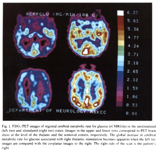
A 48-year-old woman with a stimulating electrode implanted in the right thalamic nucleus ventralis posterolateralis developed compulsive self-stimulation associated with erotic sensations and changes in autonomic and neurologic function. Stimulation effects were evaluated by neuropsychologic testing, endocrine studies, positron emission tomographic measurements of regional cerebral metabolic rate for glucose, EEG and evoked potentials. During stimulation, vital signs and pupillary diameter increased and a left hemiparesis and left hemisensory loss developed. Verbal functions deteriorated and visuospatial processing improved. Plasma growth hormone concentrations decreased, and adrenocorticotrophic hormone and cortisol levels rose. With stimulation, glucose metabolism increased in both thalami and both hemispheres, reversing baseline right-sided hypometabolism and right-left asymmetries. EEG and both somatosensory and brain-stem auditory evoked potentials remained unchanged during stimulation, while visual evoked potentials revealed evidence of anterior visual pathway dysfunction in the left eye. This case establishes the potential for addiction to deep brain stimulation and demonstrates that widespread behavioral and physiological changes, with concomitant alteration in the regional cerebral metabolic rate for glucose, may accompany unilateral thalamic stimulation.

gXRRQs1.png


Soon after insertion of the nVPL electrode, the patient noted that stimulation also produced erotic sensations. This pleasurable response was heightened by continuous stimulation at 75% maximal amplitude, frequently augmented by short bursts at maximal amplitude. Though sexual arousal was prominent, no orgasm occurred with these brief increases in stimulation intensity. Despite several episodes of paroxysmal atrial tachycardia and the development of adverse behavioral and neurological symptoms during maximal stimulation, compulsive use of the stimulator developed. At its most frequent, the patient self-stimulated throughout the day, neglecting personal hygiene and family commitments. A chronic ulceration developed at the tip of the finger used to adjust the amplitude dial and she frequently tampered with the device in an effort to increase the stimulation amplitude. At times, she implored her family to limit her access to the stimulator, each time demanding its return after a short hiatus. During the past 2 years, compulsive use has become associated with frequent attacks of anxiety, depersonalization, periods of psychogenic polydipsia, and virtually complete inactivity.

---

sekio's comment: Holy shit, her brain lights up like a christmas tree... and it's interesting to note she had prior instances of alcohol and opioid misuse. Skinner box anyone?
 
Here's a recent one that made a pretty big splash. This paper is pretty big because it was a collaboration between multiple labs including the lab that made the claim that respiratory depression is due to beta arrestin functional selectivity. This really kills the boners (non gender specific) of all the people pouring money and time into looking into mu opioid biased signalling.

The error was in the mouse strain, knockout mice are usually backcrossed (inbred) enough times to produce a true knockout.


Morphine-induced Respiratory Depression Is Independent of β-arrestin2 Signalling
Andrea Kliewer et al. Br J Pharmacol. 2020.

Abstract
Background and purpose: GPCRs can signal through both G proteins and β-arrestin2. For the μ-opioid receptor, early experimental evidence from a single study suggested that G protein signalling mediates analgesia, whereas β-arrestin2 signalling mediates respiratory depression and constipation. Consequently, for more than a decade, much research effort has been focused on developing biased μ-opioid agonists that preferentially target G protein signalling over β-arrestin signalling, as it was believed that such drugs would be analgesics devoid of respiratory depressant activity. However, the prototypical compounds that have been developed based on this concept have so far failed in clinical and preclinical development.
Experimental approach: The present study was set up to re-examine opioid-induced respiratory depression in β-arrestin2 knockout mice. To this end, a consortium was formed consisting of three different laboratories located in different countries to evaluate independently opioid-induced respiratory depression.
Key results: Our consensus results unequivocally demonstrate that the prototypical μ-opioid agonist morphine (3.75-100 mg·kg-1 s.c. or 3-30 mg·kg-1 i.p.) as well as the potent opioid fentanyl (0.05-0.35 mg·kg-1 s.c.) do indeed induce respiratory depression and constipation in β-arrestin2 knockout mice in a dose-dependent manner indistinguishable from that observed in wild-type mice.
Conclusion and implications: Our findings do not support the original suggestion that β-arrestin2 signalling plays a key role in opioid-induced respiratory depression and call into question the concept of developing G protein-biased μ-opioid receptor agonists as a strategy for the development of safer opioid analgesic drugs.
 
Disulfiram attenuates morphine or methadone withdrawal syndrome in mice
de Cordé, Annaa; Krząścik, Pawełb; Wolińska, Renataa; Kleczkowska, Patrycjaa; Filip, Małgorzatac; Bujalska-Zadrożny, Magdalena
Behavioural Pharmacology: August 2018 - Volume 29 - Issue 5 - p 393-399
doi: 10.1097/FBP.0000000000000376
Taking opioids is often accompanied by the development of dependence. Unfortunately, treatment of opioid dependence is difficult, particularly because of codependence – for example, on alcohol or other drugs of abuse. In the presented study, we analyzed the potential influence of disulfiram, a drug used to aid the management of alcoholism, on opioid abstinence syndrome, which occurs as a result of opioid withdrawal. Opioid dependence in mice was induced by subcutaneous administration of either morphine or methadone at a dose of 48 mg/kg for 10 consecutive days. To trigger a withdrawal syndrome, the opioid receptor antagonist, naloxone, was administered at a dose of 1 mg/kg (subcutaneous), and the severity of withdrawal signs was assessed individually. Interruption of chronic treatment with morphine or methadone by naloxone has led to the occurrence of opioid abstinence signs such as jumping, paw tremor, wet-dog shakes, diarrhea, teeth chattering, ptosis, and piloerection. Importantly, pretreatment with disulfiram (25, 50, and 100 mg/kg) reduced the intensity of withdrawal signs induced by naloxone in morphine or methadone-treated mice. These findings show the effectiveness of disulfiram in reducing opioid abstinence signs.

Edit: note that disulfiram can be toxic to some people and strain your liver and pancreas, as well as cause neural damage (impaired vision and hearing) due to being converted to carbon disulfide in your body.
 
Last edited:
I've got another one, this time it is a more complete explanation for the mechanism of general anesthetics that takes into account the meyer Overton correlation (the strong correlation between lipophilicity and potency with inhalational anesthetics), but proposes a better mechanism than direct dilution of the lipid membrane.

The mechanism? Disrupting the production of signaling lipids generated by phospholipase D activating TREK1 potassium channels.


Studies on the mechanism of general anesthesia


Mahmud Arif Pavel, View ORCID ProfileE. Nicholas Petersen, View ORCID ProfileHao Wang, View ORCID ProfileRichard A. Lerner, and View ORCID ProfileScott B. Hansen


PNAS first published May 28, 2020 https://doi.org/10.1073/pnas.2004259117

Significance
Anesthetics are used every day in thousands of hospitals to induce loss of consciousness, yet scientists and the doctors who administer these compounds lack a molecular understanding for their action. The chemical properties of anesthetics suggest that they could target the plasma membrane. Here the authors show anesthetics directly target a subset of plasma membrane lipids to activate an ion channel in a two-step mechanism. Applying the mechanism, the authors mutate a fruit fly to be less sensitive to anesthetics and convert a nonanesthetic-sensitive channel into a sensitive one. These findings suggest a membrane-mediated mechanism will be an important consideration for other proteins of which direct binding of anesthetic has yet to explain conserved sensitivity to chemically diverse anesthetics.

Abstract
Inhaled anesthetics are a chemically diverse collection of hydrophobic molecules that robustly activate TWIK-related K+ channels (TREK-1) and reversibly induce loss of consciousness. For 100 y, anesthetics were speculated to target cellular membranes, yet no plausible mechanism emerged to explain a membrane effect on ion channels. Here we show that inhaled anesthetics (chloroform and isoflurane) activate TREK-1 through disruption of phospholipase D2 (PLD2) localization to lipid rafts and subsequent production of signaling lipid phosphatidic acid (PA). Catalytically dead PLD2 robustly blocks anesthetic TREK-1 currents in whole-cell patch-clamp recordings. Localization of PLD2 renders the TRAAK channel sensitive, a channel that is otherwise anesthetic insensitive. General anesthetics, such as chloroform, isoflurane, diethyl ether, xenon, and propofol, disrupt lipid rafts and activate PLD2. In the whole brain of flies, anesthesia disrupts rafts and PLDnull flies resist anesthesia. Our results establish a membrane-mediated target of inhaled anesthesia and suggest PA helps set thresholds of anesthetic sensitivity in vivo.

 
Investigation of the 2,5-Dimethoxy Motif in Phenethylamine Serotonin 2A Receptor Agonists
Corresponding author: Jesper L. Kristensen (Department of Drug Design and Pharmacology, University of Copenhagen, Copenhagen, Denmark)
ACS Chemical Neuroscience 2020, Volume 11, Issue 9, Pages 1238-1244
Published online March 26th, 2020
https://doi.org/10.1021/acschemneuro.0c00129
The 2,5-dimethoxyphenethylamine (2,5-PEA) scaffold is recognized as a motif conferring potent agonist activity at the serotonin 2A receptor (5-HT2AR). The 2,5-dimethoxy motif is present in several classical phenethylamine psychedelics such as 2,4,5- trimethoxyamphetamine (TMA-2), 2,5-dimethoxy-4-methylamphetamine (DOM), 2,5-dimethoxy-4-iodoamphetamine (DOI), 2,5- dimethoxy-4-bromoamphetamine (DOB), 2,5-dimethoxy-4-bromophenethylamine (2C-B), and 2,5-dimethoxy-4-iodophenethylamine (2C-I), and it has previously been suggested that this structural motif is essential for 5-HT2AR activation. In the present study, we present data that challenges this assumption. The 2- and 5-desmethoxy derivatives of 2C-B and DOB were synthesized, and their pharmacological profiles were evaluated in vitro at 5-HT2AR and 5-HT2CR in binding and functional assays and in vivo by assessing their induction of the head-twitch response in mice. Elimination of either the 2- or 5-methoxy group leads to a modest drop in binding affinity and functional potency at 5-HT2AR and 5-HT2CR, which was more pronounced upon removal of the 2-methoxy group. However, this trend was not mirrored in vivo, as removal of either methoxy group resulted in significant reduction in the ability of the compounds to induce the head-twitch response in mice. Thus, the 2,5-dimethoxyphenethylamine motif appears to be important for in vivo potency of phenethylamine 5-HT2AR agonists, but this does not correlate to the relative affinity and potency of the ligands at the recombinant 5-HT2AR.
 

Me, myself, bye: regional alterations in glutamate and the experience of ego dissolution with psilocybin

Abstract:
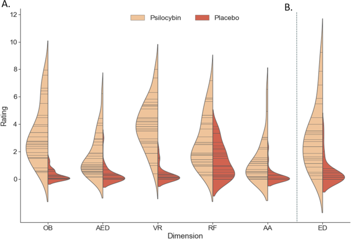
There is growing interest in the therapeutic utility of psychedelic substances, like psilocybin, for disorders characterized by distortions of the self-experience, like depression. Accumulating preclinical evidence emphasizes the role of the glutamate system in the acute action of the drug on brain and behavior; however this has never been tested in humans. Following a double-blind, placebo-controlled, parallel group design, we utilized an ultra-high field multimodal brain imaging approach and demonstrated that psilocybin (0.17 mg/kg) induced region-dependent alterations in glutamate, which predicted distortions in the subjective experience of one’s self (ego dissolution). Whereas higher levels of medial prefrontal cortical glutamate were associated with negatively experienced ego dissolution, lower levels in hippocampal glutamate were associated with positively experienced ego dissolution. Such findings provide further insights into the underlying neurobiological mechanisms of the psychedelic, as well as the baseline, state. Importantly, they may also provide a neurochemical basis for therapeutic effects as witnessed in ongoing clinical trials.
 
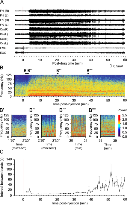
Here's another one on glutamate and consciousness, this one is on sheep k holing. Apparently very high dose (24 mg/kg) is ketamine produces complete silence in sheep corticies.

This is probably the first study of recreational ketamine using brain imaging, and while crude is fairly interesting

Characteristic patterns of EEG oscillations in sheep (Ovis aries) induced by ketamine may explain the psychotropic effects seen in humans
Scientific Reports volume 10, Article number: 9440

Abstract
Ketamine is a valuable anaesthetic and analgesic that in recent years has gained notoriety as a recreational drug. Recently, ketamine has also been proposed as a novel treatment for depression and post-traumatic stress disorder. Beyond its anaesthetic actions, however, the effects of ketamine on brain activity have rarely been probed. Here we examined the cortical electroencephalography (EEG) response to ketamine of 12 sheep. Following ketamine administration, EEG changes were immediate and widespread, affecting the full extent of the EEG frequency spectrum measured (0–125 Hz). After recovery from sedation during which low frequency activity dominated, the EEG was characterised by short periods (2–3 s) of alternating low (<14 Hz) and high (>35 Hz) frequency oscillation. This alternating EEG rhythm phase is likely to underlie the dissociative actions of ketamine, since it is during this phase that ketamine users report hallucinations. At the highest intravenous dose used (24 mg/kg), in 5/6 sheep we observed a novel effect of ketamine, namely the complete cessation of cortical EEG activity. This persisted for up to several minutes, after which cortical activity resumed. This phenomenon is likely to explain the ‘k-hole’, a state of oblivion likened to a near death experience that is keenly sought by ketamine abusers.

 
I’d like to see more research on anesthetics in general. Their mechanisms are still not fully understood, if I remember correctly.

They seem to be a good jumping off point into exploring the nature of consciousness given their ability to put it on pause
 
ketamine turns your brain off? sounds about right to me
 
Top