• N&PD Moderators: Skorpio | thegreenhand

⫸STICKY⫷ The N&PD Recent Journal ARTICLE Club

This next paper is possibly the best ketamine paper I have read to this date (no more k holing sheep).

Deep posteromedial cortical rhythm in dissociation
Nature (2020)Cite this article
Abstract
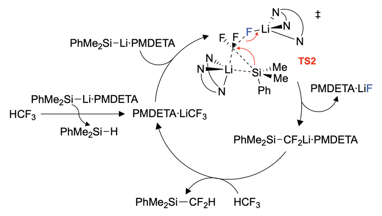
Advanced imaging methods now allow cell-type-specific recording of neural activity across the mammalian brain, potentially enabling the exploration of how brain-wide dynamical patterns give rise to complex behavioural states1,2,3,4,5,6,7,8,9,10,11,12. Dissociation is an altered behavioural state in which the integrity of experience is disrupted, resulting in reproducible cognitive phenomena including the dissociation of stimulus detection from stimulus-related affective responses. Dissociation can occur as a result of trauma, epilepsy or dissociative drug use13,14, but despite its substantial basic and clinical importance, the underlying neurophysiology of this state is unknown. Here we establish such a dissociation-like state in mice, induced by precisely-dosed administration of ketamine or phencyclidine. Large-scale imaging of neural activity revealed that these dissociative agents elicited a 1–3-Hz rhythm in layer 5 neurons of the retrosplenial cortex. Electrophysiological recording with four simultaneously deployed high-density probes revealed rhythmic coupling of the retrosplenial cortex with anatomically connected components of thalamus circuitry, but uncoupling from most other brain regions was observed—including a notable inverse correlation with frontally projecting thalamic nuclei. In testing for causal significance, we found that rhythmic optogenetic activation of retrosplenial cortex layer 5 neurons recapitulated dissociation-like behavioural effects. Local retrosplenial hyperpolarization-activated cyclic-nucleotide-gated potassium channel 1 (HCN1) pacemakers were required for systemic ketamine to induce this rhythm and to elicit dissociation-like behavioural effects. In a patient with focal epilepsy, simultaneous intracranial stereoencephalography recordings from across the brain revealed a similarly localized rhythm in the homologous deep posteromedial cortex that was temporally correlated with pre-seizure self-reported dissociation, and local brief electrical stimulation of this region elicited dissociative experiences. These results identify the molecular, cellular and physiological properties of a conserved deep posteromedial cortical rhythm that underlies states of dissociation.


The tldr by figure:

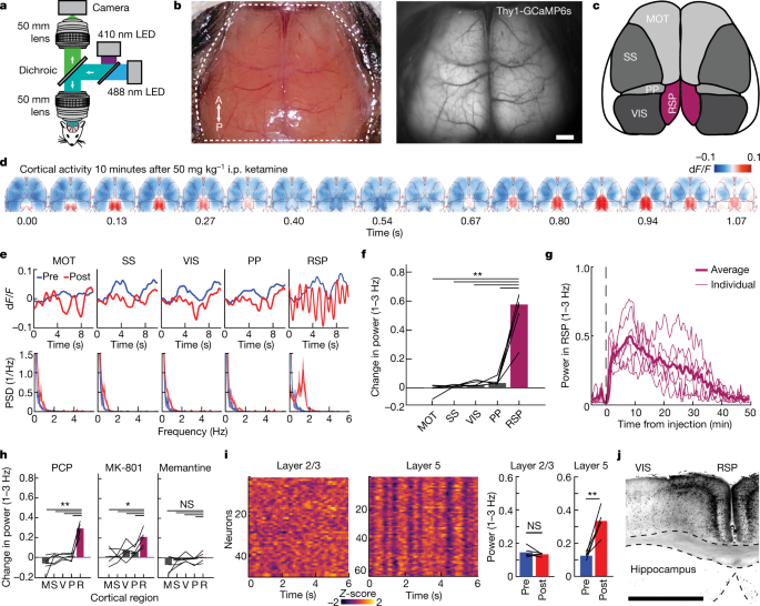
1: using brain imaging 3 hz oscillations of firing are identified in the retrosplenial cortex during high dose ketamine.

2: these waves are characterized electrophisiologically.

3: these oscillations map strongly to measures of dissociation. They are able to use optogenetics to create these waves of firing with light stimulation and they cause dissociation (3b has some of the strongest evidence I have seen of the "plateau" effect commonly cited with dxm).

4: they identify 2 sets of ion channels that drive the effect.. Nmda receptors (fairly obvious but a good internal positive control) and hcn channels which provide a pacemaker current. They abolish the effects by performing global knockouts of the channels, and then using a cre driven system, restoring the channels solely in the retrosplenial cortex.

5: finally they drop the fucking mic.

They found a patient who had seizures that cause dissociative effects, and was previously implanted with electrodes in their brain for mapping of their seizures . They found the seizures induce rhythmic activity in the retrosplenial cortex.

They briefly stimulated this person's brain and were able to induce dissociative experiences.

This is such a rare thing in neuroscience, and for it to cap off such a methodical and elegant paper is amazing. I have been popping off at my mates for like the last hour after reading this.

Definately read the paper it's a nature paper so pretty clear.
 
Structure of a Hallucinogen-Activated Gq-Coupled 5-HT2A Serotonin Receptor

Hallucinogens like lysergic acid diethylamide (LSD), psilocybin, and substituted N-benzyl phenylalkylamines are widely used recreationally with psilocybin being considered as a therapeutic for many neuropsychiatric disorders including depression, anxiety, and substance abuse. How psychedelics mediate their actions—both therapeutic and hallucinogenic—are not understood, although activation of the 5-HT2A serotonin receptor (HTR2A) is key. To gain molecular insights into psychedelic actions, we determined the active-state structure of HTR2A bound to 25-CN-NBOH—a prototypical hallucinogen—in complex with an engineered Gαq heterotrimer by cryoelectron microscopy (cryo-EM). We also obtained the X-ray crystal structures of HTR2A complexed with the arrestin-biased ligand LSD or the inverse agonist methiothepin. Comparisons of these structures reveal determinants responsible for HTR2A-Gαq protein interactions as well as the conformational rearrangements involved in active-state transitions. Given the potential therapeutic actions of hallucinogens, these findings could accelerate the discovery of more selective drugs for the treatment of a variety of neuropsychiatric disorders.

Bryan Roth, David Nichols et al. is making real strides to elucidate the true mechanism of hallucinogens. Paper of the year!

https://www.cell.com/cell/fulltext/S0092-8674(20)31066-7?utm_medium=homepage
 
New reagent discovered from CFC waste

I thought the chemists here would find this interesting.

The closest I got to working with CFCs (besides getting lifetime CFC handling certification in the state of Washington for union hall port jobs) was once experimenting with Du Pont brand Freon as an inhalant.

508409_d0cc04592ff4f_453822.png

An image showing the proposed reaction cycle for trifluoromethane defluorosilyation.

Source: © Mark Crimmin/Imperial College London
Valuable difluoromethylating agent obtained from refrigerant waste
BY HAYLEY RUSSELL8 OCTOBER 2020

UK-based researchers have devised a way to turn waste trifluoromethane into a useful reagent for introducing fluorinated building blocks into drug compounds.

Trifluoromethane (HCF3) is a waste product formed in the production of chlorofluorocarbon refrigerant gases. It is also a greenhouse gas with a global warming potential 11,700 times higher than CO2. As it poses such a high environmental risk and has few applications, it is either stored or destroyed.

Now, researchers led by Mark Crimmin at Imperial College London have developed a method for converting trifluoromethane into a reagent that can insert difluoromethyl (CF2H) groups into molecules during pharmaceutical synthesis.

Difluoromethyl groups are present in many pharmaceutical and agrochemical compounds. Scientists add C–F bonds to pharmaceutical compounds to increase drug stability and solubility. Existing methods for synthesising the difluoromethylating agent PhMe2SiCF2H use HCF2Cl, which has since been banned.

The team has proposed a reaction cycle for the defluorosilylation of trifluoromethane

Crimmin’s team has shown a simple silyl lithium reagent can break the strong C–F bond in trifluoromethane at room temperature with the reaction completing within five minutes. Researchers from Japan have demonstrated a similar reaction previously where they react the equivalent boryl lithium reagent with HCF3. They proposed a mechanism but did not test it.

The team at Imperial College conducted a mechanistic study. They say that this was the hardest part of the research, as the reaction was too fast for traditional kinetics techniques. To overcome this, the researchers turned to computational studies to gain better insight into the mechanism. Their calculations support a mechanism in which the silicon reagent seems to be acting as both a catalytic base and a nucleophile. CF3H first undergoes a deprotonation to form a trifluoromethanide intermediate, which is then susceptible to direct nucleophilic attack.

‘This type of reaction would generally require expensive or ozone depleting sources such as HCF2Cl to synthesise the R3SiCF2H reagent, for which more sustainable solutions are needed,’ explains Tatiana Besset, an organofluorine researcher at the University of Rouen in France. ‘But it’s very interesting to see how a greenhouse gas and a fluorinated byproduct coming from industry waste, was used to generate a highly valuable nucleophilic difluoromethylating source.’

‘We are looking to scale up the synthesis beyond the gram-scale,’ says Crimmin. ‘We have some concerns about potential safety issues here, as the reactions are exothermic, and so plan to use flow chemistry to do this.’ Crimmin says they also have plans to collaborate with another research group to investigate radiolabelling in the reaction.

References
1. D J Sheldon, G Coates and M R Crimmin, Chem. Commun., 2020, DOI: 10.1039/d0cc04592f (This article is open access.)

2. S Ito, N Kato and K Mikami, Chem. Commun., 2017, 53, 5546 (DOI: 10.1039/c7cc02327h)
 
Last edited:
These days machine learning is taking a steep curve proportional to how deep they can go.

This, however, is the Pandora's box & tool kit for the novices floating around our neck of the web:

Universal chemistry software can turn words into chemicals

Universal chemistry software can turn words into chemicals
BY TOM METCALFE13 OCTOBER 2020

A robotic system that can read the chemical literature, turn it into working reaction pathways and use them to automatically create specific chemicals has been developed at the University of Glasgow.

The researchers say the system is the first ‘universal’ architecture for automated chemical synthesis, and can be implemented on any capable robot to turn methods in a book or journal into the desired product. It’s already been tested on common synthesis methods, and the automated manufacture of several test compounds, including the analgesic lidocaine, the Dess–Martin oxidation reagent periodinane and the fluorinating agent AlkylFluor.1

We’ve invented the CPU [central processing unit] for chemistry

LEE CRONIN, UNIVERSITY OF GLASGOW

Lee Cronin, who leads the team behind the work, says the system is independent of robot hardware and is a breakthrough in creating a universal language for chemical methods. It’s based on a markup language that can represent the varied steps of chemical methods, called XDL – where the X is the Greek letter chi and comes from χημία, the Greek word for chemistry.


XDL will be open source and free to use, like the HTML used on webpages. It has rules for about 44 different synthesis operations to make almost any known molecule, and can be extended to recognise further operations, he says.

A scheme showing the universal system for the automatic execution of chemical synthesis from the literature

508539_1a_983809.png


Source: © Science/AAAS

The workflow of the universal system that can translate the chemistry literature into a synthesis

‘We’ve invented the CPU [central processing unit] for chemistry,’ he says. ‘That’s really important right now, because all the chemistry robots in the world are not only expensive, but they can’t be programed in the same way.’

The new system fixes that by describing chemical methods in XDL, and then using the XDL descriptions to generate instructions for a virtual machine – a theoretical device dubbed a ‘ChemPU’. They can then be run on different robots through software interpreters for the virtual ChemPU instructions, instead of creating customised steps for any particular chemistry robot. ‘This eliminates all that work,’ Cronin says.

Universal chemistry
Cronin’s team has developed the prototype on one of his fume hood-sized chemputers – there are now 12 of them with different configurations in his lab, automatically working on several projects. The next stage of the project will be to implement the system on chemistry robots at other institutions, Cronin says.

The system has three stages: the first uses a scanner to read pages from the chemistry literature – a synthesis method in a book or a journal, say – and parses it into XDL, with error correction to let a human chemist make fixes or adjustments. ‘That’s important, because then you can make sure that your robot doesn’t just halt and catch fire,’ he says.

The next stage turns the XDL into instructions for the ChemPU virtual machine, and the last stage interprets the ChemPU instructions for any given chemistry robot. The system will generate descriptive errors if a robot’s hardware is not up to the task, he says. The markup language XDL is integrated into a software development environment that can combine methods from the literature with input from a human chemist.

508538_6a_677861.png


An image showing the lidocaine synthesis

Source: © Science/AAAS

The steps the universal system planned and performed to produce lidocaine

‘Take your favourite cocktail … vodka Martini or something … and write it down,’ Cronin says. ‘The system will translate it into what reactor to use, where the pipes are, what the temperature is, the stirring rate and all that stuff.’

Cronin doesn’t see chemistry robots replacing humans, however. Instead, they will let chemists get on with important tasks, while leaving time-consuming, boring or dangerous work to robots. ‘It’s going to allow chemists to make many more molecules than they could ever have dreamed of before and discover many more drugs, because they can focus on what they are good at,’ he says.

IBM Zurich’s Teandro Laino, a researcher on RoboRXN, notes that Cronin’s system uses rules to process the chemical literature, while their system uses a method based on natural language processing (NLP). Such rule-based approaches could be hard to improve without extensive human intervention, and might prove weaker than advanced NLP methods, he adds.

But Cronin counters that the XDL rules are open, editable and ensure the scientific validity of a system to connect methods from literature with their implementations on any chemistry robot – including the proprietary robot of the RoboRXN system.

Martin Burke of the University of Illinois at Urbana–Champaign, who specialises in organic synthesis and chemical innovation, says the new system is part of a brave new world of ‘democratised’ chemistry. The paper shows the tremendous potential to revolutionise chemistry by strategically merging computer science with automated organic synthesis, he says.

References
S H M Mehr et al, Science, 2020, DOI: 10.1126/science.abc2986
 
Dopamine & Serotonin unexpected effects.

Dopamine, serotonin involved in sub-second perception, cognition.

In first-of-their-kind observations in the human brain, an international team of researchers has revealed two well-known neurochemicals — dopamine and serotonin — are at work at sub-second speeds to shape how people perceive the world and take action based on their perception.


The discovery shows researchers can continually and simultaneously measure the activity of both dopamine and serotonin — whose receptor and uptake sites are therapeutic targets for disorders ranging from depression to Parkinson’s disease — in the human brain.

Furthermore, the neurochemicals appear to integrate people’s perceptions of the world with their actions, indicating dopamine and serotonin have far more expansive roles in the human nervous system than previously known.

Carbon-Fiber-Microelectrodes-scaled.jpg


Carbon Fiber Microelectrodes
Virginia Tech researchers with the Fralin Biomedical Research Institute Center for Human Neuroscience Research construct carbon fiber microelectrodes for real-time detection of dopamine and serotonin activity in human patients. Credit: Virginia Tech


Known as neuromodulators, dopamine and serotonin have traditionally been linked to reward processing — how good or how bad people perceive an outcome to be after taking an action.

The study published in the journal Neuron on October 12, 2020, opens the door to a deeper understanding of an expanded role for these systems and their roles in human health.

“An enormous number of people throughout the world are taking pharmaceutical compounds to perturb the dopamine and serotonin transmitter systems to change their behavior and mental health,” said P. Read Montague, senior author of the study and a professor and director of the Center for Human Neuroscience Research and the Human Neuroimaging Laboratory at the Fralin Biomedical Research Institute at Virginia Tech Carilion. “For the first time, moment-to-moment activity in these systems has been measured and determined to be involved in perception and cognitive capacities. These neurotransmitters are simultaneously acting and integrating activity across vastly different time and space scales than anyone expected.”

Better understanding of the underlying actions of dopamine and serotonin during perception and decision-making could deliver important insight into psychiatric and neurological disorders, the researchers said.

Carbon-Fiber-Microelectrode-Size-Comparison.jpg


Carbon Fiber Microelectrode Size Comparison
The relative size of a microelectrode used to make recordings of dopamine and serotonin activity during deep brain stimulation procedures. Credit: Virginia Tech

“Every choice that someone executes involves taking in information, interpreting that information, and making decisions about what they perceived,” said Kenneth Kishida, a corresponding author of the study and an assistant professor of physiology and pharmacology, and neurosurgery, at Wake Forest School of Medicine. “There’s a whole host of psychiatric conditions and neurological disorders where that process is altered in the patients, and dopamine and serotonin are prime suspects.”


Lack of chemically specific methods to study neuromodulation in humans at fast time scales has impeded understanding of these systems, according to Montague, who is an honorary professor at the Wellcome Center for Human Neuroimaging at University College London and a professor of physics at the Virginia Tech College of Science.

But now, in first-ever measurements, scientists used an electrochemical method called “fast scan cyclic voltammetry,” which employs a small carbon fiber microelectrode that has low voltages ramped across it for real-time detection of dopamine and serotonin activity.

In the study, researchers recorded fluctuations in dopamine and serotonin using specially designed electrodes in five patients undergoing deep brain stimulation electrode implantation surgery to treat essential tremor or Parkinson’s disease. Patients were awake during surgery, playing a computer game designed to quantify aspects of thought and behavior while the measurements were taken.

Read-Montague-Computational-Neuroscientist-scaled.jpg


Read Montague, a computational neuroscientist at the Center for Human Neuroscience Research at Virginia Tech’s Fralin Biomedical Research Institute, said dopamine and serotonin are at work at sub-second speeds to shape how people perceive the world and take action based on their perception. Credit: Virginia Tech

On each round of the game, patients briefly viewed a cloud of dots and were asked to judge the direction they were moving. The method, designed by corresponding author Dan Bang, a Sir Henry Wellcome Postdoctoral Fellow, and Steve Fleming, a Sir Henry Dale/Royal Society Fellow, both at the Wellcome Center for Human Neuroimaging at University College London, helped indicate that dopamine and serotonin were involved in simple perceptual decisions, outside of the traditional context of rewards and losses.


“These neuromodulators play a much broader role in supporting human behavior and thought, and in particular they are involved in how we process the outside world,” Bang said. “For example, if you move through a room and the lights are off, you move differently because you’re uncertain about where objects are. Our work suggests these neuromodulators — serotonin in particular — are playing a role in signaling how uncertain we are about the outside environment.”

Montague and Kishida, along with Terry Lohrenz, a research assistant professor, and Jason White, a senior research associate, now both at the Fralin Biomedical Research Institute, started working on a new statistical approach to identify dopamine and serotonin signals while still at the Baylor College of Medicine in Houston, Texas.

“Ken rose to the challenge of doing fast neurochemistry in human beings during active cognition,” Montague said. “A lot of other good groups of scientists were not able to do it. Aside from the computation of enormous amounts of data, there are complicated issues to solve, including great, fundamental algorithmic tasks.”

Until recently, only slow methodologies such as PET scanning could measure the impact of neurotransmitters, but they were nowhere near the frequency or volume of the second-to-second measurements of fast scan cyclic voltammetry.

The measurements in the new study were taken at the Wake Forest Baptist Medical Center, and involved neurosurgical teams led by Adrian W. Laxton and Stephen B. Tatter.

“The enthusiasm the neurosurgeons have for this research is derived from the same reasons that drove them to be doctors — first and foremost, they want to do the best for their patients, and they have a real passion for understanding how the brain works to improve patient outcomes,” said Kishida, who oversaw the data collection in the operating room during the surgeries. “Both are collaborative scientists along with Charles Branch, the chair of the neurosurgery department at Wake Forest, who has been an amazing advocate for this work.”

Likewise, Montague said, “You can’t do it without the surgeons being real, shoulder-to-shoulder partners, and certainly not without the people who let you make recordings from their brains while they are having electrodes implanted to alleviate the symptoms of a neurological disorder.”

Montague had read a study in the Proceedings of the National Academy of Sciences that prompted him to approach colleagues Bang and Fleming at University College London to tailor a task for patients to perform during surgery that would reveal sub-second dopamine and serotonin signaling in real-time inference about the external world – separate from their often-reported roles in reward-related processes.

“I said I have this new method to measure dopamine and serotonin, but I need you to help with the task,” Montague said. “They ended up in the study. The research really took a lot of hard work and an integrated a constellation of people to obtain these results.”

Reference: “Sub-second Dopamine and Serotonin Signaling in Human Striatum during Perceptual Decision-Making” by Dan Bang, Kenneth T. Kishida, Terry Lohrenz, Jason P. White, Adrian W. Laxton, Stephen B. Tatter, Stephen M. Fleming and P. Read Montague, 12 October 2020, Neuron.
DOI: 10.1016/j.neuron.2020.09.015

The research was funded by grants to various researchers from the Wellcome Trust, the National Institutes of Health including the National Institute on Drug Abuse, the National Institute of Mental Health, the National Institute of Neurological Disorders and Stroke.
 
Looks quite futuristic... Automated peptide synthesizers have existed for over 20 years, but in that case there are not many precursors and reagents needed for building almost any linear peptide chain from natural amino acids.
 
Honestly the big hurdle for a lot of clandestine chemistry is getting the reagents. My only chemistry experience was a perfunctory organic chemistry lab in undergrad (where we actually did the lidocaine synthesis from above), and I feel like it would not be super hard to synthesize many common recreational drugs given a suitable lab and the precursors and enough time to read the literature.

(okay I would be fairly scared doing a grignard my first time or like a cyanogen bromide cyclization, but protocol and routine usually vanquisg fear or at least shove it into a corner where it appears in the form of caution).

The things keeping me from doing this are 1) my fear of legal repercussions, 2) my lack of a safe lab space 3) the difficulty of acquiring precursors, (which can lead to the time consuming search for legal pre precursors or even pre-pre precursors).

In my eyes this machine takes out the lack of a lab space. I would still have to source the chemicals as they are consumable, and I feel that would be fairly difficult without enlisting some Gordon Todd Skinner unsavory type.
 
Honestly the big hurdle for a lot of clandestine chemistry is getting the reagents.
Pretty much. If you have the reagents, the glassware is going to be easier and cheaper to procure than a fancy robot.
 
Nice work. I'd love to know whether I respond to SSRIs or not...perhaps if I had low serotonin that they would benefit me. I'd be willing to take an SSRI in spite of the likelihood of sexual dysfunction if it meant that I would never have to feel depressed ever again. But in all honesty I would prefer a satisfactory sexual function...which upsets me because these drugs are the mainstay of depression treatment.

If my problem is dopamine deficiency, well, we have stuff for that, they're called amphetamines.
 
I live in Washington state in the U.S., and here state insurance available to low income, for free, covers a genetics test called GeneSight. It checks to see specifically what genes relate to SSRIs, what interactions, even what serotonin receptor polymorphisms you have.

In my case I have a unusual genotype and only Selegiline & Pristiq were indicated for me, Pristiq: a drug state insurance wouldn't normally cover, but because of that test, my prescriber was able to get permission from the state insurance company to prescribe it to me at no cost, not even co-pay.

I think other states in the U.S. do the same thing, but they won't mention it, you have to bring it up to them.
 
interesting. can medicaid cover it? ill check their website, i assume its genesight.com and ill ask em. but i guess it differs by state so we shall see.
but anyway, how reliable is it really? this can be a game changer, honestly. wouldnt it change the whole antidepressant industry when you know what you should take instead of having pyschiatrists prescribe you whatever they feel like??
 
^I don't think they would know which insurance covers them; too many variables for them to know.

What do you mean by reliable? The genetic testing is legitimate, but what they consider to be efficacious for your genotype is educated guesses based on what information they have.

I will say two separate clinics vouched for it when I brought it up to them, not just the receptionists or nurses either, but the doctors.

GeneSight results

^this is a link to my results, the actual genetic information is toward the last few pages. (before anybody else brings it up, yes I am fine with this information being all over the web.)

Very interesting thanks for sharing that.

I'm really curious about your reaction to codeine. Do you get good effects from it? Since your CYP2D6 appears to be underexpressed you may not be able to convert it into morphine efficiently. According to a study I read it seems like a big proportion of codeine's effects are due to codeine-6-glucoronide, yet they don't differentiate between pain relief and the more recreational effects (euphoria). Just wondering if you get (or used to get) euphoria from it.
 
^my opioid 'honeymoon' was jumping straight to heroin. The one time I remember doing codeine, it was a whole bottle when I was in H withdrawal, and all I recall from that was a bundle of side-effects pointing to risking anaphylaxis:

..turning red and itching so badly on the bottom of my feet that I spent most of my time painfully scratching them, despite how sensitive they were.

I always wanted to try heterocodeine, though; and that should be better for me due to my genetic profile. I remember reading your theory about C6G before, though, and it sounds pretty solid to me.
 
Another recent article on the same:

CNBC: Scientists make digital breakthrough in chemistry that could revolutionize the drug industry

In June, the U.S. government purchased the vast majority of world's supply of remdesivir—a FDA-approved antiviral treatment for Covid-19—for July through September. Gilead, the company that makes the compound, recently announced that it would meet international demand by the end of October. Yet all along, digital instructions for whipping up a batch of the nearly 400-atom molecule at the push of a button have been sitting on Github, an online software repository, freely available to anyone with the hardware needed to execute the chemical "program."


A dozen such chemical computers or "chemputers" sit in the University of Glasgow lab of Lee Cronin, the chemist who designed the bird's nest of tubing, pumps, and flasks, and wrote the remdesivir code that runs on it. He's spent years dreaming of a future where researchers can distribute and produce molecules as easily as they email and print PDFs, making not being able to order a drug as archaic as not being able to locate a modern text.


"If we have standard way of discovering molecules, making molecules, and then manufacturing them, suddenly nothing goes out of print," he says. "It's like an ebook reader for chemistry."


Cronin and his colleagues described their machine's capability to produce multiple molecules last year, and now they've taken a second major step toward digitizing chemistry with an accessible way to program with the machine. Their software turns academic papers into chemputer-executable programs that researchers can edit without learning to code, they announced earlier this month in Science. And they're not alone. The team represents one of dozens of groups spread across academia and industry all racing to bring chemistry into the digital age, a development that could lead to safer drugs, more efficient solar panels, and a disruptive new industry.

Robots-inventing-drugs-Quebec-Science-780x470.jpg

H/O Leroy Cronin's chemputer
A chemical computer or "chemputer" sits in the University of Glasgow lab of Leroy Cronin, the chemist who designed the bird's nest of tubing, pumps, and flasks, and wrote the remdesivir code that runs on it. He's spent years dreaming of a future where researchers can distribute and produce molecules as easily as they email and print PDFs.

Leroy Cronin,
The Cronin team hopes their work will enable what they describe as "Spotify for chemistry"— an online repository of downloadable recipes for important molecules that they say could help developing countries more easily access medications, enable more efficient international scientific collaboration, and even support the human exploration of space.

"The majority of chemistry hasn't changed from the way we've been doing it for the last 200 years. It's very manual, artisan driven process," says Nathan Collins, the chief strategy officer of SRI Biosciences, a division of SRI International, a research company developing another automated chemistry system that's not involved in the Glasgow research. "There's billions of dollars of opportunity there."

At the heart of Cronin's new work lies what he calls a chemical description language or XDL (the "X" is pronounced "kai" after the first letter in the Greek word for chemistry). XDL is to the "chemputer" as HTML is to a browser—it tells the machine what to do. The group has also created software called SynthReader that scans a chemical recipe in peer-reviewed literature — like the six-step process for cooking up remdemisvir — and uses natural language processing to pick out verbs like "add," "stir," or "heat;" modifiers like "dropwise;" and other details like durations and temperatures. The system translates those instructions into XDL, which directs the chemputer to execute mechanical actions with its heaters and test tubes.


One of the framework's strengths, according to Cronin, is that chemists can edit the chemical protocol in plain English. This feature lets researchers operate the machine with little training, and, crucially, harness their chemistry expertise to spot bugs in the code. Chemputer crashes can be serious affairs. "The human will always need to be there to make sure you don't have a dumpster on fire," he says.

The researchers tested the system, and no dumpsters burned. The group reported extracting 12 demonstration recipes from the chemical literature, such as the numbing anesthetic lidocaine, all of which the chemputer carried out at efficiencies similar to those of human chemists.

Robotic transformation of chemistry
Cronin built a company called Chemify to sell the chemisty robots and XDL package, although he's also posted free instructions online for building and programming the machine. And already the device is making inroads in the chemical world. In May of 2019, the group installed a prototype at the pharmaceutical company GlaxoSmithKline.

"The chemputer as a concept and the work [Cronin]'s done is really quite transformational," says Kim Branson, the global head of artificial intelligence and machine learning at GSK. The company is exploring various automation technologies to help it make a wide array of chemicals more efficiently, but Cronin's work in particular, Branson says, may let GSK "teleport expertise" around the company. Once a chemist designs a promising molecular recipe, rather than writing up a report or teaching a colleague, they'll just press the share button.

Researchers say that while Chemify isn't the most sophisticated automated chemistry platform, it might be the most accessible. It's built around the traditional tools of beakers and test tubes and functions in the step-by-step "batch" paradigm that chemists have used for centuries. Cronin also intends it to be universal: compatible with any batch chemistry robot. Researchers with their own machines just need tell the software what parts they have and give it figures like how hot their heater can go.

Other groups are betting on a more dramatic break from chemistry's roots. At SRI, Collins oversees the development of a platform called AutoSyn, which uses an alternative approach called "flow" chemistry. Rather than mixing up a batch of one substance in one beaker, and then moving it to another flask, in flow chemistry reactions play out continuously. Chemicals stream together in tubing, react there, and get carried off. With more than 3,000 pathways, AutoSyn, which Collins and colleagues described in a publication in June, can recreate almost any kind of liquid based reaction.

Doing chemistry in flow requires specialized hardware and extra effort to translate chemical procedures from their batch descriptions, but that investment buys an "exquisite" control over aspects like heat transfer and mixing, Collins says. If machines like AutoSyn can automatically run hundreds of subtle variations on a published reaction, the detailed datasets they generate could highlight the best way to make a chemical.

The literature may be a good place to start, but many published experiments have flaws. Collins estimates that chemists spend 30% to 70% of their time just working out missing details in known reactions. "[A reaction] is written up by someone who sits down and bases it on their notes from something they were doing the day before, or maybe something they did six months ago," he says.

While AutoSyn and the Chemputer are both able to reproduce the majority of published reactions today, the next step will be making the machines reliable and "Apple groovy," as Cronin puts it. Collins says that AutoSyn used to need an engineer to keep it functioning for more than half of its runs, but now needs fixing less than 10% of the time. Eventually, he hopes, users will troubleshoot the system over the phone.

"This is still a very new science," he says. "It's started to explode really in the last 18 months."

One force driving that explosion has been the Defense Advanced Research Projects Agency (DARPA). It's wrapping up a four-year program called Make-It, of which both the Chemputer and AutoSyn are alumni. The long-term goal of the program's manager, Anne Fischer, is to speed up the discovery of useful molecules, which has historically involved a lot of waiting around while chemists laboriously smithed atoms into novel configurations. "The slow step is always making and testing the molecules," she says.

But now that Make-It has helped produce robotic tools to build molecules like the Chemputer, AutoSyn, and others, she's directing a new DARPA program, Accelerated Molecular Discovery, that looks to the next stage: developing smarter software to tell the robots what molecules to make, and how to make them.

Nathan Collins, Chief Strategy officer of SRI international

"We're now trying now to harness what we've done in Make-It and expand it out so we can teach computers how to discover new molecules," she says.

The secret to doing so, many believe, is machine learning. And some machines capable of rudimentary chemical learning are well underway. Connor Coley, a chemist at MIT, is a member of a team that last year paired an automated flow chemistry system with an algorithm to direct it. The algorithm trained on databases of hundreds of thousands of reactions and was able to predict recipes for new products. "It tries to understand, based on those patterns, what kind of transformations should work for new molecules it's never seen before," Coley said.

He stresses that the system has a long way to go. Its predictions were based on similar molecules and human chemists needed to flesh out details missing from the machine-generated outline. Nevertheless, the work supported the notion that software can come up with useful recipes.

MIT is collaborating with more than a dozen chemical and pharmaceutical companies to advance its molecule-predicting algorithms, and some companies have already put the software to use. Juan Alvarez, the Assistant Vice President of computational and structural chemistry at Merck, says that Coley's machine learning algorithm is one of a variety of chemistry prediction tools that the company has made available to its internal researchers. "It's absolutely being deployed to impact our timeline today," he says.

While each group approaches automation from a different angle, they're all tackling the same problem. A near infinite diversity of possible molecules exist—some of which are surely life-saving drugs or revolutionary new materials—but precious few human beings have the specialized skillset to analyze, make, and test these compounds.

They aim to keep those rare skills from going to waste. In some ways the work of chemists still resembles the work of scribes, who once painstakingly copied and corrected the writings of others. Researchers like Cronin hope that with the chemical equivalents of the printing press, word processor, and autocorrect in hand, tomorrow's chemists will spend less time recreating, and more time composing.
 
508799_16_9_rgb_604884.jpg


Engineering a handshake for proteins

IAN LE GUILLOU 26 OCTOBER 2020

Once considered undruggable, chemists are beginning to grasp protein–protein interactions, according to Ian Le Guillou

Proteins rarely work alone. From tightly bound complexes to passing transitory contacts, interactions between proteins play a central role in the function of the cell. By one estimate, there are over 650,000 protein–protein interactions in the human body. This overwhelming array of interactions represents an almost untapped resource of targets for new therapeutic drugs.

For many decades, these interactions were considered to be ‘undruggable’. High-resolution structures of protein–protein interactions (PPIs) in the 1980s and 90s showed that the interfaces were large, flat featureless surfaces. This was entirely different from the deep pockets found in typical drug targets, such as enzymes and receptors, where small molecules can easily bind.

A typical enzyme binding to its substrate is often taught at school as being like a lock and key. To find a drug to block this interaction, we simply need a new molecule that is sufficiently ‘key-shaped’ to jam the lock. By contrast, our knowledge of PPIs made them seem more like handshakes – two flat surfaces coming together firmly. But our understanding has improved to reveal that it is more complicated than that. There is a secret handshake feel to these interactions, with each side responding to the other and hidden points of contact that are of great significance.

By mutating thousands of individual amino acids at protein–protein interfaces, biochemists found that only a small subset of residues was mostly responsible for binding. These ‘hot spots’ were much more relevant to the strength of the interaction than the size of the interaction surface. This improved understanding made PPIs more tractable as targets, and the need for new, effective drugs certainly made them attractive. However, this is still easier said than done.


“I think that protein–protein interactions give you a bit more flexibility”

While some natural products approved as drugs, such as taxanes and rapamycin, act by inhibiting PPIs, this was a result of fortune rather than design. Venetoclax, a drug for chronic lymphocytic leukaemia, became the first drug to be approved for targeting a PPI in 2016 and few have followed since.

Although PPIs may no longer be viewed as undruggable, new approaches are needed to deal with this new class of targets. Inhibiting PPIs will need different libraries, assays and perspectives. Given the variety and complexity of PPIs, a range of techniques may be needed to inhibit different targets. As new approaches are developed, many researchers are turning to PPIs to address problems and therapeutic areas that were not previously manageable with traditional small molecule inhibitors.

Selective entry requirements
Most enzymes act on multiple targets in the cell and rely on their protein binding partners to be selective. An inhibitor that blocks the enzyme’s active site would prevent its activity across all of its targets, potentially causing unintended side effects. ‘I think that PPIs give you a bit more flexibility. You can target them to block subsets of enzymes’ activities in a way that’s pretty challenging to do with traditional small molecule enzyme inhibitors,’ says Louise Walport from the Crick Institute in London, UK.

She is studying a group of proteins called arginine deiminases. These are responsible for modifying arginine amino acid residues to remove their charge, which has a knock-on effect for how the proteins behave. The arginine deiminases have a wide range of targets and PPIs may be responsible for managing this. To block these PPIs, Walport screens the arginine deiminases against a library of cyclic peptides. These are molecules of 8–14 amino acid residues in a circular chain.

“We can find low nanomolar, sometimes picomolar, binders straight out of a screen”

‘I think that PPIs is where peptides have their particular niche because they’re just that bit bigger than small molecules, so they can pick up more small interactions along these featureless surfaces than you can with a small molecule,’ she says. By their cyclic nature there is less entropic cost when they bind to the protein and it helps to make them more stable against being broken down by the body.

The RaPid system, created by Hiroaki Suga from the University of Tokyo in Japan, can generate 10¹⁴ different cyclic peptides – more than the number of stars in the universe. The advantage of this approach is that Walport has been able to find ‘low nanomolar, sometimes picomolar, binders straight out of a screen with no modification, not having done any optimisation’.

The challenge, however, is that these molecules are typically too large to enter the cell, meaning that this approach can be used only for extracellular proteins – or a lot of work is needed to adapt the inhibitor to allow it to penetrate the membrane. One notable exception to this is the natural product and immunosuppressant cyclosporine. It is a cyclic peptide of 11 amino acids, but able to enter the cell. ‘No one really understands it. We’d love to be able to make a cyclosporine. It has this kind of flipping mechanism where it can have its hydrophobic face out for a while and then it goes through the membrane and then it flips round – it’s magic,’ says Walport.

A dynamic problem
If proteins often rely on small hot spots for their interactions, however, then it raises the possibility that smaller inhibitors could do the same job, and cyclic peptides with just six amino acid residues would be able to enter the cell relatively easily. This approach, pioneered by Ali Tavassoli from the University of Southampton in the UK, means that he can screen the compound library in live cells.

One of the main advantages of this approach is to study the PPI in a natural environment, says Tavassoli. ‘With assays in vitro, the protein isn’t dynamic – it’s locked in a single state in biochemical buffer. So the hidden pockets and the dynamic nature of the protein, which is one of the core components of its being, is lost.’

“It’s completely surprising, but you can make sense of it”

Tavassoli studies transcription factors, proteins that can activate or repress genes. By using genetically modified E. coli, Tavassoli can test for compounds that block a PPI responsible for repressing a particular gene. In a life–death assay, the E. coli is placed in a solution containing antibiotics and the PPI is responsible for blocking the gene for antibiotic resistance. If the E. coli survives, then it must mean that the compound has successfully inhibited the PPI.

Through taking advantage of the natural flexibility of the proteins, even very small molecules can interrupt the large interaction surfaces in PPIs. Tavassoli has even found examples where two or three amino acids can block PPIs. One of these even had a 5000Å interaction surface – approximately 100 times larger than a dipeptide.

‘It’s completely surprising, but you can make sense of it,’ says Tavassoli. ‘The fact that we’ve got di-peptides, two amino acids, that disrupt this complex – that tells me that these things can’t be working by just getting in between the interacting proteins and disrupting them.’

He suspects that instead the compound is binding to a hidden pocket in one of the proteins that only exists as part of the dynamic transition and prevents the protein from adopting the conformation needed to form the complex.

The compound library that Tavassoli uses, known as Siclopps, generates 3.2 million different cyclic peptides. While the hits may bind a thousand times less tightly than the larger cyclic peptides from the RaPid library, Tavassoli is not too concerned by this. ‘If you want to compete with the substrate of an enzyme, you are going to have to get quite a lot of your inhibitor in there, or it’s going to have to be super-duper potent for it to be effective,’ he explains. ‘But, compare that to the amount of a given protein that’s present in the cell, which is several log-orders less. Potentially you’re going to have to get a lot less of your compound into the cell and equally it doesn’t have to be quite as potent.’

Stabilisation through chemistry
Protein–protein interactions can activate biological pathways as well as repress them, so it’s not always the case that we would want to inhibit an interaction. Luc Brunsveld from Eindhoven University in the Netherlands is using his background in supramolecular chemistry to devise new compounds that can stabilise protein–protein interactions. ‘Stabilisation of protein assemblies is much more like supramolecular thinking than inhibition. Inhibition is classical med chem where you make a molecule that binds to something. But for stabilisation, you talk about bringing multiple things together and the underlying mechanisms,’ he says.

Rather than using a large library of compounds, Brunsveld favours a more structure-based approach. Using the crystal structure of the protein complex, he is looking for opportunities to design a molecule that will stabilise the complex. ‘We want to see, if you can get two proteins together, do they form a novel composite binding pocket for small molecules? That is where the individual proteins don’t have a clear pocket but the coming together of them forms a new binding pocket,’ he says.

“Every PPI can be different; you really need to adapt to it”

This approach starts with a very small molecule, or fragment, that binds very weakly, and adapting it based on where the fragment binds. There are several techniques that Brunsveld uses to study how they bind to the protein complex, such as soaking them into crystals of the protein complex and using x-ray crystallography to see where it binds or designing the fragments so that they react with the protein in a particular site to form a covalent disulfide bond. ‘For PPI stabilisation, you can’t say there’s a general mechanism; it depends very much on the type of proteins you look at,’ he says.

One of the protein–protein interactions that Brunsveld works on is between 14-3-3, a chaperone protein, and the cystic fibrosis transmembrane ion channel. The ion channel has nine binding sites that can bind to 14-3-3.

This complicated interaction raises questions about how the different binding sites are regulated in the cell and what is the impact of binding to one site over another. For investigating this type of complex, inhibition is unlikely to be successful, says Brunsveld. ‘We see in those multi-valence complexes that inhibition is a big challenge, because as soon as you inhibit one of the nine sites then the others will take over and you will hardly lose affinity [between the two proteins]. But if you specifically stabilise one of the nine binding sites, then that one interaction really becomes dominant and you get huge shifts in the stability of the complex.’

This means that if the different binding sites are important for different processes, then stabilising one will effectively inhibit the others. This provides a very different mechanism for altering biological pathways compared to inhibition. ‘Every PPI can be different; you really need to adapt to it and understand the underlying mechanisms that are acting there,’ says Brunsveld.

Links to cancer:
Stabilising the interaction between two proteins isn’t limited to natural binding partners. Bringing any two proteins together offers a powerful tool for influencing biochemical pathways and can even provide a lateral way to inhibit a protein’s activity – by removing the protein altogether.

Proteolysis-targeting chimeric molecules – Protacs – are a class of small molecules that have two binding interfaces joined by a linker chain. One end is designed to bind to proteins known as E3 ligases, which tag proteins to mark them for destruction in the cell. If the other end of the Protac is tailored to bind to your protein of interest, the target is brought into contact with the E3 ligase, tagged and destroyed.

Protacs were first suggested 20 years ago, but better understanding of the dynamic nature of these complexes has helped researchers to design better compounds. Alessio Ciulli from the University of Dundee, UK, says that the field initially pictured these complexes being like dumbbells. ‘We thought that were these two heads and then a line in the middle, and so conceptually we didn’t think that the proteins were touching. But of course, the dumbbell is flexible – it can twist and turn. Once these proteins are brought into proximity, then they can form very tight interactions,’ he says.

“The binding pocket where the Protac binds is exquisitely conserved”

This close-knit protein–protein interaction seems counter-intuitive for two proteins that would normally have little to do with one another. However, it can be used to increase the selectivity for particular proteins. One of Ciulli’s targets is a protein called BRD4, which is very similar to two other proteins, BRD2 and BRD3, making it difficult to target specifically with small molecule inhibitors. Ciulli has developed a Protac that binds to BRD2, BRD3 and BRD4 with the same affinity, and yet the degradation process is highly selective for just BRD4.

‘The reason is that the binding pocket where the [Protac] binds is exquisitely conserved. There’s no difference between the targets. In contrast, the surface around the binding pocket is much less conserved. That’s the region that forms the new protein–protein contacts with the ligase and that’s what gives us specificity,’ says Ciulli. ‘This has provided proof of concept that this is an added advantage of Protacs: that you can discriminate across highly conserved homologues of targets in ways that you can’t do simply with inhibitors.’

Recent research has shown that BRD4 in particular is linked to aggressive forms of prostate cancer. A drug that selectively targets BRD4 but not similar proteins would potentially have fewer side-effects in patients. Just as the proteins are dynamic, so too is the linker chain in the Protac. Ciulli was able to determine the first crystal structure of a ternary Protac complex, and used this to design a cyclic version that more closely resembles the active conformation. ‘This is the first demonstration of this idea of locking the Protac in a bioactive conformation by forming a macrocycle,’ he says. ‘We saw that the compound was extremely active. Interestingly, as a result of that, we lost a lot of binary binding [between the Protac and the target protein]. Despite that, it was as potent as the uncyclised one. So it clearly demonstrated that cyclisation had done something extremely favourable in the process.’

Much like the cyclic peptides used for inhibiting protein–protein interactions, this approach allows the molecule to slip seamlessly between the two proteins, only this time acting as a glue rather than a barrier.

Targeted approaches:
Proteins can sometimes be viewed as molecular machines – thousands of molecular cogs spinning to keep the cell alive. However, they are also constantly flexing and being influenced by every other molecule around them. This changing and unpredictable nature makes traditional drug discovery enough of an arduous task. But trying to work around the complex interplay of two proteins at once? It is not hard to see why PPIs were once thought to be undruggable.

But out of this complexity comes opportunity. The approaches for targeting PPIs can provide new tools for delicately manipulating biological pathways in a way that isn’t possible with small molecule inhibitors.

“People have had to innovate and invent new things”

As our understanding of these interactions has developed, so have the approaches used to manipulate them. Often the techniques are based on ideas from traditional small molecule drug discovery but with a little twist, whether it’s putting the odds in your favour with an astronomical number of potential inhibitors, looking for the hidden sweet spot or designing a molecule based on existing knowledge.

‘I think what really attracts me is the fact that your traditional approaches just haven’t been working against these targets,’ says Tavassoli. ‘And so people have had to innovate and invent new things, which if you just thought about them you would think that they wouldn’t be suitable, and yet it has taken this sort of outside the box thinking to drive the field forward.’

Ian Le Guillou is a science writer based in Paris, France
 
The Epigenetic Secrets Behind Dopamine, Drug Addiction and Depression

This is an interesting take on the "dopamine hypothesis" — that it isn't it's abundant access to the synaptic cleft once the amount is increased that causes habituation, but rather how prevalent it is to tag genes within cells.

By R. DOUGLAS FIELDS
October 27, 2020

New research links serotonin and dopamine not just to addiction and depression, but to the ability to control genes.

As I opened my copy of Science at home one night, an unfamiliar word in the title of a new study caught my eye: dopaminylation. The term refers to the brain chemical dopamine’s ability, in addition to transmitting signals across synapses, to enter a cell’s nucleus and control specific genes. As I read the paper, I realized that it completely upends our understanding of genetics and drug addiction. The intense craving for addictive drugs like alcohol and cocaine may be caused by dopamine controlling genes that alter the brain circuitry underlying addiction. Intriguingly, the results also suggest an answer to why drugs that treat major depression must typically be taken for weeks before they’re effective. I was shocked by the dramatic discovery, but to really understand it, I first had to unlearn some things.

“Half of what you learned in college is wrong,” my biology professor, David Lange, once said. “Problem is, we don’t know which half.” How right he was. I was taught to scoff at Jean-Baptiste Lamarck and his theory that traits acquired through life experience could be passed on to the next generation. The silly traditional example is the mama giraffe stretching her neck to reach food high in trees, resulting in baby giraffes with extra-long necks. Then biologists discovered we really can inherit traits our parents acquired in life, without any change to the DNA sequence of our genes. It’s all thanks to a process called epigenetics — a form of gene expression that can be inherited but isn’t actually part of the genetic code. This is where it turns out that brain chemicals like dopamine play a role.

All genetic information is encoded in the DNA sequence of our genes, and traits are passed on in the random swapping of genes between egg and sperm that sparks a new life. Genetic information and instructions are coded in a sequence of four different molecules (nucleotides abbreviated A, T, G and C) on the long double-helix strand of DNA. The linear code is quite lengthy (about 6 feet long per human cell), so it’s stored neatly wound around protein bobbins, similar to how magnetic tape is wound around spools in cassette tapes.

Inherited genes are activated or inactivated to build a unique individual from a fertilized egg, but cells also constantly turn specific genes on and off throughout life to make the proteins cells need to function. When a gene is activated, special proteins latch onto DNA, read the sequence of letters there and make a disposable copy of that sequence in the form of messenger RNA. The messenger RNA then shuttles the genetic instructions to the cell’s ribosomes, which decipher the code and make the protein specified by the gene.

But none of that works without access to the DNA. By analogy, if the magnetic tape remains tightly wound, you can’t read the information on the cassette. Epigenetics works by unspooling the tape, or not, to control which genetic instructions are carried out. In epigenetic inheritance, the DNA code is not altered, but access to it is.

“I was shocked by the dramatic discovery, but to really understand it, I first had to unlearn some things.”

This is why cells in our body can be so different even though every cell has identical DNA. If the DNA is not unwound from its various spools — proteins called histones — the cell’s machinery can’t read the hidden code. So the genes that would make red blood corpuscles, for example, are shut off in cells that become neurons.

How do cells know which genes to read? The histone spool that a specific gene’s DNA winds around is marked with a specific chemical tag, like a molecular Post-it note. That marker directs other proteins to “roll the tape” and unwind the relevant DNA from that histone (or not to roll it, depending on the tag).

It’s a fascinating process we’re still learning more about, but we never expected that a seemingly unrelated brain chemical might also play a role. Neurotransmitters are specialized molecules that transmit signals between neurons. This chemical signaling between neurons is what enables us to think, learn, experience different moods and, when neurotransmitter signaling goes awry, suffer cognitive difficulties or mental illness.

Serotonin and dopamine are famous examples. Both are monoamines, a class of neurotransmitters involved in psychological illnesses such as depression, anxiety disorders and addiction. Serotonin helps regulate mood, and drugs known as selective serotonin reuptake inhibitors are widely prescribed and effective for treating chronic depression. We think they work by increasing the level of serotonin in the brain, which boosts communication between neurons in the neural circuits controlling mood, motivation, anxiety and reward. That makes sense, sure, but it is curious that it usually takes a month or more before the drug relieves depression.

Dopamine, on the other hand, is the neurotransmitter at work in the brain’s reward circuits; it produces that “gimme-a-high-five!” spurt of euphoria that erupts when we hit a bingo. Nearly all addictive drugs, like cocaine and alcohol, increase dopamine levels, and the chemically induced dopamine reward leads to further drug cravings. A weakened reward circuitry could be a cause of depression, which would help explain why people with depression may self-medicate by taking illicit drugs that boost dopamine.

“In epigenetic inheritance, the DNA code is not altered, but access to it is.”

But (as I found out after reading that dopaminylation paper), research last year led by Ian Maze, a neuroscientist at the Icahn School of Medicine at Mount Sinai, showed that serotonin has another function: It can act as one of those molecular Post-it notes. Specifically, it can bind to a type of histone known as H3, which controls the genes responsible for transforming human stem cells (the forerunner of all kinds of cells) into serotonin neurons. When serotonin binds to the histone, the DNA unwinds, turning on the genes that dictate the development of a stem cell into a serotonin neuron, while turning off other genes by keeping their DNA tightly wound. (So stem cells that never see serotonin turn into other types of cells, since the genetic program to transform them into neurons is not activated.)

That finding inspired Maze’s team to wonder if dopamine might act in a similar way, regulating the genes involved in drug addiction and withdrawal. In the April Science paper that so surprised me, they showed that the same enzyme that attaches serotonin to H3 can also catalyze the attachment of dopamine to H3 — a process, I learned, called dopaminylation.

Together, these results represent a huge change in our understanding of these chemicals. By binding to the H3 histone, serotonin and dopamine can regulate transcription of DNA into RNA and, as a consequence, the synthesis of specific proteins from them. That turns these well-known characters in neuroscience into double agents, acting obviously as neurotransmitters, but also as clandestine masters of epigenetics.

Maze’s team naturally began exploring this new relationship. First they examined postmortem brain tissue from cocaine users. They found a decrease in the amount of dopaminylation of H3 in the cluster of dopamine neurons in a brain region known to be important in addiction: the ventral tegmental area, or VTA.

“That turns these well-known characters in neuroscience into double agents.”

That’s just an intriguing correlation, though, so to find out if cocaine use actually affects dopaminylation of H3 in these neurons, the researchers studied rats before and after they self-administered cocaine for 10 days. Just as in the human cocaine users’ brains, dopaminylation of H3 dropped within the neurons in the rats’ VTA. The researchers also found a rebound effect one month after withdrawing the rats from cocaine, with much higher dopaminylation of H3 found in these neurons than in control animals. That increase might be important in controlling which genes get turned on or off, rewiring the brain’s reward circuitry and causing an intense drug craving during withdrawal.

Ultimately, it looks as though dopaminylation — not just typical dopamine functioning in the brain — may control drug-seeking behavior. Long-term cocaine use modifies neural circuits in the brain’s reward pathway, making a steady intake of the drug necessary for the circuits to operate normally. That requires turning specific genes on and off to make the proteins for those changes, and this is an epigenetic mechanism driven by dopamine acting on H3, not a change in DNA sequence.

To test that hypothesis, the researchers genetically modified H3 histones in rats by replacing the amino acid that dopamine attaches to with a different one it doesn’t react with. This stops dopaminylation from occurring. Withdrawal from cocaine is associated with changes in the readout of hundreds of genes involved in rewiring neural circuits and altering synaptic connections, but in the rats whose dopaminylation was prevented, these changes were suppressed. Moreover, neural impulse firing in VTA neurons was reduced, and they released less dopamine, showing that these genetic changes were indeed affecting the brain’s reward circuit operation. This might account for why people with substance use disorder crave drugs that boost dopamine levels in the brain during withdrawal. Finally, in subsequent tests, the genetically modified rats exhibited much less cocaine-seeking behavior.

“It looks as though dopaminylation … may control drug-seeking behavior.”

To put it plainly, the discovery that monoamine neurotransmitters control epigenetic regulation of genes is transformative for basic science and medicine. These experiments show that the tagging of H3 by dopamine does indeed underlie drug-seeking behavior, by regulating the neural circuits operating in addiction.

And, equally exciting, the implications likely go well beyond addiction, given the crucial role of dopamine and serotonin signaling in other neurological and psychological illnesses. Indeed, Maze told me that his team’s latest research (not yet published) has also found this type of epigenetic marking in the brain tissues of people with major depressive disorder. Perhaps this connection even explains why antidepressant drugs take so long to be effective: If the drugs work by activating this epigenetic process, rather than just supplying the brain’s missing serotonin, it can take days or even weeks before these genetic changes become apparent.

Looking ahead, Maze wonders if such epigenetic changes might also occur in response to other addictive drugs, including heroin, alcohol and nicotine. If so, medicines based on this newly discovered epigenetic process could eventually lead to better treatments for many types of addiction and mental illnesses.

In a commentary accompanying the research, Jean-Antoine Girault of Sorbonne University in Paris made a final, intriguing observation. We know that typical neural impulse firing works by causing a ripple effect of dynamic changes in calcium concentration inside neurons that eventually reach the nucleus. But Girault noted that the enzyme that catalyzes the attachment of dopamine to H3 is also regulated by levels of intracellular calcium. In this way, electrical chatter between neurons is relayed to the nucleus, suggesting that neural activity — driven by a behavior — could attach the dopamine epigenetic marker to genes responsible for drug-seeking behavior. That’s how the experiences one has in life can select which genes get read out, and which do not. Lamarck would be proud.
 
It’s a really interesting article, I can definitely see what he’s getting at. I really do hate rat studies though, especially in regards to drugs as they are definitely manipulated in regards to what the study should show. I would be interested in some more post mortem brain results though.

Definitely something I will be researching and looking into. Great post!
 
Can lab-grown brains become conscious?

27 OCTOBER 2020
Sara Reardon

A handful of experiments are raising questions about whether clumps of cells and disembodied brains could be sentient, and how scientists would know if they were.

d41586-020-02986-y_18498284.jpg

Illustration by Fabio Buonocore

In Alysson Muotri’s laboratory, hundreds of miniature human brains, the size of sesame seeds, float in Petri dishes, sparking with electrical activity.

These tiny structures, known as brain organoids, are grown from human stem cells and have become a familiar fixture in many labs that study the properties of the brain. Muotri, a neuroscientist at the University of California, San Diego (UCSD), has found some unusual ways to deploy his. He has connected organoids to walking robots, modified their genomes with Neanderthal genes, launched them into orbit aboard the International Space Station, and used them as models to develop more human-like artificial-intelligence systems. Like many scientists, Muotri has temporarily pivoted to studying COVID-19, using brain organoids to test how drugs perform against the SARS-CoV-2 coronavirus.

But one experiment has drawn more scrutiny than the others. In August 2019, Muotri’s group published a paper in Cell Stem Cell reporting the creation of human brain organoids that produced coordinated waves of activity, resembling those seen in premature babies. The waves continued for months before the team shut the experiment down.

This type of brain-wide, coordinated electrical activity is one of the properties of a conscious brain. The team’s finding led ethicists and scientists to raise a host of moral and philosophical questions about whether organoids should be allowed to reach this level of advanced development, whether ‘conscious’ organoids might be entitled to special treatment and rights not afforded to other clumps of cells and the possibility that consciousness could be created from scratch.

The idea of bodiless, self-aware brains was already on the minds of many neuroscientists and bioethicists. Just a few months earlier, a team at Yale University in New Haven, Connecticut, announced that it had at least partially restored life to the brains of pigs that had been killed hours earlier. By removing the brains from the pigs’ skulls and infusing them with a chemical cocktail, the researchers revived the neurons’ cellular functions and their ability to transmit electrical signals.

Other experiments, such as efforts to add human neurons to mouse brains, are raising questions, with some scientists and ethicists arguing that these experiments should not be allowed.

The studies have set the stage for a debate between those who want to avoid the creation of consciousness and those who see complex organoids as a means to study devastating human diseases. Muotri and many other neuroscientists think that human brain organoids could be the key to understanding uniquely human conditions such as autism and schizophrenia, which are impossible to study in detail in mouse models. To achieve this goal, Muotri says, he and others might need to deliberately create consciousness.

Researchers are now calling for a set of guidelines, similar to those used in animal research, to guide the humane use of brain organoids and other experiments that could achieve consciousness. In June, the US National Academies of Sciences, Engineering, and Medicine began a study with the aim of outlining the potential legal and ethical issues associated with brain organoids and human–animal chimaeras.

The concerns over lab-grown brains have also highlighted a blind spot: neuroscientists have no agreed way to define and measure consciousness. Without a working definition, ethicists worry that it will be impossible to stop an experiment before it crosses a line.

The current crop of experiments could force the issue. If scientists become convinced that an organoid has gained consciousness, they might need to hurry up and agree on a theory of how that happened, says Anil Seth, a cognitive neuroscientist at the University of Sussex near Brighton, UK. But, he says, if one person’s favoured theory deems the organoid conscious whereas another’s doesn’t, any confidence that consciousness has been attained vanishes. “Confidence largely depends on what theory we believe in. It’s a circularity.”

Sentient states
Creating a conscious system might be a whole lot easier than defining it. Researchers and clinicians define consciousness in many different ways for various purposes, but it is hard to synthesize them into one neat operational definition that could be used to decide on the status of a lab-grown brain.

Physicians generally assess the level of consciousness in patients in a vegetative state on the basis of whether the person blinks or flinches in response to pain or other stimuli. Using electroencephalogram (EEG) readings, for instance, researchers can also measure how the brain responds when it is zapped with an electrical pulse. A conscious brain will display much more complex, unpredictable electrical activity than one that is unconscious, which responds with simple, regular patterns.

But such tests might not adequately probe whether a person lacks consciousness. In brain-imaging studies of people who are in a coma or vegetative state, scientists have shown that unresponsive individuals can display some brain activity reminiscent of consciousness — such as activity in motor areas when asked to think about walking.

d41586-020-02986-y_18520130.jpg

Micrograph of a developing brain organoid.
In developing human brain organoids, pre-neuronal cells (red) turn into neurons (green), which wire up into networks (white).

In any case, standard medical tests for consciousness are difficult to apply to brain cells grown in dishes, or disembodied animal brains. When Muotri suggested that his organoids’ firing patterns were just as complex as those seen in preterm infants, people were unsure what to make of that. Some researchers don’t consider the brain activity in a preterm infant to be complex enough to be classed as conscious. And organoids can’t blink or recoil from a painful stimulus, so they wouldn’t pass the clinical test for consciousness.

By contrast, it’s much more likely that an intact brain from a recently killed pig has the necessary structures for consciousness, as well as wiring created by memories and experiences the animal had while it was alive. “Thinking about a brain that has been filled with all this, it is hard to imagine that brain would be empty,” says Jeantine Lunshof, a philosopher and neuroethicist at Harvard University in Cambridge, Massachusetts. “What they can do in terms of thinking I don’t know, but it’s for sure not zero,” says Lunshof. Bringing a dead brain back to a semblance of life, as the Yale team did, might have the potential to restore a degree of consciousness, although the scientists took pains to avoid this by using chemical blocking agents that prevented brain-wide activity.

Researchers agree that they need to take the possibilities raised by these studies seriously. In October 2019, UCSD held a conference of about a dozen neuroscientists and philosophers, together with students and members of the public, with the intention of establishing and publishing an ethical framework for future experiments. But the paper has been delayed for months, partly because several of the authors could not agree on the basic requirements for consciousness.

Increasingly complex
Almost all scientists and ethicists agree that so far, nobody has created consciousness in the lab. But they are asking themselves what to watch out for, and which theories of consciousness might be most relevant. According to an idea called integrated information theory, for example, consciousness is a product of how densely neuronal networks are connected across the brain. The more neurons that interact with one another, the higher the degree of consciousness — a quantity known as phi. If phi is greater than zero, the organism is considered conscious.

Most animals reach this bar, according to the theory. Christof Koch, president of the Allen Institute for Brain Science in Seattle, Washington, doubts that any existing organoid could achieve this threshold, but concedes that a more advanced one might.

Other competing theories of consciousness require sensory input or coordinated electrical patterns across multiple brain regions. An idea known as global workspace theory, for instance, posits that the brain’s prefrontal cortex functions as a computer, processing sensory inputs and interpreting them to form a sense of being. Because organoids don’t have a prefrontal cortex and can’t receive input, they cannot become conscious. “Without input and output, the neurons may be talking with each other, but that doesn’t necessarily mean anything like human thought,” says Madeline Lancaster, a developmental biologist at the University of Cambridge, UK.

Connecting organoids to organs, however, could be a fairly simple task. In 2019, Lancaster’s team grew human brain organoids next to a mouse spinal column and back muscle. When nerves from the human organoid connected with the spinal column, the muscles began to spontaneously contract.

d41586-020-02986-y_18520132.jpg

Developmental biologist Madeline Lancaster works with organoids to study brain organization and disorders in her lab at the University of Cambridge, UK.

Most organoids are built to reproduce only one portion of the brain — the cortex. But if they develop long enough and with the right kinds of growth factor, human stem cells spontaneously recreate many different parts of the brain, which then begin coordinating their electrical activity. In a study published in 2017, molecular biologist Paola Arlotta at Harvard University coaxed stem cells to develop into brain organoids composed of many different cell types, including light-sensitive cells like those found in the retina5. When exposed to light, neurons in the organoids began firing. But the fact that these cells were active doesn’t mean the organoids could see and process visual information, Arlotta says. It simply means that they could form the necessary circuits.

Arlotta and Lancaster think their organoids are too primitive to be conscious, because they lack the anatomical structures necessary to create complex EEG patterns. Still, Lancaster admits that for advanced organoids, it depends on the definition. “If you thought a fly was conscious, it’s conceivable that an organoid could be,” she says.

However, Lancaster and most other researchers think that something like a revitalized pig brain would be much more likely to achieve consciousness than an organoid. The team that did the work on the pig brains, led by neuroscientist Nenad Sestan, was trying to find new ways to revitalize organs, not to create consciousness. The researchers were able to get individual neurons or groups to fire and were careful to try and avoid the creation of widespread brain waves. Still, when Sestan’s team saw what looked like coordinated EEG activity in one of the brains, they immediately halted the project. Even after a neurology specialist confirmed that the pattern was not consistent with consciousness, the group anaesthetized the brains as a precautionary measure.

Sestan also contacted the US National Institutes of Health (NIH) for guidance on how to proceed. The agency’s neuroethics panel, including Lunshof and Insoo Hyun, a bioethicist at Case Western University in Cleveland, Ohio, assessed the work and agreed that Sestan should continue to anaesthetize the brains. But the panel hasn’t settled on more general regulations, and doesn’t routinely require a bioethics assessment for organoid proposals because its members think that consciousness is unlikely to arise. The NIH hasn’t arrived at a definition of consciousness, either. “It’s so flexible, everyone claims their own meaning,” Hyun says. “If it’s not clear we’re talking about the same thing, it’s a big problem for discourse.”

d41586-020-02986-y_18520128.jpg

Neuroscientist Nenad Sestan used the BrainEx platform to restore neural activity in disembodied pig brains.

Fuzzy definitions
Some think it is futile to even try to identify consciousness in any sort of lab-maintained brain. “It’s just impossible to say meaningful things about what these bunches of brain cells could think or perceive, given we don’t understand consciousness,” says Steven Laureys, a neurologist at the University of Liège in Belgium, who pioneered some of the imaging-based measures of consciousness in people in a vegetative state. “We shouldn’t be too arrogant.” Further research should proceed very carefully, he says.

Laureys and others point out that the experience of an organoid is likely to be very different from that of a preterm infant, an adult human or a pig, and not directly comparable. Furthermore, the structures in an organoid might be too small to have their activity measured accurately, and similarities between the EEG patterns in organoids and preterm baby brains could be coincidental. Other scientists who work on brain organoids agree with Laureys that the question of whether a system is conscious could be unanswerable. Many avoid the idea entirely. “I don’t know why we would try to ask that question, because this system is not the human brain,” says Sergiu Pasça, a neuroscientist at Stanford University in California. “They’re made out of neurons, neurons have electrical activity, but we have to think carefully about how to compare them.”

Muotri wants his organoid systems to be comparable, in at least some ways, with human brains, so that he can study human disorders and find treatments. His motivation is personal: his 14-year-old son has epilepsy and autism. “He struggles hard in life,” Muotri says. Brain organoids are a promising avenue, because they recapitulate the earliest stages of brain wiring, which are impossible to study as a human embryo develops. But studying human brain disorders without a fully functioning brain, he says, is like studying a pancreas that doesn’t produce insulin. “To get there, I need a brain organoid model that really resembles a human brain. I might need an organoid that becomes conscious.”

Muotri says he is agnostic about which definition to use to decide whether an organoid reaches consciousness. At some point, he says, organoids might even be able to help researchers answer questions about how brains produce conscious states. For instance, mathematician Gabriel Silva at UCSD is studying neural activity in Muotri’s organoids to develop an algorithm that describes how the brain generates consciousness6. The goal of his project, which is partially funded by Microsoft, is to create an artificial system that works like human consciousness.

At the moment, there are no regulations in the United States or in Europe that would stop a researcher from creating consciousness. The National academies panel plans to release a report early next year, outlining the latest research and making a judgement on whether regulations are needed. Members plan to weigh in on questions such as whether to obtain people’s consent to develop their cells into brain organoids, and how to study and dispose of organoids humanely. The International Society for Stem Cell Research is also working on organoid guidelines, but is not addressing consciousness because it doesn’t think the science is there yet.

Hyun says that the NIH neuroethics panel has not yet seen any proposals to create complex, conscious organoids that would necessitate new guidelines. And Muotri says he doesn’t know of anyone else deliberately trying to create conscious organoids either, although a sufficiently complex organoid could, by some definitions, reach that status accidentally.

Still, Muotri and others say they would welcome some guidelines. These could include requiring scientists to justify the number of human brain organoids they use, to use them only for research that cannot be done in any other way, to restrict the amount of pain that can be inflicted on them, and to dispose of them humanely.

Having such advice in place ahead of time would help researchers weigh up the costs and benefits of creating conscious entities. And many researchers stress that such experiments have the potential to yield important insights. “There are truly conscious people out there with neurological disorders with no treatments,” Lancaster says. “If we did stop all of this research because of the philosophical thought experiment,” she adds, “that would be very detrimental to actual human beings who do need some new treatment.”

Treatments could still, however, be tested in brain organoids made using mouse stem cells , or in regular animal models. Such experiments could also inform discussions about the ethical use of human organoids. For instance, Hyun would like to see researchers compare the EEG patterns of mouse brain organoids with those of living mice, which might indicate how well human organoids recapitulate the human brain.

For his part, Muotri sees little difference between working on a human organoid or a lab mouse. “We work with animal models that are conscious and there are no problems,” he says. “We need to move forward and if it turns out they become conscious, to be honest I don’t see it as a big deal.”

Nature 586, 658-661 (2020)

doi: https://doi.org/10.1038/d41586-020-02986-y
 
Top