• Psychedelic Medicine

MS | +50 articles

banner-itin-CA-SF-to-LAX-via-the-coast-Big_Sur_Coast_California.jpg


Gut immune cells may help send MS into remission

UCSF | Neuroscience News | 20 Nov 2020

Study finds signs of IgA antibodies in the cerebrospinal fluid of patients with multiple sclerosis during a flare-up of the disease, but not when the patients are in remission. The findings suggest gut immune cells are involved in relapse episodes of MS.

An international research team led by UCSF scientists has shown, for the first time, that gut immune cells travel to the brain during MS flare-ups in patients. These gut cells seem to be playing a protective role, helping drive MS symptoms back into remission.

Scientists know that in MS, other types of immune cells go haywire and attack myelin, crucial insulation material that helps nerve cells communicate with one another quickly and reliably. The resulting damage leads to periodic MS attacks that can leave patients struggling with vision loss, memory problems, pain and other symptoms. These “relapse” symptoms often subside on their own after days or weeks, but medical experts still don’t have a good understanding of what flips the switch from flare-up to remission and back again.

The new findings, published November 20, 2020 in Science Immunology, suggest that an unexpected new player might help bring flare-ups under control: immune cells from the gut that express a type of antibody called IgA. In the gut, these cells serve as a critical first line of defense against foreign invaders and, scientists think, help keep the teeming bacteria of our gut microbiome from growing out of control.

Recently, a UCSF-led international research team made the surprising discovery that, in animal models of MS, these gut immune cells leave the digestive system and travel to the brain where they appear to help cut inflammation.

“It was a very new idea,” said Sergio Baranzini, PhD, a professor of neurology and member of the UCSF Weill Institute for Neurosciences, lead author on the new study. “Nobody thought to look for this type of immune cell.”

Now the team, including scientists in Canada, Germany, Sweden and Switzerland, has gone a step further, finding traces of the IgA antibody in the cerebrospinal fluid of MS patients during flare-ups, but not when episodes are in remission. They also found signs of IgA-producing immune cells in donated postmortem brain tissue that had been damaged during MS attacks. The findings confirm for the first time that gut immune cells are involved in MS relapses in humans.

“Only at the time of an attack was there an increase in these cells and the antibodies they produce,” Baranzini said. “That really caught our attention.”

In the hopes of determining what these gut immune cells were doing in the brain, the team then looked to see what kinds of molecules the IgA antibody reacted to. Recent research has provided evidence that an unhealthy gut microbiome plays a role in MS, when certain potentially damaging species of bacteria proliferate.

While the team found that IgA did not bind to myelin protein, it did bind to some of these harmful bacteria species, suggesting that, unlike other immune cells, which are known to cause damage in MS, IgA-expressing immune cells play a protective role, possibly chasing these harmful bacteria to the brain and mounting a defense against them there.

“This opens up a whole new line of research,” said Anne-Katrin Pröbstel, MD, a former UCSF postdoctoral researcher, now at the University of Basel in Switzerland and first author on the paper. “I think it has huge potential for therapeutics.”

Collaborations within the UCSF Benioff Center for Microbiome Medicine allowed researchers to work with the various bacteria thought to be hallmarks of the MS microbiome, and the work relied heavily on data and biological samples collected through the multidisciplinary UCSF EPIC Study, which has followed hundreds of MS patients over 16 years.

“I think UCSF is one of the only places where we could have done this, because of the access to patient samples that allow us to look at bacteria in the gut, immune cells from the blood, immune cells from the spinal fluid and brain tissue,” said Pröbstel. “It’s really a unique resource.”

 
Last edited:
This shows the outline of a brain and a head


New compound protects myelin and nerve fibers*

Oregon Health & Science University | Neuroscience News | 14 Jan 2021

A newly engineered compound prevents damage to myelin and nerve fibers by stimulating a protective response in cells that produce and maintain myelin. The compound has positive implications for the treatment of Multiple Sclerosis.

The discovery, published in the Journal of Neuroimmunology, could be important in treating or preventing the progression of multiple sclerosis and other central nervous system disorders.

The new research in a mouse model advances earlier work to develop the compound – known as sobetirome – that has already showed promise in stimulating the repair of myelin.

“Sobetirome and related drugs are effective at stimulating myelin repair after damage has occurred. Our new findings now suggest that these drugs could also prove to be beneficial for preventing damage from occurring,” said senior author Dennis Bourdette, M.D., former chair and professor emeritus of neurology in the OHSU School of Medicine. “It means that these drugs have a dual effect that we didn’t know about before.”

Nerve fibers carry electrical impulses between nerve cells, and myelin is an insulation-like protective sheath covering nerve fibers.

Myelin and nerve fibers become damaged in multiple sclerosis, slowing or blocking electrical signals required for us to see, move our muscles, feel sensations and think. Researchers previously developed sobetirome as a compound that mimics the effect of the thyroid hormone in stimulating the maturation of precursor cells known as oligodendrocytes, which generate myelin.

OHSU scientists developed a strategy to greatly increase the delivery of sobetirome into the brain of mice – remyelinating nerve fiber sheaths after damage had occurred.

The OHSU technology related to these findings is licensed to a startup biotechnology company committed to developing new medications for demyelinating diseases such as MS. Co-founders of the company include Bourdette along with two other co-authors on the new study: Tom Scanlan, Ph.D., professor of physiology and pharmacology in the OHSU School of Medicine, and Ben Emery, Ph.D., associate professor of neurology in the OHSU School of Medicine.

In the new research, scientists tested the compound by inducing an autoimmune disease in a mouse model of MS, causing inflammation damage to myelin and nerve fibers.

Lead author Priya Chaudhary, Ph.D., assistant professor of neurology in the OHSU School of Medicine who is focused on developing therapies for neurodegenerative diseases, said that the technique is a common step in drug discovery.

Myelin and nerve fibers become damaged in multiple sclerosis, slowing or blocking electrical signals required for us to see, move our muscles, feel sensations and think.

“It is important to show the effectiveness of potential drugs in a model that is most commonly used for developing new therapies,” Chaudhary said.

The researchers discovered that they were able to prevent damage to myelin and nerve fibers from occurring, by stimulating a protective response in the cells that make and maintain myelin. They also reduced the activity of migroglia, a type of inflammatory cell in the brain and spinal cord that’s involved in causing damage in multiple sclerosis and other diseases.

“The effects are impressive and are at least in part consistent with a neuroprotective effect with particular inhibition of myelin and axon degeneration, and oligodendrocyte loss,” the authors write.

The discovery, if proven in clinical trials involving people, could be especially useful for people who are diagnosed with multiple sclerosis early in the disease’s progression.
“The drug could protect the nervous system from damage and reduce the severity of the disease,” Bourdette said.

*From the article here :
 
Last edited:
This shows an avocado


Oleic Acid may help combat Multiple Sclerosis

Yale University | Neuroscience News | January 19, 2021

Dietary changes to include more foods containing oleic acid, a monounsaturated fatty acid, may help to increase the number of regulatory T cells in patients with Multiple Sclerosis.

The abnormal immune system response that causes multiple sclerosis (MS) by attacking and damaging the central nervous system can be triggered by the lack of a specific fatty acid in fat tissue, according to a new Yale study. The finding suggests that dietary change might help treat some people with the autoimmune disease.

The study was published Jan. 19 in The Journal of Clinical Investigation.

Fat tissue in patients diagnosed with MS lack normal levels of oleic acid, a monounsaturated fatty acid found at high levels in, for instance, cooking oils, meats (beef, chicken, and pork), cheese, nuts, sunflower seeds, eggs, pasta, milk, olives, and avocados, according to the study.

This lack of oleic acids leads to a loss of the metabolic sensors that activate T cells, that mediate the immune system’s response to infectious disease, the Yale team found. Without the suppressing effects of these regulatory T cells, the immune system can attack healthy central nervous system cells and cause the vision loss, pain, lack of coordination and other debilitating symptoms of MS.

When researchers introduced oleic acids into the fatty tissue of MS patients in laboratory experiments, levels of regulatory T cells increased, they found.

Fat tissue in patients diagnosed with MS lack normal levels of oleic acid, a monounsaturated fatty acid found at high levels in, for instance, cooking oils, meats (beef, chicken, and pork), cheese, nuts, sunflower seeds, eggs, pasta, milk, olives, and avocados, according to the study. Image is in the public domain

“We’ve known for a while that both genetics and the environment play a role in the development of MS,” said senior author David Hafler, William S. and Lois Stiles Edgerly Professor of Neurology and professor of immunobiology and chair of the Department of Neurology. “This paper suggests that one of environmental factors involved is diet.”

Hafler noted that obesity triggers unhealthy levels of inflammation and is a known risk factor for MS, an observation that led him to study the role of diet in MS.

He stressed, however, that more study is necessary to determine whether eating a diet high in oleic acid can help some MS patients.

 
Last edited:
This shows a toy laying in a gutter

Mice that developed EAE after stress in their childhoods did not respond to treatment with
interferon beta, one of the therapies most widely prescribed to those with MS


Childhood trauma could affect development and treatment of Multiple Sclerosis

University of Illinois | Neuroscience News | 29 Jan 2021

Childhood trauma could affect the trajectory of multiple sclerosis development and response to treatment in adulthood, a new study in mice found.

Mice that had experienced stress when young were more likely to develop the autoimmune disorder and less likely to respond to a common treatment, researchers at the University of Illinois Urbana-Champaign found. However, treatment that activated an immune-cell receptor mitigated the effects of childhood stress in the mice.

Multiple sclerosis is a progressive autoimmune disease in which the body attacks and strips away the protective coating around neurons, resulting in a wide range of neurological symptoms. Both genetic and environmental factors play a role in MS development.

Previous work has shown that early-life trauma increases susceptibility to developing more severe MS, but researchers have not been able to determine how, said Makoto Inoue, a professor of comparative biosciences at Illinois. In the new study, published in Nature Communications, Inoue’s group studied a mouse model of MS. The mice were genetically susceptible to experimental autoimmune encephalomyelitis, the model most widely used for studying MS.

The researchers watched the development and progression of EAE in mice that had been briefly separated from their mother and given a salene injection while young and compared it with mice that had not experienced the same stress.

“Mice that had early-life trauma were more susceptible to EAE disease development and suffered prolonged motor paralysis with severe neuronal damage in the central nervous system, which we found was caused by a heightened immune response,” said graduate student Yee Ming Khaw, the first author of the study.

The researchers traced the EAE triggers to the immune system – in particular, a receptor on immune cells that binds to the stress hormone norepinephrine. The researchers found that childhood stress in the mice triggered a prolonged release of norepinephrine. The receptor was activated for long periods of time, which led the cells to decrease its expression – leaving the immune system less equipped to respond to the stress and inflammation of EAE.

"Importantly, mice that developed EAE after stress in their childhoods did not respond to treatment with interferon beta, one of the initial therapies most widely prescribed to individuals with MS. Meanwhile, the drug effectively prevented EAE progression in mice without childhood stress," Khaw said.

Next, the researchers treated the mice with a compound that boosts the receptor’s response. The treatment prevented paralysis and slowed damage to the spinal cord. In addition, mice that received the treatment responded to interferon beta treatment, though they had not responded before.

“This work suggests that individuals with experience of childhood trauma develop autoimmune disease with symptoms and mechanisms that greatly differ from their peers with no history of childhood trauma, and may need different medical treatment,” Inoue said. “This receptor activator may be a therapeutic drug for MS patients with a history of childhood trauma.”

Next, the researchers plan to verify the mechanisms of the receptor, and to perform translational studies to verify whether boosting the receptor in human patients with MS gives the same benefits as it did for the mice with EAE.

“We believe that the best approach to addressing autoimmune diseases in individuals with a history of childhood trauma or other risk factors is a comprehensive and personalized medicine approach that addresses the whole person,” Inoue said.

 
Last edited:
mrna-vaccine-ms-neuroscinees-public.jpeg

The researchers found that when the mRNA was injected into mice with MS-like disease,
the mice developed less severe disease than would normally occur.

mRNA vaccine developed to treat MS-like condition in mice

Multiple Sclerosis Trust | Neuroscience News | 5 Mar 2021

Researchers have developed a potential treatment for multiple sclerosis which uses technology similar to two of the Covid-19 vaccines. When injected with the new mRNA vaccine, mice with multiple sclerosis-like symptoms developed less severe symptoms than would normally occur.

German researchers injected messenger RNA (mRNA) into mice with an MS-like condition. The mRNA had been amended to instruct certain cells to produce substances similar to myelin, the fatty protein which acts as insulation for nerve cells. In MS, the immune system mistakenly attacks and destroys the myelin. The aim of this study was to induce the immune system to tolerate myelin, rather than attack it.

The researchers found that when the mRNA was injected into mice with MS-like disease, the mice developed less severe disease than would normally occur.

The research uses similar technology to two of the Covid-19 vaccines (Pfizer/BioNTech and Moderna) but used in a different way. Instead of using a vaccine to prime the immune system to recognise and fight off an infection, this approach uses the vaccine to teach the immune system to tolerate (or ignore) myelin.

So far, this approach has only been tested in mouse models of MS. The results are encouraging but translating a treatment from the MS-like condition in mice is not straightforward. Further research will need to study whether it is effective and safe in humans. However, the technology shows potential, and we will be keeping a close watch for any further developments.

 
Last edited:
remyelination-neuroscienews-public.png


Restoring myelin on damaged nerve cells

Children’s Hospital of Boston | Neuroscience News | 6 Nov 2020

Loss of myelin—the fatty substance that surrounds the axons of nerve cells—is one of the reasons nerve cells fail to recover after injury and in some diseases. Myelin acts like insulation, covering the long axon threads that enable high-speed communication between neurons. Without myelin, the neurons may not be able to coordinate well, leading to less than optimal function.

In new research from the laboratory of Zhigang He, Ph.D., of the F.M. Kirby Neurobiology Center, scientists discovered a two-pronged approach to restore myelin on regenerated axons in a mouse model of optic nerve injury. Their findings may have implications for disease associated with myelin loss, like multiple sclerosis (MS).

“We believe that this work represents an important step towards functional restoration of cells in the adult central nervous system,” says He.

Axons fail to remyelinate after injury

In earlier research, He’s lab discovered several treatments that could promote the regeneration of damaged axons in the optic nerve, but do not restore nerve function. The reason? These regenerated axons are not myelinated. In this new study, published in Neuron, He explains why those axons fail to remyelinate after injury.

In the adult brain, myelination is carried out by cells called oligodendrocyte precursor cells (OPCs).

“We found that in injured optic nerves, OPCs fail to differentiate into mature myelination-competent oligodendrocytes,” says He, meaning they do not develop into cells capable of producing myelin and functioning normally.

He’s team discovered two reasons why. The first is that OPCs in injured nerves produce a protein known as GPR17, which blocks the first step of OPC differentiation into mature cells. Second, inflammatory cells in the injured nerves interfere with another step of OPC differentiation.

Combination treatment restores myelin

After testing a set of available compounds, co-first author Jing Wang, Ph.D., of the He lab, discovered that montelukast, an anti-inflammatory used for treatment of asthma and seasonal allergies, blocked development (or action) of GPR-17. Some axon remyelination was restored but only in about approximately 15 percent of treated nerve cells.

However, myelination rates were boosted significantly after removing immune cells, called microglia, from the damaged nerve cells with a drug called PLX3397. By itself, PLX3397 increased re-myelination in 21 percent of axons. In combination with montelukast, the combination lead to remyelination in about 60 percent of damaged axons.

Typically, microglia act as scavengers in the central nervous system, looking for damaging inflammatory cells and infectious compounds and removing them from the site of the injury. However, in this case, the presence of microglia stopped the OPCs from developing into normal oligodendrocytes. Further studies with microglia showed that when they were removed later after injury—in this case two weeks after injury to the optic nerve—OPC development followed more normally.

“In a mice model of optic nerve injury, we found that when we combined treatment of montelukast and removing microglia later after injury, the majority of axons could be remyelinated,” He says.

Relevance to Multiple Sclerosis

In the advanced stage of multiple sclerosis (MS), injured nerves suffer from myelination failure. While this research was not conducted in a MS mouse model, the fact that He’s team discovered a way to remyelinate regenerated axons is encouraging since no currently available MS treatments currently work by rebuilding the myelin sheath.

“It is similar to what we see in the optic nerve after injury,“ says He. “Both suffer from myelination failure so we believe our results could have some implications for new MS treatments, particularly for progressing MS.”

 
Last edited:
inflammation-ms-neurosinces-public.jpg


New treatment reduces inflammation in MS mice models*

by Maria Jesus Delgado | UAB | Neuroscience News | 30 Apr 2022

Administering a lipid that mediates inflammation reduced chronic inflammation associated with multiple sclerosis in mouse models. Researchers found these mediator lipids are reduced in humans with multiple sclerosis.

A team led by the Institut de Neurociències at the Universitat Autònoma de Barcelona (INc-UAB) managed to reduce chronic inflammation associated with multiple sclerosis in mice thanks to the administration of a type of lipid that mediates inflammation.

The team found that these types of mediator substances, responsible for resolving the inflammatory process when it is no longer beneficial, are minimized in people with multiple sclerosis as well as in animal models of the disease.

The use of these mediators could become a good strategy for the treatment of this autoimmune disease.

Acute inflammation is a protective response to infection that promotes tissue regeneration after injury. Once its function has been performed, a series of mechanisms regulated by lipids acting as mediators are responsible for resolving it.

An error in the resolution response results in uncontrolled inflammation that is detrimental for the tissues.

In multiple sclerosis, an autoimmune disorder in which the body’s defense cells attack the lining of the tail of neurons (myelin), the inflammation is persistent and plays a key role in the development of the disease.

A research team led by Rubén López-Vales, Professor of Physiology at the UAB and researcher at the Neuroplasticity and Regeneration Group, INC-UAB, has managed to reduce the chronic inflammation associated with multiple sclerosis in a mice model of the disease, by administering one of the resolving lipid mediators of inflammation, Maresin-1.

The substance exerted a therapeutic effect on mice, drastically reducing the amount of proteins promoting inflammation (cytokines), as well as the number of cells in the immune system in both the spinal cord and the blood.

A continuous administration of the lipid over time also protected neurons from demyelination and improved the effects of neurological deterioration caused by the disease.

In the study, published in the Journal of Neuroinflammation, researchers looked at samples from patients with multiple sclerosis and from mice models, and found that there was insufficient production of Maresin-1 and other lipid mediators that end inflammation.

The levels of these immunosuppressive substances, which were almost undetectable, prevented the inflammatory process from stopping.

“Our results suggest that one of the body’s mechanisms for resolving inflammation is not working properly in patients with multiple sclerosis, which could partly explain the episodes of autoimmunity they experience”, says Dr. López-Vales.

The study, conducted in collaboration with the University of Montreal and the Universidad de La República in Uruguay, points to therapy with inflammatory-resolving mediators as an innovative and promising strategy for the treatment of multiple sclerosis and other autoimmune diseases needing further research.

Finally, López-Vales explains that the next steps will be a series of tests and experiments to demonstrate the safety of the administration of this lipid, which could allow them to address possible efficacy studies in humans.

Original Research: Open access.
Administration of Maresin-1 ameliorates the physiopathology of experimental autoimmune encephalomyelitis” by Rubén López-Vales et al. Journal of Neuroinflammation

*From the article here :
 
Last edited:
whiplash.jpg


Tiny cannabis capsules could help treat Alzheimer’s, MS, and TBI

Curtin University | Neuroscience News | 18 Jun 2021

A newly developed cannabidiol capsule can be absorbed by the body faster and penetrate the brain more quickly in mouse models of neurological disorders such as Alzheimer’s Disease, Multiple Sclerosis and TBI.

A team of researchers led by Curtin University has discovered a new way to improve the absorption rate of medicinal cannabis when taken orally, which could potentially be used to treat neurological disorders such as Alzheimer’s Disease, Multiple Dclerosis and Traumatic Brain Injuries in the future.

Published in the journal PLOS ONE and funded by industry partner Zelira Therapeutics, the researchers were able to create tiny capsules containing cannabinoids which, when taken orally, were absorbed by the body faster and penetrated the brain quicker in mice models with neurological diseases, than when it was delivered in liquid form.

Lead researcher, Associate Professor Ryu Takechi from the Curtin Health Innovation Research Institute (CHIRI) and the School of Population Health at Curtin University, said there has been a growing interest in the use of cannabidiol to treat various neurological diseases, but there are limitations due its poor absorption and sensitivity to light and stomach acid when consumed orally.

“Cannabidiol is found in medicinal cannabis and is a popular natural remedy for people living with neurological and metabolic diseases. Due to limitations in absorption, we aimed to design and test a new drug delivery method,” Associate Professor Takechi said.

“Our team was able to significantly improve the absorption and brain delivery of cannabidiol by administering it in a novel microcapsule form, in combination with a naturally occurring bile acid."

“With this new capsulated form, we were able to improve the brain delivery of cannabidiol remarkably by 40 times in animal models and we were also able to protect the drug from oxidation and degradation by light, which helps extend product shelf-life.”


Associate Professor Takechi said the findings may be helpful in supporting the clinical use of medicinal cannabis in the treatment of neurological disorders.

“In this study, we were able to show for the first time that a bile acid actually increased the uptake and retention of cannabidiol within the brain. This shows that bile acids, could be used to enhance the delivery of cannabidiol when taken orally, particularly when treating neurological disorders,” Associate Professor Takechi said.

“Further research is needed to test whether this type of drug delivery method could be successful in human studies, but our findings are very promising.”

Zelira CEO Dr Oludare Odumosu said he was delighted with the outcome of the collaboration with Associate Professor Takechi and his team.

“The new encapsulation technology appears to significantly improve the efficiency with which cannabinoid-based drugs can be delivered into the brain. This could lead to improvements in the effectiveness of cannabinoid therapies to treat neurological disorders while reducing cost and enhancing safety,” Dr Oludare Odumosu said.

 
Last edited:
princ_rm_photo_of_mri_scan_showing_ms_lesions.jpg


Novel agent significantly reduces MS Relapse*

by | Pauline Anderson | Medscape | 30 Jun 2021

Patients with multiple sclerosis (MS) may soon have another less expensive, more convenient treatment option compared with other agents in the same drug class, new research suggests.

Results from two new phase 3 trials show that the investigational drug ublituximab (TG Therapeutics) significantly reduced the annualized relapse rate (ARR) and MRI parameters compared with teriflunomide in patients with relapsing forms of MS.

"The positive results suggest another strong and reasonably safe medication might be available to increase the repertoire of effective medicines that we can offer MS patients," lead author Lawrence Steinman, MD, professor of neurology, Stanford University, Stanford, California, told Medscape Medical News.

The findings were presented at the virtual Congress of the European Academy of Neurology (EAN) 2021.

"Glycoengineered" anitbody

If approved by the US Food and Drug Administration (FDA), ublituximab would become the only glycoengineered anti-CD20 monoclonal antibody for MS. Glycoengineering involves changing protein-associated carbohydrates to alter pharmacokinetic properties.

There are currently two approved anti-CD20 agents for MS, but both require 4-hour infusions. For many patients, this means "at least half their day is shot," Steinman said. "A lot of people don't want to or can't miss a half day of work." Ublituximab can be infused more rapidly, he noted.

For the study, the investigators analyzed data from the ULTIMATE 1 and ULTIMATE II studies, which included a total of 1089 mostly White patients with MS. Almost all participants had the relapsing-remitting form of the disease and were between 18 and 55 years of age (average age, 36 years). Their scores on the Expanded Disability Status Scale (EDSS) were from 0 to 6, and they had been neurologically stable for at least 30 days prior to screening.

Participants were required to have experienced two or more relapses within the previous 2 years or one or more relapses in the year prior and/or had one gadolinium-enhancing lesion in the year prior to screening.

The study population was mostly from the Ukraine and Russia. It is more difficult to recruit patients into MS drug studies in the United States and Western Europe because many patients in these countries are already receiving approved drugs, which deters enrollment, explained Steinman.

Investigators randomly assigned the participants to receive the investigational drug or 14 mg of oral teriflunomide, a drug that blocks the proliferation of immune cells, once daily. The ublituximab group received an initial infusion of 150 mg over 4 hours and then a 1-hour infusion of 450 mg every 6 month over the course of the 96-week study.

One way of interpreting the data is that patients are likely to have only one relapse in 10 years, said Steinman. "So that was very good news."

From MRI scans, the total number of relevant lesions was reduced by 97% with ublituximab compared with teriflunomide in ULTIMATE I and by 96% in trial II.

Another "piece of really good news" from the studies is that the drug led to a significant improvement in disability, rather than "just slowing it down," Steinman noted.

There was a 116% increased chance of confirmed disability improvement (CDI) with ublituximab vs teriflunomide in the first trial, and a 103% increased chance of CDI in the second trial.

The percentage of patients who had no evidence of disease activity was 198% for the patients who received the trial drug in comparison with the control group in trial I and 277% in trial II (P < .0001 for both trials).

A life changer?

Steinman said the "robust" findings suggest that patients with MS "won't have a relapse and will improve. Those are two pretty good messages for somebody with this wretched disease."

The investigational drug was generally well tolerated. The percentage of adverse events (AEs) with the study drug was about the same as with the comparator. About 10 pecent of the ublituximab group had a serious AE, compared with 6 percent of the teriflunomide group.

The ublituximab group had more infections, which Steinman said is not surprising because the drug is a potent immune suppressant.

"It's an unfortunate consequence of this kind of strong biologic that knocks down a whole arm of the immune system. The wonder to me is that these are still rather infrequent," he said.

"If approved, it will be interesting to see how regulatory agencies handle this in terms of risk mitigation," said Steinman. He added that a warning label might be a consideration.

However, the safety of this drug "is certainly acceptable," said Steinman. "In general, this drug is not that different from the other drugs in the class of anti-CD20s."

Steinman noted that he understands why some patients prefer an oral drug and may have an "aversion to getting stuck with a needle," but he pointed out that teriflunomide has some drawbacks. For example, it tends to thin hair.

"For people who have had relapses, people who are unable to do what they want to in life ― attend school, hold down jobs, exercise ― this new drug could really be life changing," he said.

He added that he would "strongly urge" his own family and relatives, if they had MS, to take one of the anti-CD20 drugs.

Ublituximab also has a number of advantages over the others agents in the same class. Not only does it work well, have an acceptable safety profile, and require a shorter infusion time, but it could also be be less costly, Steinman noted. "The company has said it intends to come in at a lower price point," he said.

The company is now planning to prepare a biological licence application for use in MS. Interestingly, the drug, in combination with umbralisib (Ukoniq), is already under review by the FDA for use in chronic lymphoctytic leukemia and small lymphocytic lymphoma.

Striking improvement

When session chair Marcello Moccio, MD, Multiple Sclerosis Clinical Care and Research Center, Federico II University, Naples, Italy, asked Steinman to elaborate on the "very strong effect" of the drug with regard to improving disability, Steinman said the improvement was "striking."

"Being able to talk to patients about possible improvement rather than about delaying disability is really gratifying" and provides a much more constructive and optimistic outlook," he said.

He noted "as physicians improve their management of patients with MS "and are paying attention to things that we haven't over the years, like vitamin D and even mental health," disability progression management is getting better."

*From the article here :
 
Last edited:
shutterstock_173814428-1400x480@2x.jpg


Ketamine may help treat MS-related fatigue*

by Marisa Wexler, MS | MS Today | July 24, 2020

Low doses of the anesthetic ketamine could help treat fatigue in people with multiple sclerosis (MS), according to results from a small Phase 1/2 clinical trial.

Trial findings were published in the study, Pilot randomized active-placebo-controlled trial of low-dose ketamine for the treatment of multiple sclerosis–related fatigue, in the Multiple Sclerosis Journal.

Fatigue is one of the most common symptoms associated with MS. Currently no medications are approved for the management of MS-related fatigue in the U.S.

Previous research has indicated that people with MS treated with medications that block the activity of glutamate — a molecule called a neurotransmitter used in the communication between neurons — experience increased fatigue. This suggests that increasing glutamate activity might have the opposite effect — that is, reducing fatigue.

Ketamine is a medication used for anesthesia. Among its effects, ketamine increases the amount of glutamate in certain regions of the brain.

Researchers at Johns Hopkins University School of Medicine conducted a randomized Phase 1/2 pilot trial to evaluate whether low doses of ketamine could be used to treat fatigue in people with MS.

A total of 18 MS patients with reported fatigue were recruited. Of these, 12 were randomly assigned to receive a single low-dose infusion of ketamine (0.5 mg/kg). The remaining six participants were given a single infusion of midazolam, a short-acting benzodiazepine.

Midazolam has no known effects on fatigue, but some of its other known effects are similar to those of ketamine. Thus, midazolam was used as an active placebo, “so the sedative and psychomimetic effects of ketamine would not unblind the staff and the participants,” the researchers wrote.

The two treatment groups were similar demographically, being predominantly white and female. The only significant difference noted was that participants in the ketamine group had significantly lower depression scores, indicating fewer depressive symptoms.

The study’s primary endpoint — its main measurement of effectiveness — was the change in daily fatigue severity, rated on a scale from zero (no fatigue) to 10 (extreme fatigue), over the first week after the infusion.

Secondary endpoints were other measures of fatigue, including the fatigue-specific part of the Quality of Life in Neurological Disorders questionnaire, the Fatigue Severity Scale (FSS), and the Modified Fatigue Impact Scale (MFIS), at various time points after the infusion. Broadly, lower scores on these assessments indicate less fatigue.

Results showed no significant differences between the ketamine and midazolam groups in daily fatigue scores after one week of treatment — the study’s primary endpoint.

Regarding secondary endpoints, no significant differences were seen between the groups in NeuroQoL scores. For FSS scores, the ketamine group trended toward lower scores one week after infusion. However, FSS results did not reach statistical significance, so it is impossible to confidently exclude the possibility that the difference was just due to random chance.

In the ketamine group, average MFIS scores decreased about a month after infusion. In contrast, no change was observed in average MFIS scores in the midazolam group.

The difference between the groups’ MFIS scores was statistically significant, and remained so after adjusting for the differences between the groups in terms of depression. This decrease in MFIS score was likely clinically relevant and thus noticed by the patients.

“The effects of ketamine on the fatigue severity were not only statistically significant … the effects might have been clinically meaningful too. Also, the difference in MFIS at day 28 (which was the most robust and significant finding) may point to the prolonged anti-fatigue effects of ketamine,” the researchers wrote.

Ketamine treatment was generally safe and well tolerated, with no serious adverse events reported. The most commonly reported ketamine-associated adverse events were euphoria, dizziness, numbness, impaired concentration, and drowsiness. All these events were transient, resolving within an hour of the infusion.

Adverse events reported in the first week after infusion included depression, impaired concentration, stiffness, and feeling feverish.

Some participants experienced a spike in blood pressure during ketamine infusion. For these participants, the infusion was slowed or stopped until blood pressure normalized, and then continued with no further incidents reported.

“Although the primary outcome of this study was negative, because ketamine infusion was associated with large, clinically significant and long-lasting changes in well-validated and multidimensional fatigue measures, we think these results can be the basis for performing a larger study of ketamine or other glutamate modulating agents for MS-related fatigue,” the researchers concluded.

Author Marisa Wexler holds an MS in Cellular and Molecular Pathology from the University of Pittsburgh, where she studied novel genetic drivers of ovarian cancer. She specializes in cancer biology, immunology, and genetics. Marisa began working with BioNews in 2018, and has written about science and health for SelfHacked and the Genetics Society of America.

*From the article here :
 
wallpaper%2Bpemandangan%2Balam%2B1-min.jpg


Treating MS with cannabinoids

CORNERSTONE WELLNESS

Neuroinflammation is known to play a significant role in essentially all neurodegenerative processes. Diseases such as Alzheimer’s, Multiple Sclerosis (MS), Huntington’s Disease, and Parkinson’s Disease all involve hyperactive microglia, which are the live-in macrophages of the brain, spinal cord, and central nervous system. Macrophages are immune cells that capture and dissolve foreign substances, germs, and cancer cells within the body. The microglia in the brain and spinal cord form the first line of immune defense in the central nervous system. Unfortunately, in the case of aforementioned diseases, these cells have become overactive causing them to secrete excess substances, such as cytokines (cell signals that regulate cell group growth and response), glutamate, and harmful free radicals. This excessive production of chemicals causes inflammation, which leads to further cell death.

Cannabis and the family of chemicals it produces are known to act on two major cell receptor types named CB1 and CB2 respectively. The CB1 receptor is most commonly found in neurons throughout the brain. The psychedelic effects of cannabis come from this receptor’s function, which re-wires the way neurons signal each other. The CB2 receptor on the other hand, is found throughout the body, especially within the immune system cells. The effects of activating the CB2 receptor are more myriad, but within the immune system specifically four groups of effects have been identified:

1. Induction of apoptosis or forced cell death
2. Suppression of cell proliferation
3. Induction of regulatory T cells
4. Inhibition of pro-inflammatory cytokine/chemokine production and increase in anti-inflammatory cytokines

The last of these effects is the basis upon exploring using cannabinoids to halt the progress of neurodegenerative disorders. The idea is that if cannabinoids can prevent excess production of cytokine, inflammation will decrease, and the resultant cell death around that inflammation will not occur. This would go a long measure toward slowing progression of neuro-inflammatory diseases. However, it is important to note that tempering inflammation would still not allow the brain to recover to its pre-disease state and slow neural damage would inevitably continue to occur. Likewise, modern medicine currently utilizes a variety of treatments in these diseases as more or less palliative care.

Although the cause of MS is unclear, researchers have determined that both genetic susceptibility and environmental trigger play a role. Some patients develop their symptoms of MS after contracting a virus. Likewise, testing rodents injected with a virus that intentionally lead to animal models of MS provided ground to investigate the effects that CB2 agonists might have on MS in humans. As is, cannabis concentrate is already prescribed under the name Sativex to alleviate neuropathic pain, spasticity, and overactive bladder symptoms associated with MS. Although some agonists did increase symptoms, several, including THC, delayed onset and reduced severity of symptoms. Three agonists, WIN55,212-2, ACEA, and JWH-015 were shown to improve motor function by attenuating microglia and immune cell infiltration into the spinal cord.

Exactly how these effects are achieved is still unknown. Although it is presumed that the effects of the CB2 agonists (any molecules that can activate CB2 receptors, including cannabinoids) stem from CB2 receptor activation, other theories have been proposed. One research group at the University of Bari has explored the extra-cannabinoid receptor binding activity of cannabidiol (CBD). Researchers there found that CBD can surprisingly communicate with the nucleus of the cell directly through interaction with nuclear hormone receptors. There are several possible chemical pathways through which cannabinoids can achieve their effects, beyond CB2 receptors. Further research will illuminate these pathways.

https://cornerstonecollective.com/ho...der-treatment/
 
Last edited:
https%3A%2F%2Fspecials-images.forbesimg.com%2Fimageserve%2F1911795e3ae5da11af9f0014c2589dfb%2F640x0.jpg%3Ffit%3Dscale


Why did my grandmother try LSD for MS in the 1960s?

by Emily Willingham

Last year, LSD took a tentative, presumably groovy step back into the medical limelight when it was the featured performer in a controlled human trial for the first time in 40 years. Its re-debut involved a role as an anti-anxiety drug in 12 patients in Switzerland, most of them with cancer, and marked a new interest in LSD and other psychedelics for things beyond having a long, strange trip.

This resurrection of a drug that was made illegal in 1966, putting the brakes on a fairly robust research interest, reflects the fact that history can repeat itself. After all, the ‘60s and earlier were a great time to try these not-yet-illegal, mind-altering substances for a variety of human ills, or at least to try to figure out how they work.

The Journal of Nervous and Mental Disease, where results of this most recent LSD trial appeared, offers up four pages of… hits … on LSD, from the 1950s through the 1970s, covering everything from therapeutic uses in psychotherapy to basic discoveries about how it works to why people like it.

But I don’t find much about using LSD for multiple sclerosis (MS), which is interesting because my grandmother took the trouble to visit a clinic in the 1960s on several occasions for experimental LSD treatments for her MS. Why is unclear, although some research had looked at LSD’s effects on spinal reflexes, and possibly something to do with myelin, the target of the neurodegenerative processes that result in MS. It was definitely something that researchers like Antonio Balestrieri, working in Italy in the 1950s, tried out in patients with MS and other neurodegenerative diseases, and I assume my grandmother was one of the “human subjects” of similar experimentation.

The physician who treated her was one T.T. Peck*, who had a clinic at San Jacinto Memorial Hospital in Baytown, TX. This self-described 'country doctor' came to his interest in LSD by way of peyote, which intrigued him based on a "Latin-American patient" having told him that he stayed healthy by chewing peyote buttons. Peck also tried LSD on a 5-year-old girl because she was "completely rebellious about everything." That intervention does not seem to have been successful.

Given her typical non-tripping behavior, the mix of my grandmother and LSD must have been ...interesting, even under controlled clinical conditions. As with the rebellious 5-year-old, LSD did not, however, do much for her MS, which progressed until she was in a wheelchair, where she stayed, ruling the world around her for 5 more decades.

According to reports, therapies like this in controlled clinical spaces didn’t carry a risk of permanent psychosis, but concerns about that popular perception were very much to the fore in the mid-1960s, and justifiably so, given the 1966 ban.

But before those rumors about brain damage and psychosis associated with LSD and those darned college students who would try anything, the drug carried a sort of cachet that made it rather popular in a certain set. Around the time my grandmother was taking her clinically controlled acid trips, people like conservative Congresswoman Clare Boothe Luce were also giving it a spin as research subjects in a government-sponsored study of LSD.

Perhaps the most famous legacy of Luce’s meticulously journaled acid trips is a line that encapsulates the feeling of heightened consciousness associated with hallucinogens: “Capture green bug for future reference.” What did the green bug mean? What would secrets would it unlock? That part remains obscure."

Although some see the potential return of LSD, marijuana, and other currently illegal drugs as triumphant and hopeful, with therapeutic potential for anxiety and pain—which is a key feature of multiple sclerosis—others are a tad dismissive. One expert quoted in a Bloomberg piece published when the Swiss study was just enrolling in 2008 was particularly down on the renewed interest and super satisfied with the effectiveness of current pharmaceuticals:

Detachment from reality isn't a good way to address illness, said Ken Checinski, a fellow of the U.K.'s Royal College of Psychiatrists. New antidepressants and psychological techniques make LSD irrelevant to modern medicine, while the potential side effects and findings of previous studies don't justify renewed research, he said.

“Sometimes if patients take drugs such as LSD, they perceive benefit, maybe because they become detached from reality, but we all have to come back and live in the real world,'' Checinski said.

That assertion, of course, begs some philosophical questions about perception and the real world and biological questions about the role that our sensory systems play in both. But it also elides the very real physiological effects that any psychoactive drugs can have that might be worth exploring. For now, they’re effects that we don’t understand well enough in part because of the four decades that they were the noli me tangere of research chemicals.

 
Last edited:
genetics-multiple-sclerosis-neurosinces-public.jpg

MS is driven by immune cells attacking oligodendrocytes and the myelin they produce.

New genetic clues for Multiple Sclerosis risk

Karolinska Institute | Neuroscience News | 4 Feb 2022
An international team of researchers led by Karolinska Institutet in Sweden reports that cells in the central nervous system known as oligodendrocytes might have a different role in the development of multiple sclerosis (MS) than previously thought.

The findings, published in the journal Neuron, could open for new therapeutical approaches to MS.

MS is driven by immune cells attacking oligodendrocytes and the myelin they produce. Myelin is an insulating sheath around nerve cells. These attacks disrupt information flow in the brain and spinal cord and causes nerve damage that triggers symptoms associated with MS such as tremors and loss of gait.

Understanding which mechanisms influence the risk of MS is central to finding effective therapies. Previous genetic studies have found regions in the human genome that contain mutations (single nucleotide polymorphisms) associated with increased risk of MS. Many of these regions are localized near genes that are active in immune cells.

Open configuration of the genome

In this study, the researchers show in mice and human brain samples that oligodendrocytes and their progenitors have an open configuration of the genome near immune genes and at MS-risk associated regions.

This suggests that the MS risk mutations may have a role in the activation of nearby genes in oligodendrocytes and their progenitors, meaning they could play a more important part than previously thought in the development of MS.

“Our findings suggest that the risk for multiple sclerosis might manifest by misfunction not only of immune cells, but also of oligodendrocytes and their precursor cells,” says Gonçalo Castelo-Branco, professor at the Department of Medical Biochemistry and Biophysics, Karolinska Institutet, who conducted the study with co-first authors Mandy Meijer, a Ph.D. student, and Eneritz Agirre, a researcher.

“These findings indicate that these cells can also be targeted for therapeutical approaches for MS, to prevent misfunction that might be caused by these mutations.”

 
Last edited:
Screenshot-2021-08-23-at-08.53.37-1920x498.png


BioNTech applies COVID-19 vaccine’s mRNA technology to MS*

Biotechnology and Biopharma Community

The new vaccine technology mRNA is making waves these days as COVID-19 shots based on it deliver efficacy that’s unrivaled by other platforms. One of the successful shots, Comirnaty (BNT162b2), was developed with BioNTech’s technology and is being rolled out in the U.S. and EU. Now BioNTech’s CEO, Ugur Sahin, M.D., Ph.D., has led new research showing that an mRNA vaccine might also work in multiple sclerosis (MS).

In several mouse models of MS, Sahin’s team showed that an mRNA vaccine encoding a disease-related autoantigen successfully ameliorated MS symptoms in sick animals and prevented disease progression in rodents showing early signs of MS. The results were published in Science.

MS occurs when the immune system mistakenly attacks the protective myelin sheath that covers nerve cells in the brain and spinal cord. Existing treatments work by systemically suppressing the immune system. That can control MS, but it also leaves patients vulnerable to infections.

Sahin, together with colleagues at BioNTech and scientists at the Johannes Gutenberg University of Mainz, hypothesized that an mRNA vaccine could work in a targeted fashion to help the immune system tolerate specific MS-related proteins without compromising normal immune function.

The team came up with an mRNA candidate that wrapped the genetic information coding for MS-causing self-antigens in fatty substances. A similar lipid nanoparticle is used in Comirnaty to protect the COVID-19 mRNA material until it reaches target cells, where it produces the antigen protein.

In mice with autoimmune encephalomyelitis, a model for human MS, the team found that the vaccine was processed by lymphoid antigen-presenting cells without triggering a systemic inflammatory immune response, even when delivered at very high antigen concentrations. It did not impair the animals’ ability to launch a protective immune response.


pexels-rfstudio-3825529-900x500.jpg


The vaccine blocked all clinical signs of MS in mice, while control animals experienced the typical symptoms of the disease. In mice that started on the mRNA vaccine when small signs of disease such as paralysis of the tail were noted, the treatment prevented further disease progression and restored motor functions, the team reported.

In treated mice, the researchers observed lower levels of infiltrating and antigen-specific CD4+ T cells in the brain and spinal cord, and the T cells in the spleen showed low expression of certain markers that are critical for the immune cells to be able to enter the central nervous system.

What’s more, the treatment led to the expansion of regulatory T cells, or Treg cells. This is important because MS is a complex disease in which the specific self-antigens may differ from one patient to the next. But Treg cells offer a more general “bystander tolerance,” which suppresses T cells against other antigens in the inflamed tissue, the researchers explained in the paper.

The mRNA technology is being hailed as a revolution in the vaccine space. BioNTech’s Pfizer-partnered Comirnaty demonstrated 95% efficacy in preventing COVID-19 in its phase 3 trial, leading to one industry watcher to predict the success will “open the floodgates” of mRNA application especially in infectious disease.

Sahin originally founded BioNTech to translate the mRNA idea to cancer immunotherapy, but the firm rose to the challenge of COVID-19 amid the pandemic. Now, Sahin and colleagues believe their research shows mRNA vaccines also hold promise in treating MS.

As COVID-19 has shown, mRNA vaccines can be designed quickly and mRNA can code for virtually any autoantigen. “Thus, tailoring the treatment for the disease-causing antigens of individual patients is conceivable, similar to that which has been successfully executed in the setting of personalized cancer vaccines,” the researchers wrote in the study. The combination of mRNAs may enable control of even more complex autoimmune diseases, they suggested.

 
Last edited:
MS.png
MS and tripping on psychedelic mushrooms*

LSD THERAPY NETHERLANDS | LEGAL LSD AND PSILOCYBIN THERAPY IN HOLLAND

Yesterday I was the tripsitter during a psychedelic trip of a Londoner with MS. The past of this gentleman has not been easy for him and of course that does something to this person. The goal of the trip was to accept the past and to live more in the present. A life without fear can help people with various diseases. Fear and anxiety is a catalyst for many diseases.

MS is an autoimmune disorder in which the nerve cells of the brain and spinal cord are broken down by macrophages attacking myelin. As a result, communication from the brain to other areas will be more difficult. This also makes it more difficult to control muscles. There is still a lot of confusion around MS. We do not yet know the exact cause.

If you assume that MS is a response of the body to clean up damaged cells, it could be that if you make sure that there is less damage, then there is less response of the immune system. Less inflammation also means less work for the macrophages. Inflammation can be drastically reduced with a change in lifestyle. The image below shows the things we need to prevent and thus get less inflammation.
Vrije-radicalen-1024x649-1.png

Vitamin D against MS

Around the equator there is the smallest chance that someone will get MS. This may be due to sunshine hours and therefore the formation of vitamin D. Temperate sunbathing might just help. Taking supplements with vitamin D or moderate sunbathing can also help.

Diet against MS

A diet can do so much good. We’ve given diet tips against stress and depression before. So the same diet can also help against MS because it helps in three ways. Firstly, this diet reduces inflammation in the intestines and other locations in the body. Secondly, it indirectly reduces stress. Less stress and cortisol promotes body recovery, which is badly needed in MS. Thirdly, this diet increases serotonin and thus the release of BDNF.

Read more about the antidepressant diet

BDNF may play a key role in the fight against MS

BDNF stands for Brain Derived Neurotrophic Factor and is a hormone (protein) that stimulates nerve cell repair and the production of new nerve cells. In MS, BDNF can therefore play a key role in the recovery of damaged areas. Perhaps the increased concentration of BDNF can restore the body faster than the immune system could break it down. If you could combine this technique with a healthy lifestyle and thus fewer inflammatory reactions, this might very well help.

Read here how to raise BDNF

psilocybine-en-5htp-receptor.png


Psychedelics against MS

The highest possible stimulation of the 5HT2a receptor to release BDNF is the use of psychedelics. So these are really mind-altering drugs. Psilocin from magic mushrooms, DMT from ayahuasca or LSD stimulates the 5HT2a receptor in such a way that the concentration of BDNF increases significantly. This combination provides neurogenesis, which means the creation of new neurons, at a level that is unprecedented.

Psilocybin, Ayahuasca, and LSD increase BDNF in a very effective way, repairing brain and nerve cells.
We believe in the beneficial effects of magic mushrooms and truffles

Whether magic mushrooms and increasing BDNF can help cure MS has yet to be proven. To me it sounds very plausible that progress can be made with these techniques. So on the question if magic mushrooms can help cure MS we can only answer that we think they can.​

What do you think?

Do you have MS yourself? How do you think it came about? Have you experienced a past with a lot of stress and anxiety? Does vitamin D and little sunlight play a role for you? Do you think our trip therapy can help? Please let us know!
Any questions?

Would you like to first find out more about what we do and who we are? You can ask us what we of the head. You can do this via the options below. Choose a medium you feel comfortable with.​


Public questions via forum: Click here
Phone: +31640898455
Email: [email protected]
Contact form: Click here
Facebook: Clickhere
Twitter: Click here

*From the article here :
 
Last edited:
c8853884-43fe-474d-b32e-061d9fcfe61c-GettyImages-593319838.jpg


Researchers focus on use of psilocybin to treat Parkinson’s and MS*

by Clara Furlong | Silo Pharmaceuticals | USA Today | 17 Feb 2021

In 2006, Johns Hopkins researchers published a groundbreaking study on the effects of a single dose of psilocybin, a naturally occurring psychedelic compound found in more than 200 species of mushrooms. The team had to set up “unusually rigorous scientific conditions and measures” amid highly restrictive regulatory policies, which were partially due to unfavorable media coverage from the recreational use of “shrooms” in the 1960s and ‘70s that had resulted in misperceptions of risk.

The landmark study provided a scientific framework to better understand how hallucinogenic drugs target the brain’s serotonin receptors, and the researchers announced future plans to investigate how psilocybin might be used to treat conditions like cancer-related anxiety and depression, and other mental health conditions. Since then, more than 60 peer-reviewed articles in scientific journals further validated the safety and enduring positive effects of a unique class of pharmacological compounds known as psychedelics.

The US Food and Drug Administration (FDA) has since granted breakthrough therapy designation for psilocybin to determine its viability for treating major depressive disorder, opening enrollment to patients for clinical studies in 2019.

5c1ea3a0-855d-4f64-abc0-2e274f015b5f-GettyImages-1211251035_1.jpg


“Psilocybin therapy studies have had a promising start, but research is still in its infancy,” said Dr. Josh Wooley, an Associate Professor in the Department of Psychiatry and Behavioral Sciences at the University of California, San Francisco (UCSF). His Bonding and Attunement

in Neuropsychiatric Disorders (BAND) Laboratory is studying why people with mental illnesses have trouble with social connection.

Dr. Wooley recently joined the scientific advisory board of Silo Pharmaceuticals, a development stage biopharmaceutical company that focuses on merging traditional therapeutics with psychedelic research.

“Our mission here at Silo Pharmaceuticals is to identify assets to license and fund the research which we believe will be transformative to the well-being of patients and the health care industry,” said Eric Weisblum, its Chairman and Chief Executive Officer.

18628214-f2a8-499b-95fd-5e03469cf60e-GettyImages-1266746971.jpg


Silo Pharmaceuticals is addressing an enormous need to develop and test novel treatments for major depressive disorder, bipolar depression, chronic pain and mood symptoms associated with Parkinson's disease. Researchers believe Psilocybin therapy under supervised care may be a more effective treatment and offer patients an avenue to mainstream back into society.

“Our approach is unique in that we have already filed four provisional patents for the delivery of anti-inflammatory therapeutics couples with psilocybin,” added Weisblum. He is actively guiding his company through the different stages of research, licensing, partnerships and eventually clinical trials.

Silo Pharmaceuticals currently has the exclusive rights to license patents owned by the University of Maryland Baltimore for the treatment of neuroinflammatory disease and can utilize the university’s research for potential multiple sclerosis treatments by delivering psilocybin to the central nervous system.

2acd4537-c045-4ac7-9610-1b5e8191168b-GettyImages-1182436133.jpg


Silo Pharmaceuticals has also entered into an investigator-sponsored study agreement with Maastricht University of the Netherlands for the use of psilocybin and LSD to treat Parkinson’s disease.

For Weisblum, working in this industry has been particularly personal.

“I’ve watched one of my best friends’ father-in-law suffer through the diagnosis, getting support and education from the community, and living life to the fullest before eventually losing the battle," he said. “A close friend of mine who is a doctor lost both of his parents to Parkinson’s. With so many lives impacted by this disease, I was moved to do my part and to shepherd the financial resources of my company to improve quality of life.”

“Every member of our advisory board and management team is fully committed to this vision,”
said Weisblum. “The positive reception that we’ve been getting from both the scientific community and the investment community validates our mission of taking psilocybin therapy studies out of the lab and turning them into real-world applications and treatments.”

To learn more about Silo Pharmaceuticals, please visit the website at silopharma.com
*From the article here :
 
VXPJX7DE7FCUZJOMVMQKE435SI.jpg


Psychedelics as prospective therapeutics for neurodegenerative disorders*

Urszula Kozlowska, Charles Nichols, Kalina Wiatr, Maciej Figiel | https://doi.org/10.1111/jnc.15509 | 13 September 2021

The studies of psychedelics, especially psychedelic tryptamines like psilocybin, are rapidly gaining interest in neuroscience research. Much of this interest stems from recent clinical studies demonstrating that they have a unique ability to improve the debilitating symptoms of major depressive disorder (MDD) long-term after only a single treatment. Indeed, the Food and Drug Administration (FDA) has recently designated two Phase III clinical trials studying the ability of psilocybin to treat forms of MDD with "Breakthrough Therapy" status. If successful, the use of psychedelics to treat psychiatric diseases like depression would be revolutionary. As more evidence appears in the scientific literature to support their use in psychiatry to treat MDD on and substance use disorders (SUD), recent studies with rodents revealed that their therapeutic effects might extend beyond treating MDD and SUD. For example, psychedelics may have efficacy in the treatment and prevention of brain injury and neurodegenerative diseases such as Alzheimer's Disease. Preclinical work has highlighted psychedelics’ ability to induce neuroplasticity and synaptogenesis, and neural progenitor cell proliferation. Psychedelics may also act as immunomodulators by reducing levels of proinflammatory biomarkers, including IL-1β, IL-6, and tumor necrosis factor-α (TNF-α). Their exact molecular mechanisms, and induction of cellular interactions, especially between neural and glial cells, leading to therapeutic efficacy, remain to be determined. In this review, we discuss recent findings and information on how psychedelics may act therapeutically on cells within the central nervous system (CNS) during brain injuries and neurodegenerative diseases.
------------------------------------------------------------------------------​

INTRODUCTION

We are in the midst of a renaissance of research into a class of drugs named psychedelics. This class of drugs was made illegal to use or possess worldwide in the late 1960s, but is now making a comeback as a possible clinical therapy for treating psychiatric conditions such as treatment-resistant depression (TRD), post-traumatic stress disorder (PTSD), and other neuropsychiatric diseases. There is no doubt that psychedelics influence essential functions of the Central Nervous System (CNS). Therefore, they are increasingly recognized and being studied as therapeutic agents for psychiatric disorders. In modern pharmacology, the term "psychedelic" refers to a class of CNS active drugs that primarily produce their effects through serotonin 5-HT2A receptor activation. Classic psychedelics are the natural products: psilocybin, DMT, 5-MeO-DMT, mescaline, and the semi-synthetic ergot derivative lysergic acid diethylamide (LSD). Non-classic psychedelics are newer derivatives of these classic compounds and also include DOx and 2C compounds such as (R)-DOI and 2C-B. CNS active drugs that can produce similar perceptual alterations such as ketamine, MDMA, and THC are not pharmacologically considered psychedelics because their effects are not mediated primarily through the 5-HT2A receptor. However, recent phase-III-clinical trial data on MDMA-assisted psychotherapy demonstrated therapeutic benefits in patients with severe PTSD.

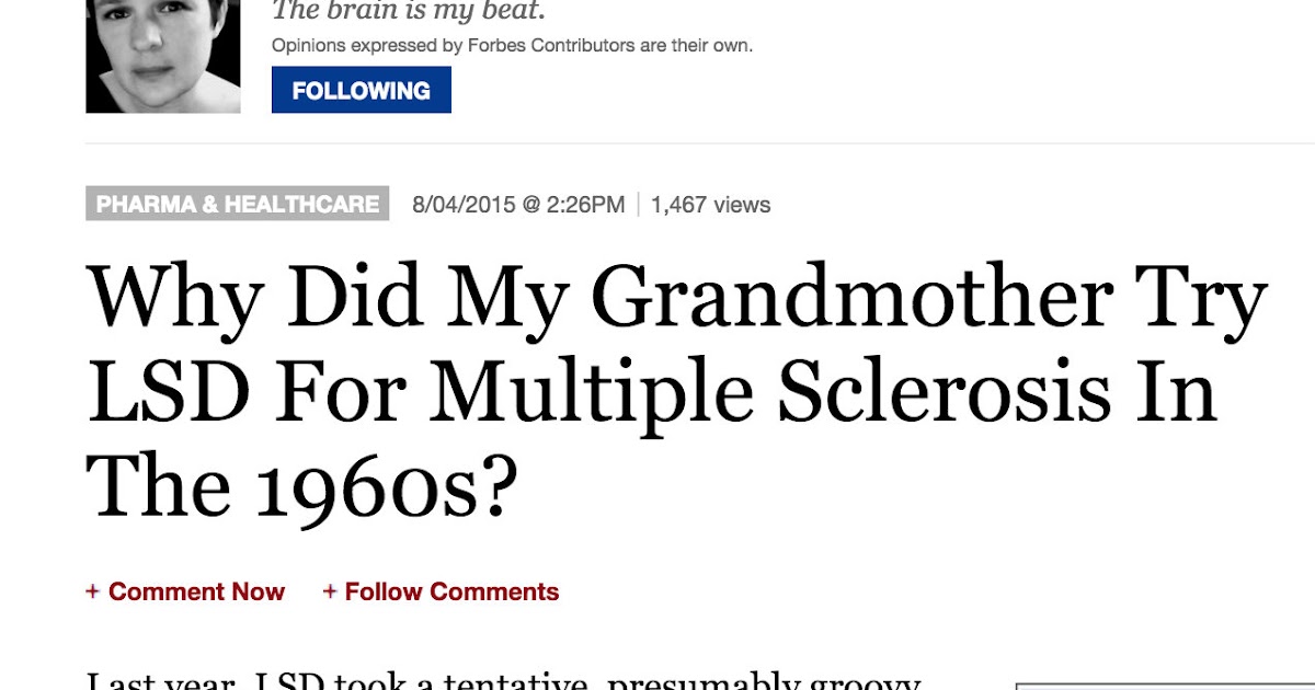
Research using psychedelics was essentially banned worldwide in the late 1960s and early 1970s, and this class of drug labeled dangerous with no medical value. Fortunately, research in this field has gained interest in recent years, and clinical trials in several areas show promise for these drugs as potential new therapeutics. For instance, so-called "magic mushrooms" are a well-known natural source of the classic psychedelic tryptamine psilocybin. Although known and used for millennia, psilocybin itself was not isolated until 1957 by Albert Hoffman from Psylocibe mexicana, who first synthesized it in 1958. Psilocybin itself is a prodrug, rapidly converted to the active form, psilocin, in the body. Another classic psychedelic compound, DMT, is found in significant concentrations in several plants such as Mimosa tenuiflora, Psychotria viridis, and Diplopterys cabrerana, among others. It is also produced in the mammalian body but at low levels. DMT was first synthesized in 1931 and isolated in 1942 from M. tenuiflora by Oswaldo Gonçalves de Lima. Its psychoactive properties, however, were not confirmed until 1956. The β-carboline and monoamine oxidase inhibitors (MAOi) harmine, tetrahydroharmine, and harmaline in Banisteropsis caapi are often used to facilitate the oral activity of DMT in the Amazonian brew ayahuasca, which has also recently been studied for therapeutic benefits.

This review will discuss the current state of the art of how psychedelics influence neural tissue homeostasis and activity. We hypothesize that psychedelics can also be used as therapeutics in the treatment of neurodegenerative diseases and brain injuries. We will mainly focus on neuroimmunology and how data from recent research in the context of neuroinflammation support the hypothesis that psychedelics may have a beneficial outcome in restoring the balance of neural tissue function. In this context, we will also discuss psychedelic-induced neuroplasticity, neurogenesis, and gliogenesis. We propose that psychedelic research in studies of neurodegeneration may be beneficial for future development in this field. We hope that this review will provide information useful to support future psychedelic research in the area of regenerative medicine and the treatment of neurodegenerative disorders and brain injuries.

PSYCHEDELIC INDUCED NEUROPLASTICITY AND NEUROGENESIS

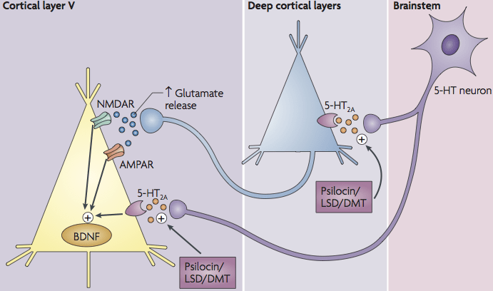
The term "neural plasticity" describes changes in functional neural connectivity. The mechanisms are mostly associated with neural cells, but the process reaches beyond the plasticity of neural synapses. The adaptive changes in the fMRI-measurable macro-scale come from changes in local micro-scales within multicellular interactions, involving neurons, astrocytes, microglia, and oligodendrocytes. These cellular interactions are characterized by complicated homeostatic processes employing both paracrine and direct cell-to-cell communication. Neural plasticity is still poorly understood, but some mechanisms have already been described. Psychedelics may induce a so-called elevated brain entropy state, resulting in an increased ability to learn and "unlearn" certain information. Such action may be therapeutic, and is likely associated with increased neural plasticity mechanisms at the cellular level. Acute changes in the density and complexity of synaptic architecture induced by psychedelics and 5-HT2A receptor activation have been shown by several investigators in both in vitro and in vivo models. These changes involve multiple mechanisms. For example, increases in spine density and morphology can involve direct signaling downstream of 5-HT2A receptor stimulation by psychedelics through serotonylation and activation of Rac1 and kalirin-7, or indirect modulation of synaptic architecture by elevated glutamate levels acting through BDNF/TrkB and mTOR signaling (Figure 1). A feature of psychedelic therapy is the long-lasting effect after only a single treatment. The reason for this is unclear, but likely involves changes in gene expression and/or epigenetic factors underlying the maintenance of neural processes normalized by treatment. There are several known genes involved in synaptic plasticity whose expression is changed in response to psychedelics. Neurogenesis may also be a factor; the administration of DMT induces neural progenitor cells proliferation and adult hippocampal neurogenesis in vivo via activation of Sigma-1 receptors in C57BL/6 mouse. Taken together, neurotrophic signaling and neuroplasticity promoting pathways activated by psychedelics are hypothesized to be key to the mechanism(s) of action for therapeutic effect(s).

A potential key mechanistic component not taken into account for nearly all proposed models is the involvement of microglia for therapeutic effect, as most if not all attention has been focused on neurons in the mechanism of action of psilocybin and other psychedelics. Microglia are tissue-specific, self-renewable CNS macrophage-like cells that are different from other cell types since they appear in the brain and spinal cord during fetal development in the process of primitive hematopoiesis. During their life-long residency inside the CNS environment, microglia assume specific immune cell characteristics and functions. Microglia are very mobile, continually scanning the environment, ready to respond to injury and infections, and take an active part in synaptic rearrangement and neural tissue regeneration. They modulate the deletion of unnecessary connections and the formation of new ones. It is tempting to speculate that psychedelics may stimulate neural plasticity through microglia regulation, especially since many receptors targeted by certain psychedelics like psilocybin and LSD are also present on microglia, including 5-HT2A, 5-HT2B, and 5-HT7 receptors, and the Sigma-1 receptor. Interestingly, in vitro application of DMT and 5-MeO-DMT to monocyte-derived dendritic cells (moDCs) reduce mRNA and protein expression of IL-1β, IL-6, TNF-α, IL-8, and increase expression of regulatory and tolerogenic IL-10.

Another interesting phenomenon is the reciprocity in the dynamics of neuron–microglia interactions. For instance, activation of NMDA receptors on a single neuron's dendrites can stimulate the growth of microglial extensions. Further research may help better understand how neuronal–microglia interactions affect learning and memory, neurodegeneration, and possibly the progression of certain mental illnesses. Microglia may be regulating synaptic pruning or growth by signals from neurons themselves. These regulatory signals may rely on the electrochemical transmission or the complement system, which is also involved in the process of synaptic pruning. During this process, unnecessary synapses are tagged with specific complement proteins that are detected and phagocytized by microglia. Errors in this process during childhood may lead to the development of autism, schizophrenia, or mental retardation, and in adult also result in degenerative diseases. The cellular phenotype and activity of microglia, the involvement of the complement system, and the neuronal signals relevant to synaptic plasticity upon psychedelics stimulation are probably critical aspects of the psychedelic therapeutic mechanism.

Our unpublished data suggest that psilocin increases the protein expression of triggering receptor expressed on myeloid cells 2 (TREM2) on microglia while reducing p65, TLR4, and CD80 proinflammatory markers. TREM2 is involved in the regulation of several microglial functions, including phagocytosis and synaptic refinement. Microglia deficient in TREM2 expression results in synaptic pruning defects, increased excitatory neurotransmission, and reduced long-range functional connectivity. The down-regulation of TREM2 was also observed in brain samples of patients suffering refractory epilepsy. According to our pilot data, psychedelics may prevent neuronal damage in microglia-neuron co-culture, however, it is presently unknown if the protective mechanisms of psychedelics are mediated by microglial TREM2.

PATHOLOGICAL MECHANISMS IN NEURODEGENERATION—A POTENTIAL TARGET FOR PSYCHEDELICS

Because the brain is very fragile and hardly an accessible organ, only limited therapeutic approaches can be proposed for the treatment of brain-specific neurodegeneration. These can be pharmacological, stem cell, or gene therapy approaches. Unfortunately, results to date with these approaches have not been very successful. Interestingly, one recently proposed solution is the application of traditional psychiatric drugs because they have been shown to prevent neural loss and stimulate neurogenesis, Neuroprotection and induction of neurogenesis may be a fruitful avenue to treat MDD as the histopathology of depressed individuals sometimes show signs of subtle neurodegeneration. For example, post-mortem brain studies have revealed neural loss and atrophy in the prefrontal cortex (PFC) and the hippocampus. Neural protective mechanisms (e.g., neuroprotection, neurogenesis, neuroplasticity) have been shown to be induced by psychedelics, which are effective in the treatment of MDD. Here, we discuss pathologies that occur in neurodegenerative disorders that may potentially be targeted by psychedelics for therapeutic effect. We speculate that their application at early disease stages may result in the delay of pathological symptoms.

Oxidative stress

Oxidative cell damage is often a reported in brain-specific neurodegeneration. This damage usually occurs because of an imbalance between free radicals, reactive oxygen species (ROS), and reactive nitrogen species (RNS), and the presence of antioxidants and antioxidative proteins, such as superoxide dismutases (SOD), hioredoxin peroxidases (TRXPs), glutathione peroxidases (GPXs). In a typical situation, if reactive species are held in balance, they play an essential role in regulating essential cellular processes including phagocytosis, apoptosis, and cellular signaling. However, when cells are unable to neutralize excesses of reactive molecules, these molecules may induce damage to mitochondria, cellular and nucleolar membrane, and DNA, and over time result in organ and/or tissue degeneration. Elevated oxidative stress and disruption in redox balance are observed in many psychiatric conditions such as MDD, schizophernia, bipolar disorder, anxiety disorder. Importantly, disruption of redox homeostasis occurs in the pathology of ALS, PD, AD, and DNA repeat expansion disorders such as HD and SCAs.

In the psychedelic brew ayahuasca, two components, harmine and harmaline, are monoamine oxidase inhibitors with antioxidant properties and have the capability to induce gliogenesis and neural progenitor cell migration. Another anti-oxidative effect of psychedelics such as psilocybin and/or DMT may come as a result of 5-HT1A receptor activation. The 5-HT1A receptor agonist 8-OH-DPAT induces expression of the anti-oxidative factor metallothionein-1/-2 (MT-1/-2) and Nfr2. In retina pigment epithelial cell line (ARPE-19), 8-OH-DPAT reduces damage caused by paraquat, an oxidative herbal agent, through elevation of MT1, heme oxygenase-1 (HO1), NAD(P)H: quinone acceptor oxidoreductase 1 (NqO1), superoxide dismutase 1 and 2 (SOD1, SOD2), and catalase (Cat) mRNA expression. 8-OH-DPAT also reduces oxidative stress damage in retinal pigment epithelium/choroid in Sod2 knockout mice.

Endoplasmatic reticulum stress (ERS)

Some psychedelics (e.g., DMT) target the Sigma-1 receptor, which is reported to protect cells from various insults. ER stress induces up-regulation of Sigma-1 expression and modulates the action of PERK, IRE1α, and ATF6 proteins in mitochondria-associated membrane (MAM). Stimulation of Sigma-1 may prevent ERS-mediated cellular apoptosis by regulation of ATF4, ATF6/ C/EBP homologous protein (CHOP), and the balance between Bax and Bcl-2 in granulosa cells. Because ERS damage is reported in MDD and several neurodegenerative disorders, targeting Sigma-1 receptors with psychedelics is proposed as a novel therapeutic strategy.

Blood–brain barrier disruption

The vasculature system in the brain is equipped with a special feature called the blood–brain barrier (BBB). The BBB is composed of a tight layer of astrocytes, is selectively permeable, and separates the intracerebral circulatory system from the peripheral blood to protect the brain against chemical and biological insults. The BBB also contains other cells types, such as microglia cells, perivascular macrophages, and pericytes. The whole structure is embedded in the basal membrane, with extracellular matrix secreted by endothelial cells and pericytes. The endothelial cells inside the blood capillaries form tight junctions (TJ), multi-protein complexes composed of occludins, claudins, and tight junction proteins ZO-1, -2, -3. Breakdown of this system is associated with brain-specific damage and neurodegeneration, and may be the cause of serious illness. Breakdown can originate from prolonged exposure to oxidative stress and/or immune cell activity. For example, microglia inflammatory cytokines acting via IL-1β on Sonic Hedgehog (SHH) can down-regulate tight-junction proteins in astrocytes, resulting in BBB leakage. Moreover, suppressing SHH in astrocytes leads to increased secretion of proinflammatory chemotactic proteins and immune cell activation. Microglia also secrete IL-1β via inflammasome-dependent mechanisms in response to proinflammatory cytokines, DAMPS, β-amyloid, or other toxic protein aggregates. In a C57BL/6 healthy male mouse model, chronic social stress causes BBB disruption via claudin-5 down-regulation, which leads to the infiltration of proinflammatory factors and depression-like behaviors. Disruption of the BBB is also observed in a genetic mouse model of schizophrenia, and may be involved in bipolar disorder pathology. BBB disruption is also observed in multiple neurodegenerative disorders including ALS, AD, and SCA3.

Conceptually, inflammation-based BBB leakage could be prevented to some degree by the presence of psychedelics. The drugs N,N-DMT and 5-MeO-DMT, applied into LPS and polyI:C – activated dendritic cells in vitro, result in down-regulation of expression of IL-1β, IL-6, IL-8, TNF-α, and up-regulation of IL-10 as measured by mRNA and protein expression through stimulation of Sigma-1 receptors. This observation has also been confirmed in vivo in a Wistar rat model of stroke, where N,N-DMT administration significantly decreased IL-1β, IL-6, and TNF-α, but increased IL-10, measured by mRNA and protein expression. Furthermore, N,N-DMT-treated rats demonstrate improved motor skills post-stroke. Although not validated yet in brain tissues, several psychedelics, including DOI, LSD, and psilocybin, have been shown to have potent anti-inflammatory effects of suppressing many of these same proinflammatory biomarkers in peripheral tissues, and they may represent effective therapies for inflammation-related neuropathologies.

Oligodendrocyte pathology

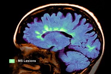
Oligodendrocytes protect and support neurons and their axons by providing myelin that improves electric signal transmission. Unfortunately, their active role in immune response and neural regeneration has long been overlooked. Microglia and oligodendrocytes actively work to regulate each other's functions. Moreover, oligodendrocytes’ pathology occurs in many neurodegenerative diseases, including Alzheimer's and Parkinson's Disease, ALS, Multiple Sclerosis, Spinal Cord Injury, but also non-degenerative psychiatric conditions including MDD, schizophrenia, and Alcohol Use Disorder. For example, in MDD, abnormalities in oligodendrocyte density are observed in the PFC and amygdala. Interestingly, oligodendrocytes are extremely vulnerable to oxidative stress and prolonged exposure to proinflammatory factors secreted by microglia. The previously discussed psychedelic-mediated reduction in cytokine secretions may play a protective role in myelin and oligodendrocyte cell survival. Certain psychedelics can target Sigma-1 receptors, which are essential in stimulating OPC differentials. Together, these findings indicate that more attention should be paid to the influence of psychedelics on oligodendrocyte biology.
PSYCHEDELICS AS IMMUNOMODULATORS

The study of psychedelics at target receptors and the activation of effector pathways have brought new, but still limited, insights into their immunomodulatory potential (see Table 1). Classic psychedelics like LSD, DMT, 5-Meo-DMT, and psilocin have the potential to interact with several 5-HT receptor subtypes, Sigma-1R, and TAAR, which are present in CNS and other tissues, including cells of innate and adaptive immunity like macrophages, monocytes, dendritic cells, and T cells. These receptors are mediators of immunological response, and serotonin is considered a critical factor in immune homeostasis. Therefore, psychedelics can regulate both adaptive and innate immune responses. A review of putative molecular mechanisms in which psychedelics may act as immunomodulators was published by Szabo, emphasizing cross-talk between pattern recognition receptors (PRR), such as Toll-Like Receptor 4 (TLR4), 5-HTRs, and Sigma-1R, and regulation of inflammatory response via NFκB/IRF signal transduction pathways. Although these modulations result in changes, Sigma-1, and TAAR seem to play a crucial role in immune response, and all three of them can be stimulated by psychedelics.
Psychedelic ligandsReceptorEffectLiterature
(R)- DOI5-HT2AIn smooth muscle cells in vitro:

Prevention of Nf-κβ nuclear translocation and inhibition of nitric-oxide synthase activity

In aortic arch and small intestine in vivo:

Down-regulation of TNF-α-mediated Cx3CL1, Icam-1, Vcam-1, MCP-1, IL-6, IL-1β

In OVA-treated asthma-model lung in vivo:

Suppression of Th2-related genes: Mcp-1, Il-13, Il-5, and Gm-csf,

Inhibition of neutrophil infiltration,

Inhibition of mucus hyperproduction
Yu et al. (2008)

Nau et al. (2013)

Nau et al. (2015
DMT,

5-MeO-DMT
Sigma-1Down-regulation of: IL-1 β, IL-6, TNF-α, IL-8

Up-regulation of: IL-10

(gene and protein expression)

Decrease of Th1 and Th17 activation after E. coli or H1N1 co-culture
Szabo et al. (2014)
5-MeO-DMT5-HT2A (?)

5-HT2c (?)
In human cerebral organoid in vitro model:

Down-regulation of Nf-κβ pathways (involved in the immune response)

Down-regulation of Nuclear Factor of Activated T cells (NFAT) (T-cell activation, stem cell differentiation)

Modulation of Gaq-Rho-ROCK pathway (cytoskeletal rearrangement, phagocytosis)
Dakic et al. (2017)
DOI, DMT, Psilocin5-HT2B(Reported with BW723C86 5-HT2B ligand)

In CD1+ moDC down-regulation of: TNF-α, IL-6, IL-8/CXCL8/ IP-10/CXCL10 after TLR2, and TLR6/7 activation,

In TLR3-activated CD1+ moDC: down-regulation of CD80, CD83, CD86 (anti-inflammatory, tolerogenic)

Prevention of Th1, Th17 lymphocyte polarization
Szabo et al. (2018)
DMTSigma-1In in vivo model of stroke:

Down-regulation of: IL-1β, IL-6, IL-8, TNF-α, NOS, APAF-1 (proinflammatory, proapoptotic)

Up-regulation of BDNF and IL-10 (tolerogenic, neurogenic)
Nardai et al. (2020)
(Hypothetically) DOI, DMT, Psilocin, LSD, mescalineTAAR1, TAAR2(Not tested yet with psychedelics)

Lymphocyte migration, increase in IL-4 secretion, Th1/Th2/Th3 phenotype modulation, mediation of IgE-secretion
Babusyte et al. (2013)

5.1 5-HT receptors

Serotonin is one of the most critical factors during fetal brain development and neurogenesis, and is responsible for the formation of axons and dendrites, and adult axonal regeneration. Serotonin receptors are present on most, if not all, types of cells in the CNS. In neurons, for example, their activation can influence cellular membrane polarization states through multiple mechanisms. Serotonin also plays significant roles aside from being a neurotransmitter. There are several receptor subtypes expressed in mammalian peripheral tissues and cells outside the CNS, including adaptive and innate immune cells. Serotonin itself has an endocrine effect on the regulation of whole-body homeostasis, such as heart rate, intestinal motility, and last but not least: the immune response.

Although 5-HTRs are primarily described as activators of proinflammatory pathways, they surprisingly have anti-inflammatory properties when activated by certain, but not all, psychedelics. The selective 5-HT2 receptor agonist (R)-DOI, reduces mRNA expression of proinflammatory adhesion molecules ICAM-1 and VCAM-1 as well as mRNA levels for proinflammatory cytokines MCP1, IL-1β, and IL-6 in various tissues like intestine and aorta, and circulating levels of IL-6 in TNF-α treated mice. Several of these findings were confirmed in a high fat-fed ApoE−/− knockout moue model of cardiovascular and metabolic disease. An increase in levels of VCAM-1, IL-6, and MCP-1 mRNA expression was observed in animals fed a high-fat and -cholesterol "Western diet" compared to control mice fed regular food, and this increase was prevented in mice fed the Western diet and treated with (R)-DOI. The precise mechanism underlying why 5-HT2A receptors, which are widely described as inflammation inducers, induce anti-inflammatory processes after activated by (R)-DOI and some other psychedelics is not known. The hypothesis proposed by Flanagan and Nichols involves the concept of functional selectivity, in which different ligands induce slightly different conformations of the receptor to recruit different sets of effector pathways. Psychedelics are hypothesized to recruit and activate anti-inflammatory effector signaling pathways, whereas serotonin itself recruits proinflammatory pathways. In rodent models of allergic asthma, nasal administration of (R)-DOI at a very low dose (EC50: ~0.005 mg/kg) completely prevents symptoms, including airways hyperresponsiveness, pulmonary inflammation, and mucus overproduction in response to allergen. Further examination of the lung tissue revealed prevention of eosinophilia and a reduction in Th2 cell recruitment. Interestingly, the behavioral potency of different psychedelics does not correlate with anti-asthma efficacy. Significantly, therapeutic drug levels in these models are orders of magnitude lower than the levels necessary to induce measurable behavioral responses. These findings suggest that subperceptual levels of some psychedelics may be an exciting alternative to currently available steroid drugs in the treatment of asthma and other inflammatory-related disease.

Given the high level of expression of 5-HT2A receptors in the brain on multiple cell types, it may be that psychedelics have similar anti-inflammatory properties against neuroinflammation. In vitro application of 5-MeO-DMT in human cerebellar organoids results in down-regulation of NF-κβ and nuclear factor of activated T cells (NFAT) pathways, as well as modulation of the Gαq-RhoA-ROCK pathway involved in cytoskeleton rearrangement and phagocytosis.

Classical psychedelics have mid affinity for and efficacy at 5-HT2B receptors. Interestingly, the activation of this receptor type with the agonist (BW723C86) regulates immune responses in CD1+ monocyte-derived dendritic cells (moDC). The application of BW723C86 resulted in down-regulation of CD80, CD83, and CD86 proinflammatory molecules on CD1+ moDC. Furthermore, stimulation of 5-HT2B down-regulates TLR2, TLR3, and TLR7/8-mediated proinflammatory cytokine protein expression. It also prevents moDC-mediated activation of T cells toward inflammatory Th1 and Th-17 phenotypes. Furthermore, certain immune stimulators such as the molecule polyI:C, which is a TLR3 agonist, up-regulate the expression 5-HT2B receptor protein. Together, these observations suggest that 5-HT2B agonism may participate in some aspects of their anti-inflammatory mechanism. However, in the allergic asthma model, (R)-DOI was not effective in reducing pulmonary inflammation in the 5-HT2A receptor knockout mouse indicating that for at least asthma and pulmonary inflammation 5-HT2A receptor activity is necessary and sufficient for therapeutic effect.

5.2 Sigma-1 receptor

The sigma-1R is a transmembrane protein located in mitochondria and the endoplasmatic reticulum (ER). It is abundantly present within the CNS in neurons, astrocytes, oligodendrocytes, and microglia, where it mediates a neuroprotective effect. Sigma-1R activity promotes neural function and survival via modulation of Ca2+ homeostasis, mitigation of oxidative stress, regulation of gliosis, neuroplasticity, and glutamate activity.

Stimulation of Sigma-1R in oligodendrocyte progenitor cells (OPC) results in oligodendrocyte differentiation, and stimulation in astrocytes improves the BDNF secretion. This suggests that targeting Sigma-1R may be a promising therapeutic strategy for psychiatric and neurodegenerative conditions. Interestingly, Sigma-1R activity is involved in the transition between M1-like proinflammatory and M2-like proregenerative and tolerogenic microglia phenotypes. These mechanisms are not very well understood. Moreover, as microglia are cells of complicated biology and are somewhat difficult to study, the concept of M1/M2 polarization may be too simplistic to address many aspects of microglia function. It has been recently proposed that microglia displaying a proinflammatory phenotype are crucial for their role in neural tissue reorganization and regeneration.

DMT is an agonist of Sigma-1Rs, and found to be produced by specific tissues in the brain. Szabo et al. observed that the application of DMT into human iPSC-derived cortical neuron cultures in vitro resulted in better survival under hypoxia conditions, but that the protective effect vanished after Sigma-1R gene knockdown with siRNA. This suggests that DMT can protect cells from hypoxia-induced apoptosis via Sigma-1 receptor stimulation. This observation was later confirmed in an in vivo rat model of stroke, where continuous administration of DMT reduced the size and number of lesions, and decreased levels of IL-1β while up-regulating IL-10, and BDNF protein and mRNA levels. Similar results were found with another Sigma-1 agonist (PRE-084) after embolic stroke to significantly reduce the size of lesions, improve neuronal deficits, and reduce concentrations of some proinflammatory cytokines while elevating levels of some anti-inflammatory cytokines like IL-10. Interestingly, application of the Sigma-1R selective antagonist (MR309), had similar neuroprotective effects. It is tempting to speculate that elevated anti-inflammatory cytokine levels after DMT administration in these stroke models may be caused by Sigma-1R mediated changes in microglia phenotypes. For example, in a study by Moritz et al., stimulation of Sigma-1R "switched off" activated microglia and made them migrate away from the location of damaged tissue. Moreover, in the LPS-treated microglial BV2 cell line, application of Sigma-1R agonist SKF83959 results in the prevention of M1-like phenotype switching by microglia, and a decrease in TNF-α, IL-1β, and inducible NOS levels. A similar effect was reported in a model of traumatic brain injury (TBI), and Parkinson's Disease.

TAAR

Trace amine-associated receptors (TAARs) are G-protein-coupled receptors abundantly present in the CNS. In most vertebrates, they exist in nine isoforms. Only TAAR1 has been studied in-depth, however. This receptor is relatively non-selective and has an affinity for endogenous trace amines as well as the classical neurotransmitters serotonin and dopamine, and multiple psychoactive drugs, including amphetamines, ergoline derivatives, psilocin, DMT, and mescaline. TAAR1 is a modulator of neurotransmission induced by canonical dopamine, serotonin, and glutamine receptors, and its aberrations and rare variants may contribute to the etiology of schizophrenia. TAAR1 is also expressed in non-CNS tissues such as the thyroid, stomach, pancreas, and intestine, where it may regulate body functions in an endocrine manner.

Although abnormalities in TAARs expression or function may be related to the development of schizophrenia, data suggest involvement in additional neuropsychiatric conditions. For example, stimulation of TAAR1 in an experimental model of Parkinson's Disease results in L-DOPA-related dyskinesias, and TAAR1 knockout mice are reported to be more vulnerable to various substance addiction. Targeting TAAR1 has also been suggested as a possible therapeutic target for the treatment of bipolar depression, fibromyalgia syndrome, and diabetes.

TAAR are found in immune cells and can elicit immunomodulatory effects; however, our knowledge about TAARs and immune responses is limited. TAAR1 is expressed in polymorphonuclear leukocytes (PMN), T cells, and B cells, whereas TAAR2 is also abundant on NK cells and monocytes. In T cells, stimulation of TAAR1 and TAAR2 receptors induce IL-4 production and modulation of Th1, Th2, and Th3 markers, whereas silencing of these receptors reduces IgE secretion in B cells after induction with trace amines. TAAR1 and TAAR2 are also reported to be involved in PMN chemotactic migration. DMT, (R)-DOI, d-LSD, and 5-MeO-DMT are TAAR1 agonists, and it is, therefore, possible that psychedelics may regulate immune cells to respond and regulate neural tissue homeostasis via TAAR1 activation.

RESEARCH PERSPECTIVES FOR PSYCHEDELICS IN PREVENTING NEURODEGENERATION

In this review, we have highlighted the beneficial outcomes of psychedelic treatment for MDD. Our primary focus was on processes in neural tissue microenvironments which can be affected by psychedelics. These include the induction of neurogenesis and neuroplasticity and reduction of inflammation and oxidative stress. These characteristics of psychedelics may play crucial roles in restoring long-term healthy homeostasis in depressed patients. We also emphasized potential areas of therapeutic actions in brain-specific neurodegeneration in which psychedelics may be beneficial. These include oxidative stress, inflammation, BBB disruption, and loss of oligodendrocytes and myelin.

In Europe, around 7 million people suffer from dementia-related disorders, and the aging of society is expected to double this number by 2040. Around 15 mln people worldwide and 1,12 mln in Europe experience a stroke every year, with 5 mln of those being fatal incidents (650 000 in Europe), and another 5 mln of patients suffer post-stroke severe disability. Each year 10 mln new dementia cases are being diagnosed globally, and 60–70% of them are Alzheimer's Disease. In 2015 ALS was diagnosed in 222 801 people worldwide, and that number is predicted to grow by 69%, reaching 376 674 new ALS cases annually by 2040. Therefore, neurodegenerative diseases are a serious and growing burden for modern societies. Moreover, the development of effective therapeutics lags behind other fields such as cardiovascular diseases and cancer. Therefore it is a top priority to search for novel candidates for therapeutic approaches to revert these dire statistics.

Psychedelics represent such a novel approach. Certain psychedelics, with demonstrated therapeutic efficacy for psychiatric disorders in clinical trials, have been used safely for centuries by indigenous populations. The beneficial therapeutic dosages of these substances have been shown to be well-tolerated, and they present a favorable safety profile in treating a variety of disorders. Clinical trials are investigating therapeutic efficacy for anorexia nervosa, the early stage of Alzheimer's Disease, and traumatic brain injury, among other CNS disorders.

Psychedelics (e.g., DOI, DMT, LSD) promote structural plasticity via BDNF signaling and are thus proposed as potential therapeutics for MDD and related disorders. Neuropsychiatric disorders are well known to be associated with atrophy of neurons and abnormal neuronal circuits. Among neurodegenerative disorders, commonalities in pathological characteristics may be seen in polyQ disorders such as spinocerebellar ataxia type 3 (SCA3).

Similar to many other neurodegenerative disorders, the down-regulation of BDNF is observed in SCA3 cells and in dentate neurons of SCA3 patients. Moreover, several essential proteins belonging to the BDNF signaling pathway are also down-regulated in mouse models. For example, Rac1, acting downstream of BDNF and TrkB, is down-regulated in the cerebral cortex and cerebellum of young SCA3 mice. Together with another down-regulated protein, RhoA, Rac1 mediates proplasticity properties evoked by BDNF by facilitating sLTP (structural long-term potentiation) and regulating actin cytoskeleton in dendritic spines. Furthermore, MAPK1 (a.k.a. Erk) and MAP2K1 are also down-regulated in the young SCA3 mice, and Erk signaling is down-regulated in MDD. Importantly, BDNF is an immediate upstream regulator of the MAPK (mitogen-activated protein kinase) cascade. Activation of MAPK (ERK) signaling by neurotrophins is involved in long-term synaptic plasticity and the structural remodeling of the spines in the excitatory synapses.

Another protein necessary for BDNF signal transduction to the nucleus is Pea15, which is also down-regulated in SCA3 mice. Silencing of Pea15 results in inhibition of BDNF retrograde signaling. Pea15 acts as a scaffolding protein for PLD1, RSK2, and ERK1/2, and the formation of this complex is triggered by BDNF in cortical neurons. Therefore, the potential use of psychedelics could affect the levels of several proteins, which are down-regulated in the SCA3 model probably by an increase in BDNF which may promote synaptic plasticity. The role of BDNF signaling in the survival of neurons has been well documented in other neurodegenerative disorders, such as Huntington's, Parkinson's, and Alzheimer's disease.

Psychedelics have also been shown to reduce oxidative stress, which is a significant issue in neurodegenerative disorders. Several oxidative stress biomarkers are elevated in models of neurodegenerative disorders. Other anti-oxidative proteins, which play an essential role in reducing oxidative stress by breaking down ROS, are down-regulated in the SCA3 model. Moreover, Txn down-regulation in SCA3 mice might increase the vulnerability of neurons to ROS. Therefore, psychedelics’ anti-oxidative properties could be a beneficial component of a therapeutic strategy for SCA3 and other neurodegenerative disorders.

The strategy of serotonergic signaling modulation by psychedelics in SCA3 is also strongly supported by studies showing a therapeutic effect for SCA3 through 5-HT1AR activity, which is activated by several psychedelics including psilocybin and LSD. Targeting of the 5-HT1A serotonin receptor orthologue SER-4 in C. elegans ameliorates motor dysfunction and reduced mutant ATXN3 aggregation. Furthermore, treatment with partial agonists of 5-HT1A receptors has been demonstrated to reduce ataxia, pain, insomnia, and depressive symptoms in patients with SCA3 and other forms of SCA. The SSRI citalopram has beneficial therapeutic effects in animal models of SCA3 and preclinical trials. Thus, activation of serotonergic signaling in SCA3 patients with psychedelic agents is a promising therapeutic strategy.

Non-hallucinogenic psychedelics approach

The “hallucinogenic” effects of psychedelics have been proposed to be directly associated with their therapeutic potential in psychedelic-assisted psychotherapeutical approach as the subjective peak intensity has a high correlation with therapeutic efficacy. However, in patients suffering brain-specific neurodegeneration, psychotropic effects of psychedelics may be a serious limitation, especially if because of the disease physiology, the medicine would have to be administrated more often and in higher doses. Furthermore, correlation is not causation, and subjective peak experiences may merely indicate that sufficient drug has been administered to produce therapeutic efficacy at cellular and molecular targets and circuits. Although ibogaine is not classified as a psychedelic, it is a type of hallucinogen that may have therapeutic efficacy to treat substance use disorder. Recently, analogs of ibogaine have been reported by two different investigators to attenuate behaviors associated with substance abuse in rodent models, in a similar way to ibogaine. One is peer reviewed, and the other awaiting peer review. Because of the low toxicity and lack of hallucinogenic properties of these new molecules, they represent potential non-hallucinogenic derivatives of hallucinogenic parent molecules with therapeutic effect. With regard to psychedelics, their demonstrated potency in multiple animal models of disease suggests it may not be necessary to eliminate hallucinogenic behaviors from effective molecules because the dose is so low that effects on behaviors would not be seen at relevant therapeutic levels. Regardless, work by Flanagan et al. suggest that it may be possible to engineer hallucinogenic effects away from therapeutic effects to develop non-hallucinogenic 5-HT2A receptor agonists with anti-inflammatory potential. Development of a 5-HT2A receptor agonist therapeutic devoid of hallucinogenic effects would conceivably allow higher levels to be used, especially for those with weaker potencies, which may allow for greater efficacy in certain circumstances.

Prospect implications for regenerative medicine

Besides the prevention and regulation of pathology in neurodegenerative disease, the immunomodulatory properties of psychedelics may also be relevant to regenerative medicine. Neural Stem/Progenitor Cell (NSC/NPC) transplantation is a recently developed and promising therapeutic tool. However, the limitation of such a strategy is poor graft survival because of immune response. Our recent study revealed that DMT and psilocin down-regulate CD80 co-stimulatory molecule expression on the surface of microglial cells, with and without LPS stimulation. The co-stimulatory signal is crucial for recruiting adaptive immune cells; therefore, blockade of co-stimulatory molecules an attractive immunosuppressive strategy. The unique properties of psychedelics in suppressing inflammatory responses, and promoting neural survival and plasticity, could be a strong rationale for the hypothesis that psychedelics might support grafted cells and facilitate their survival for therapeutic benefits.

Therapeutic perspectives for microdosing

One dosing method of psychedelics is the use of so called “microdoses”—very low concentrations of various psychedelics that do not reach the threshold of perceivable behavioral effects. This is usually 10% of active recreational doses taken up to three times per week. This regimen is popular in underground settings without medical guidance. Microdosing is believed to improve the creative thinking, cognitive function, and overall psychological well-being, and is described mostly in the context of self-application by healthy enthusiasts. There have been few rigorously controlled studies of microdosing, and the therapeutic effects of psychedelic microdoses for the treatment of psychiatric disorders are questionable. According to a self-blinding study involving 191 healthy volunteers, in which the mood changes were measured using various questionaries, the authors concluded that anecdotal psychological improvements are more likely associated with the placebo effect rather than drug effect. Furthermore, Family et al. reported that repeated administration of LSD (5–20 µg) in healthy individuals in a blinded placebo-controlled clinical trial produced no significant changes in several cognitive outcome measures. However, a recent study using fMRI showed that 13 µg of LSD changes connectivity inside the limbic circuits 90 min after drug administration compared to the placebo control, which was associated with positive mood changes as measured with a Positive and Negative Affect Schedule (PANAS). Each of these reports has been in healthy individuals, and there have been no rigorous and controlled studies to date on microdosing in patients with diagnosed depressive disorder. The application of psychedelic microdosing in the context of the treatment of brain-specific neurodegenerative disorders has not been yet directly investigated, however, researches speculate that it may influence the hippocampal neurogenesis. Importantly, a Phase I feasibility and safety study on repeated low-dose LSD administration has been conducted in an elderly healthy population in preparations for later phase clinical trials to treat Alzheimer's Disease.

CONCLUSION

Psychedelics stimulate neuro- and gliogenesis, reduce inflammation, and ameliorate oxidative stress. Therefore, they are promising candidates for future therapeutics for psychiatric, neurodegenerative, and movement disorders. Importantly, psychedelics hold the promise of being disease-modifying therapeutics, and not simply just providing symptomatic relief. Current clinical trials have demonstrated both safety and efficacy for their therapeutic use in controlled clinical settings, and psilocybin, has even been designated with “Breakthrough Therapy” status by the FDA in the United States for two different Phase III clinical trials. Often only just one or two therapeutic administrations produce profound and persistent effects. Preclinical research has shown promise in several disease models for both psychiatric and non-psychiatric diseases. Therefore, the use of psychedelics as therapeutics is very promising and should be further developed, paying special attention in the future to prospect applications in neurodegenerative diseases.

*From the article (including references) here :
 
Last edited:
uci_jobs-landing-v28_pic-5.jpg
University of California, Irvine

Simple sugar found in human breast milk possible therapy for repairing myelin in MS

UC Irvine | Neuroscience News | 7 Oct 2020

N-acetylglucosamine, a simple sugar found in human breast milk and sold as an over-the-counter dietary supplement in the United States, promotes myelin repair in mouse models and correlates with myelination levels in multiple sclerosis patients according to a new University of California, Irvine-led study.

Published in the Journal of Biological Chemistry, the study also demonstrates that in mice, delivering N-acetylglucosamine orally to lactating mothers drove primary myelination in their nursing offspring. N-acetylglucosamine is a simple sugar that is metabolically attached to proteins at the cell surface to control cellular function.

“We found that N-acetylglucosamine activates myelin stem cells to promote primary myelination and myelin repair,” said Michael Demetriou, MD, PhD, FRCP(C), professor of neurology, microbiology and molecular genetics at UCI School of Medicine and leader of the study. “Our data raises the intriguing possibility that N-acetylglucosamine may be a simple therapy to promote myelin repair in multiple sclerosis patients.” Formal human studies will be required to test this theory.

The failure of robust re-myelination following inflammatory demyelination in multiple sclerosis leads to chronic disability and neurodegeneration. Myelin insulates the long, cable-like nerve cell branches called axons, and serves to increase the speed of electrical signal conduction between neurons. Myelination in the central nervous system also plays an important role in cognitive development during childhood.

“Interestingly, since N-acetylglucosamine is a major component of human breast milk but not baby formula, it may explain some of the cognitive function and myelination benefits realized by children fed breast milk as opposed to formula.” said Michael Sy, MD, PhD, assistant professor of neurology at UCI School of Medicine, co-director of the regional MS program at the VA Long Beach Healthcare System, and first author of the study.

Dr. med. Alexander Brandt, MD, who led the clinical parts of the study together with Dr. med. Friedemann Paul, MD, added, “The association of reduced N-acetylglucosamine serum levels with white matter changes in the brain of patients with multiple sclerosis suggests that N-acetyglucosamine deficiency may contribute to disease severity.” At the time of the study, Brandt has been the head of the Translational Neuroimaging laboratory in the Clinical Neuroimmunology group at Charité – Universitätsmedizin Berlin, which is led by Paul. Brandt has since transitioned to the UCI School of Medicine as associate professor of neurology.

Formal clinical trials are required to test the applications identified in this study and are currently being pursued by the investigators.

 
2-smartphoneus.jpg

The way patients with MS use cellphones could provide tools for their treatment.

Smartphone use offers tool to treat Multiple Sclerosis

by American Institute of Physics | Medical Xpress | 17 Nov 2020

Monitoring how patients with multiple sclerosis or other degenerative diseases use their smartphones could provide valuable information to help get them better treatment.

In an article published in Chaos, researchers used a mobile app to record the keystroke dynamics of a control group and those of subjects in various stages of multiple sclerosis treatment over the course of a year.

Keystroke dynamics show how quickly or slowly someone is typing on a touch screen, the amount of time between letters typed, the number of mistakes made and corrected while typing, and other behaviors. As part of the study, researchers at Amsterdam University Medical Center used a mobile app that tracks how a user is typing on their phone's keyboard.

In doing so, they observed changes over time in the way people with MS typed that were not seen in subjects who did not have the disease.

"The clinically relevant changes in keystroke dynamics can be seen as early warning signals for changes in disease activity of the patient prior to the change occurring," the authors wrote.

James Twose, one of the authors, called the study's findings a "first promising step" toward using keystrokes to help diagnose changes in patients with chronic diseases like MS.

"The dream is prediction," said Twose. "If there is some semblance of predictability, the joy would be to forecast the disease in a similar way you do with weather."

Multiple sclerosis patients generally make clinical visits every 3-12 months, according to the authors, and MRIs are the best way to measure changes in damage to the brain from the disease.

If doctors were able to use something like keystrokes to monitor patients on a more ongoing basis, they could better predict when new treatments are needed.

"In chronic diseases like MS and Alzheimer's disease and Parkinson's disease, there is inherent worsening over time," Twose said. "When it comes to typing, you need all your faculties to do this well. We notice when you have problems with that."

Such a system would be helpful to chronic disease sufferers for a variety of reasons. Consistent monitoring would give doctors a fuller picture of their condition than occasional clinic visits, which can only provide a snapshot of how they are doing at that moment. It also would be a boon for patients whose conditions make traveling to and from doctor appointments difficult.

 
Top