• Psychedelic Medicine

ALZHEIMER'S | +80 articles

tingri-everest-base-camp-trek.jpg


In vivo evidence of therapeutic properties of CBD for Alzheimer's Disease

A review and summary of animal subject studies of CBD in relation to neuroprotective effects and Alzheimer's disease. The studies demonstrate the ability of CBD to reduce the change caused by damage to neural cells and reduce the neuroinflammatory response. CBD was also shown to promote neurogenesis. Importantly, CBD also reverses and prevents the development of cognitive deficits in Alzheimer's rodent models. Interestingly, combination therapies (of CBD and THC) showed that CBD can antagonize the psychoactive effects associated with THC and possibly mediate greater therapeutic benefits than either phytocannabinoid alone.

https://www.ncbi.nlm.nih.gov/pmc/articles/PMC5289988/

---

CBD modulates the expression of Alzheimer's Disease-related genes

Stem cells, mesenchymal stem cells to be exact, have emerged as a promising tool for the treatment of neurodegenerative disorders, including Alzheimer's disease. Current therapies for Alzheimer's that utilize mesenchymal stem cells have shown limited effectiveness. This study evaluates whether pre-treatment with cannabidiol modulates mesenchymal stem cells in order to improve their therapeutic potential. The results showed that pre-treatment with CBD prevented the expression of proteins and suggested that mesenchymal stem cells preconditioned with CBD possess a molecular profile that might be more beneficial for the treatment of Alzheimer's.

https://www.ncbi.nlm.nih.gov/pubmed/28025562

---

Safety and efficacy of CBD for behavioral and psychological symptoms of dementia

With a given premise of THC as a potential treatment for Alzheimer's, this clinical trial looked at whether CBD oil was effective in relieving behavioral and psychological symptoms of dementia. CBD oil was given in addition pharmaceutical drugs already prescribed for this group of 11 trial participants. The trial lasted for 4 weeks. Results showed that delusions, agitation/aggression, irritability, apathy, sleep distress and caregiver distress were all reduced.

https://www.ncbi.nlm.nih.gov/pubmed/26757043

---

Cannabinoids for the treatment of agitation and aggression in Alzheimer's Disease

Utilizing the endocannabinoid system and synthetic cannabinoids for the modulation of neuroinflammation that is related to Alzheimer's disease was a given for this review. They looked at many studies, case studies and trials. Findings from 6 studies showed significant benefits from synthetic cannabinoids, specifically dronabinol and nabilone. It was noted that conclusions were limited by small sample sizes, short trial duration, and lack of placebo control in some of these studies.

https://www.ncbi.nlm.nih.gov/pubmed/26271310

---

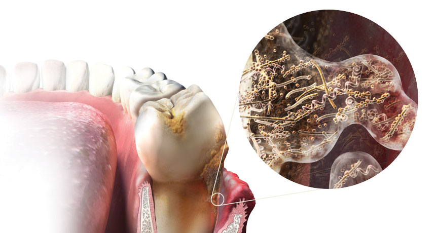
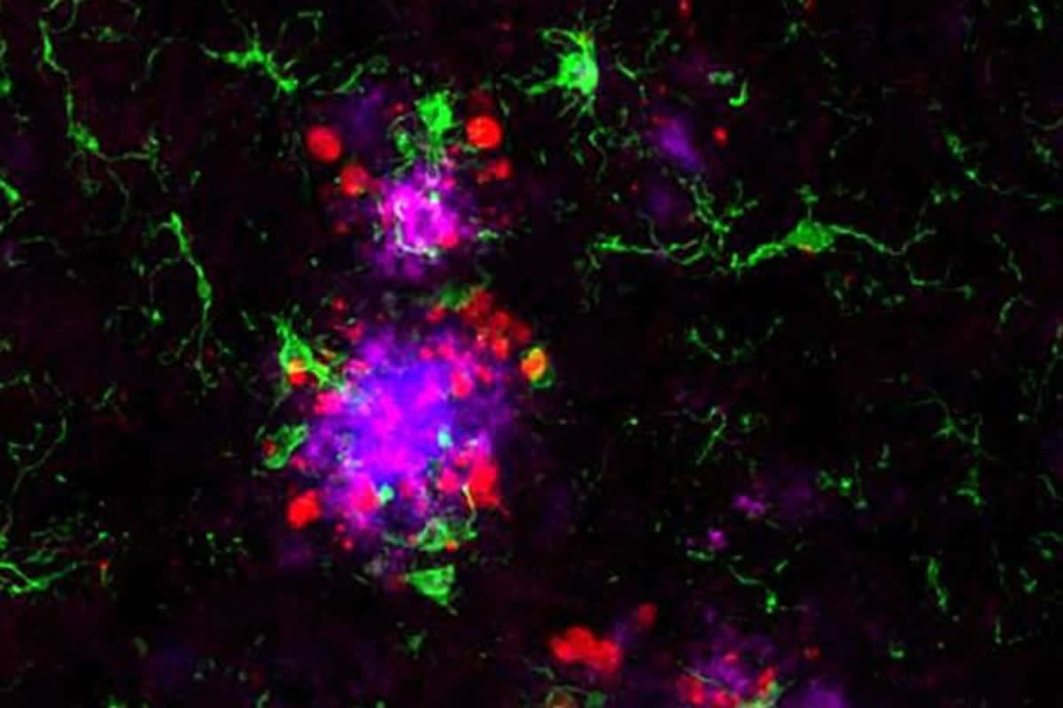
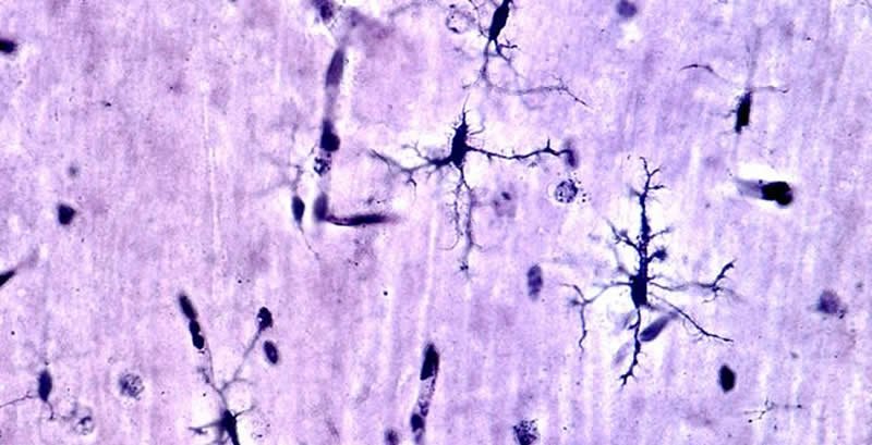
CBD and other cannabinoids reduce microglial activation in Alzheimer's Disease

This study suggests that microglial activity has been shown as a feature of Alzheimer's disease. In the present study, they compared the effects of cannbidiol with those of other cannabinoids on microglial cell functions, learning behavior and cytokine expression after beta amyloid administration in mice. The study found that CBD was able to modulate microglial activity, and produce beneficial effects in animal subjects model of Alzheimer's.

https://www.ncbi.nlm.nih.gov/pubmed/21350020

---

CBD in vivo blunts meta-amyloid-induced neuroinflammation

To confirm the results of a previous study showing that cannabidiol inhibited gliosis in vivo in mice, mice were inoculated with human peptide into the right dorsal hippocampus, and treated daily with CBD and a non-CBD substance for 7 days. The dosage of CBD used was 2.5 or 10 mg per kg of body weight. CBD significantly inhibited markers of neuroinflammation in the animal subjects. This was also dose dependent. The results of this study confirm the in vivo anti-inflammatory actions of CBD.

https://www.ncbi.nlm.nih.gov/pubmed/17592514

---

Neuroprotective effect of CBD on Beta-amyloid-induced toxicity in PC12 cells

This study looked at the effect of cannabidiol on peptide toxicity in rat cells in culture. The toxic effect was increased reactive oxygen species, lipid peroxidation, cell death signaling cascade, increased intracellular calcium, and DNA fragmentation. Treatment of the cells with cannabidiol prior to toxic peptide exposure significantly elevated cell survival while it decreased reactive oxygen species production, lipid peroxidation, cell death signaling levels, DNA fragmentation and intracellular calcium. Results indicated that CBD exerts a combination of neuroprotective, anti-oxidative and anti-apoptotic (anti cell death) effects against peptide toxicity.

https://www.ncbi.nlm.nih.gov/pubmed/15030397

---

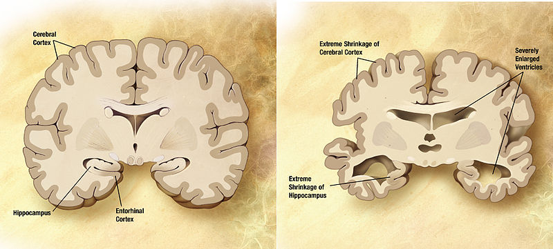
CBD reverses deficits in hippocampal LTP in a model of Alzheimer's Disease

Synaptic plasticity protection by cannabidiol is demonstrated for the first time in an in vitro model. The study looked at the effect of CBD on amyloid peptides. Amyloid peptides are known to reduce persistent increase in synaptic strength in the hippocampus part of the brain. CBD alone did not alter persistent increase in synaptic strength in the hippocampus. But, pre-treatment with CBD protected the persistent increase in synaptic strength. Essentially, CBD protected the hippocampus and memory from the peptide that is known to cause Alzheimer's disease.

https://www.ncbi.nlm.nih.gov/pubmed/29574668
.
 
Last edited:
b3829b961b9baf8e4618b05c10db459a.jpg


Gum disease bug ‘central’ to developing Alzheimer’s

Evidence infectious bacteria drives the disease’s progression

A common gum disease bug plays a “central role” in the development of Alzheimer’s, scientists claim.

The new evidence that infectious bacteria drive Alzheimer’s progression could potentially transform current thinking about how the disease should be tackled.

Porphyromonas gingivalis is one of the chief causes of gum disease and tooth loss in humans, and has also been associated with artery damage.

There have been previous suggestions that it may play a role in Alzheimer’s, but the latest study by a US-led international team of scientists appears to put the link beyond question.

Researchers made the discovery after analysing brain tissue, spinal fluid, and saliva from dead and living patients with diagnosed and suspected Alzheimer’s.

They found evidence of toxic enzymes, known as gingipains, that are released by P. gingivalis, as well as DNA from the bacterium.

In addition, the bug was found to spread from the mouths to the brains of mice.

Tests on the animals confirmed that gingipain enzymes destroyed brain neurons.

However, there was also good news. The scientists tested drugs that blocked gingipains and found they were able halt the neurodegeneration.

Memory neurons

One drug, given to mice in food, effectively treated P. gingivalis brain infections and prevented the loss of memory neurons.

The team has now developed a new drug, COR388, that better penetrates the central nervous system and could form the basis of a human Alzheimer’s treatment.

A large-scale clinical trial that will involve giving the drug to patients with mild to moderate Alzheimer’s is planned for later this year.

Dr Stephen Dominy, one of the study authors and co-founder of the US company Cortexyme, which developed COR388, said: “Infectious agents have been implicated in the development and progression of Alzheimer’s disease before, but the evidence of causation hasn’t been convincing."

“Now, for the first time, we have solid evidence connecting the intracellular gram-negative pathogen P. gingivalis and Alzheimer’s pathogenisis, while also demonstrating the potential for a class of small molecule therapies to change the trajectory of the disease.”


The findings are published in the latest edition of the journal Science Advances.

The scientists tested more than 50 brain tissue samples and found that 96 per cent contained one type of gingipain enzyme and 91 per cent another.

Further testing of DNA uncovered the P. gingivalis gene hmuY in three brains from dead Alzheimer’s patients.

The gene was also found in cerebro-spinal fluid from seven out of 10 living patients diagnosed with Alzheimer’s.
Positive

Saliva samples from 10 patients with suspected Alzheimer’s all proved positive for the P. gingivalis gene.

The mouse studies found that blocking gingipains effectively protected the hippocampus, a part of the brain vital to memory, from P. gingivalis infection.

It also reduced inflammation and levels of beta-amyloid, a brain molecule strongly linked to Alzheimer’s.

Writing in the journal, the scientists concluded: “The findings of this study offer evidence that P. gingivalis and gingipains in the brain play a central role in the pathogenesis development of Alzheimer’s disease, providing a new conceptual framework for disease treatment.”

The bacteria may access the brain by infecting immune system cells or spreading through cranial nerves passing through the head and jaw, said the researchers.

Laboratory tests indicated that gingipain enzymes may trigger tau tangles — knots of proteins within neurons that are thought to drive the damage caused by Alzheimer’s.

The research also lent support to the controversial idea that the peptide beta-amyloid is actually part of the brain’s anti-microbial defence system, said the team.

P. gingivalis infection was thought to fuel high levels of beta-amyloid that eventually became toxic.

Traditional broad-spectrum antibiotics would probably be ineffective against P. gingivalis in the brain, according to the research. In laboratory experiments, antibiotics did not prevent cell death induced by P. gingivalis.

Resistance

The bug was also shown rapidly to develop resistance against the broad-spectrum antibiotic moxifloxacin, but not to COR388.

Dr David Reynolds, chief scientific officer at charity Alzheimer’s Research UK, pointed out that Alzheimer’s was likely to have multiple causes, one of which may be gum disease bacteria.

He added: “Maintaining good dental health is an important part of a healthy lifestyle, and while we don’t yet fully know the extent to which it can affect our dementia risk, the presence of a single type of bacteria is extremely unlikely to be the only cause of the condition.”

Gum disease affects an estimated 45 per cent of the UK population, according to the British Dental Association (BDA).

Many types of bacteria contributed to gum disease, but P. gingivalis was one of the most important.

BDA scientific adviser Professor Damien Walmsley said: “This study offers a welcome reminder that oral health can’t remain an optional extra in our health service."

“Everyone’s life can be improved by regular appointments and good oral hygiene, reducing the bacterial load that’s ever present in our mouths to a level that’s unlikely to cause tooth decay, gum disease or tooth loss.”


Dr James Pickett, head of research at Alzheimer’s Society, said: “The laboratory work does suggest that this infection could cause damage to cells of the brain but there isn’t yet clear evidence that it can cause this damage in people or result in Alzheimer’s."

“Success of this new drug depends on whether the infection really does play an important role in Alzheimer’s disease."

“It’s important to pursue that as there hasn’t been a new drug for dementia in 15 years. The upcoming clinical trial will be a crucial test to see if this can be a potential treatment for Alzheimer’s.”


https://www.irishtimes.com/life-and...velopment-of-alzheimer-s-says-study-1.3769122
 
Last edited:
vacation-ideas-bigsur-ca-banner.jpg


Molecular study clarifies the link between Alzheimer's and high blood sugar


A new molecular study reveals for the first time that high blood sugar or glucose damages an important enzyme that is involved with how the immune system responds in the early stages of Alzheimer's disease. The researchers say the findings will help map the progression of the devastating disease to better identify those at risk and perhaps find new ways to treat or prevent it.

Abnormally high blood sugar, or hyperglycemia, is a well-known characteristic of diabetes and obesity.

However, apart from the fact that people with diabetes have a higher risk of developing Alzheimer's disease, the link between hyperglycemia and this common cause of dementia has been less clear.

Now, researchers show that macrophage migration inhibitory factor (MIF) - an enzyme that plays an important role in immune function and insulin regulation - undergoes damage associated with high glucose in early Alzheimer's disease.

The team - from the University of Bath and King's College London, both in the United Kingdom - describes the findings in a paper published in the journal Scientific Reports.

Toxic changes in the brain

Alzheimer's is a progressive brain-wasting disease that erodes people's ability to remember, think, perform daily tasks, and lead an independent life.

Among older adults, Alzheimer's disease is the most common cause of dementia, a condition that affects 46 million people worldwide.

As more studies are done, scientists are gradually unraveling the complex changes that happen in the brain during the onset and development of the disease.

Many experts believe the damage caused by Alzheimer's disease starts 10 years or more before the cognitive decline becomes apparent.

During this preclinical stage, when people appear symptom-free, toxic changes are taking place in the brain.

One of the main changes occurring in the brain is the accumulation of abnormal proteins into toxic plaques and tangles, causing once-healthy cells to stop working, lose connections with other cells, and die.

Scientists already knew that glucose and its metabolic byproducts can damage proteins through a reaction called glycation, which has also been linked to Alzheimer's disease, and is a known feature of the hyperglycemia induced by diabetes.

For the new study, the researchers used a sensitive technique to detect glycation in brain samples from people with and without Alzheimer's disease.

Sugar damage to the enzyme "MIF" could be 'tipping point'

The team found evidence that the enzyme MIF undergoes glycation damage in the early stages of the disease. It also seems that the extent of MIF glycation increases as the disease progresses.

MIF is involved in how brain cells called glia respond to the buildup of the abnormal proteins during Alzheimer's disease.

The team suggests that sugar damage to MIF reduces some of the enzyme's functions and blocks others completely, and this could be the "tipping point" that allows Alzheimer's to develop.

"We've shown that this enzyme is already modified by glucose in the brains of individuals at the early stages of Alzheimer's disease," says Jean van den Elsen, one of the senior investigators and professor in biology and biochemistry at Bath.

He and his colleagues are now investigating if they can detect similar changes in the blood.

"Excess sugar is well known to be bad for us when it comes to diabetes and obesity, but this potential link with Alzheimer's disease is yet another reason that we should be controlling our sugar intake in our diets." -Dr. Omar Kassaar

https://www.medicalnewstoday.com/art...oday_TrendMD_1
 
Last edited:
Enhanced-MRI-scan-of-the-brain-by-Wellcome-Images-Creative-Commons.jpg


Vicious circle leads to loss of brain cells in old age: THC may help reverse the process

The so-called CB1 receptor is responsible for the intoxicating effect of cannabis. However, it appears to act also as a kind of sensor with which neurons measure and control the activity of certain immune cells in the brain. A recent study by the University of Bonn at least points in this direction. If the sensor fails, chronic inflammation may result - probably the beginning of a dangerous vicious circle. The publication appears in the journal Frontiers in Molecular Neuroscience.

The activity of the so-called microglial cells plays an important role in brain aging. These cells are part of the brain's immune defense: For example, they detect and digest bacteria, but also eliminate diseased or defective nerve cells. They also use messenger substances to alert other defense cells and thus initiate a concerted campaign to protect the brain: an inflammation.

This protective mechanism has undesirable side effects; it can also cause damage to healthy brain tissue. Inflammations are therefore usually strictly controlled. "We know that so-called endocannabinoids play an important role in this," explains Dr. Andras Bilkei-Gorzo from the Institute of Molecular Psychiatry at the University of Bonn. "These are messenger substances produced by the body that act as a kind of brake signal: They prevent the inflammatory activity of the glial cells."

Endocannabinoids develop their effect by binding to special receptors. There are two different types, called CB1 and CB2. "However, microglial cells have virtually no CB1 and very low level of CB2 receptors," emphasizes Bilkei-Gorzo. "They are therefore deaf on the CB1 ear. And yet they react to the corresponding brake signals - why this is the case, has been puzzling so far."

Neurons as 'middlemen'

Scientists at the University of Bonn have now been able to shed light on this puzzle. Their findings indicate that the brake signals do not communicate directly with the glial cells, but via middlemen - a certain group of neurons, because this group has a large number of CB1 receptors. "We have studied laboratory mice in which the receptor in these neurons was switched off," explains Bilkei-Gorzo. "The inflammatory activity of the microglial cells was permanently increased in these animals."

In contrast, in control mice with functional CB1 receptors, the brain's own defense forces were normally inactive. This only changed in the present of inflammatory stimulus. "Based on our results, we assume that CB1 receptors on neurons control the activity of microglial cells," said Bilkei-Gorzo. "However, we cannot yet say whether this is also the case in humans."

This is how it might work in mice: As soon as microglial cells detect a bacterial attack or neuronal damage, they switch to inflammation mode. They produce endocannabinoids, which activate the CB1 receptor of the neurons in their vicinity. This way, they inform the nerve cells about their presence and activity. The neurons may then be able to limit the immune response. The scientists were able to show that neurons are similarly regulatory for the other major glial cell type, the astroglial cells.

During ageing the production of cannabinoids declines reaching a low level in old individuals. This could lead to a kind of vicious circle, Bilkei-Gorzo suspects: "Since the neuronal CB1 receptors are no longer sufficiently activated, the glial cells are almost constantly in inflammatory mode. More regulatory neurons die as a result, so the immune response is less regulated and may become free-running."

It may be possible to break this vicious circle with drugs in the future. It is for instance hoped that cannabis will help slow the progression of dementia. Its ingredient, tetrahydrocannabinol (THC), is a powerful CB1 receptor activator, even in low doses free from intoxicating effect. Last year, the researchers from Bonn and colleagues from Israel were able to demonstrate that cannabis can reverse the aging processes in the brains of mice. This result now suggest that an anti-inflammatory effect of THC may play a role in its positive effect on the ageing brain.

https://neurosciencenews.com/apoptosis-aging-9783/
 
Last edited:
1200px-Faculdade_de_Direito_da_UFRJ.jpg

Federal University of Rio de Janeiro

Ayahuasca component Harmine found to inhibit Alzheimer's

Ayahuasca exhibits anxiolytic and antidepressant effects in humans. One of the main substances present in the beverage is harmine, a beta-carboline which potential therapeutic effects for depression has been recently described in mice.

"It has been shown in rodents that antidepressant medication acts by inducing neurogenesis. So we decided to test if Harmine, an alkaloid with the highest concentration in the psychotropic plant decoction ayahuasca, would trigger neurogenesis in human neural cells," said Vanja Dakic, PhD student and one of the authors in the study.

In order to elucidate these effects, researchers from the D'Or Institute for Research and Education (IDOR) and the Institute of Biomedical Sciences at the Federal University of Rio de Janeiro (ICB-UFRJ) exposed human neural progenitors to this beta-carboline. After four days, Harmine led to a 70% increase in proliferation of human neural progenitor cells.

Researchers were also able to identify how the human neural cells respond to Harmine. The described effect involves the inhibition of DYRK1A, which is located on chromosome 21 and is over activated in patients with Down syndrome and Alzheimer's Disease.

"Our results demonstrate that harmine is able to generate new human neural cells, similarly to the effects of classical antidepressant drugs, which frequently are followed by diverse side effects. Moreover, the observation that Harmine inhibits DYRK1A in neural cells allows us to speculate about future studies to test its potential therapeutic role over cognitive deficits observed in Down syndrome and neurodegenerative diseases," suggests Stevens Rehen, researcher from IDOR and ICB-UFRJ.

-D'Or Institute​
 
Last edited:
Dec22_2016_AJCity_Alzheimer1381861247-1.jpg


Alzheimer’s trial finds favorable safety and tolerability of LSD in older volunteers

by Barb Bauer | Psychedelic Review | 23 Jan 2020

There were no abnormalities or deviations in safety or cognitive outcome measures compared to the baseline data.

In December 2019, Eleusis Benefit Corporation announced the results of their Phase I clinical trials studying the use of LSD (lysergic acid diethylamide) in therapy for treating Alzheimer’s disease. The results of the trial were published in a paper in the journal Psychopharmacology.

The double-blind, placebo-controlled, and randomized trial used 48 volunteers with an average age of 64 years. They were randomly divided into four dose groups consisting of 5, 10, or 20 µg of LSD or a placebo. The volunteers were dosed six times over three weeks (every four days). Then, they had a follow-up visit after one month.

Study results

The data showed that plasma levels of LSD were undetectable for anyone in the 5 µg dose group. Peak blood plasma levels for the 10 µg and 20 µg groups occurred 30 minutes after dosing.

There were no abnormalities or deviations in safety or cognitive outcome measures from the baseline readings. There were some mild to moderate headaches reported in the treatment groups. However, this is a known side-effect of LSD and was expected. The authors explained that because the headaches were mild to moderate in intensity, they likely would not interfere with or impede daily activities.

The results also indicated a dose-dependent increase in vigilance reduction, i.e., statements such as “feeling bad drug effects,” “feeling dizzy,” and “sleepy.” Possible explanations for this reduction include the results being highly influenced by the setting of the study. All the volunteers were sitting in their beds the whole day after dosing, which may have made them feel more fatigued. Also, being in bed all day can enhance the suggestion of feeling sleepy. The researchers said that both of these explanations are supported by historical and current LSD research.

Dr. Charles Nichols, a co-author of the study and professor of pharmacology at Louisiana State University, said,

"LSD’s unique polypharmacology may serve to enhance its capacity to simultaneously modulate multiple key pathological processes in the brain associated with Alzheimer’s disease, including neuroinflammation, that are implicated in its progression from mild cognitive impairment."

Dr. Robin Carhart-Harris, head of the Centre for Psychedelic Research at Imperial College London, commented on the impact of the study results:

"The study provides reassuring safety data and opens the door for larger scale clinical trials to evaluate the potential therapeutic effects of LSD."

Shlomi Raz, chairman and founder of Eleusis, said,

"We are committed to unlocking the therapeutic potential of psychedelics at subperceptual, non-psychoactive doses, to safely address the most urgent unmet needs in public health."

Founded in 2013, Eleusis is a private life sciences company that studies and develops psychedelic drugs for therapeutic use. They accomplish this by understanding how to mitigate and manage their psychoactivity.

 
Last edited:
Enhanced-MRI-scan-of-the-brain-by-Wellcome-Images-Creative-Commons.jpg


New early detection blood test for Alzheimer’s

Neuroscience News | May 5, 2019

Using current techniques, Alzheimer’s disease, the most frequent cause of dementia, can only be detected once the typical plaques have formed in the brain. At this point, therapy seems no longer possible. However, the first changes caused by Alzheimer’s take place on the protein level up to 20 years sooner. A two-tier method developed at Ruhr-Universität Bochum (RUB) can help detect the disease at a much earlier stage. The researchers from Bochum published their report in the March 2019 edition of the journal Alzheimer’s and Dementia: Diagnosis, Assessment and Disease Monitoring.

“This has paved the way for early-stage therapy approaches, where the as yet inefficient drugs on which we had pinned our hopes may prove effective,” says Professor Klaus Gerwert from the Department of Biophysics at RUB.

Protein folds incorrectly

In Alzheimer’s patients, the amyloid beta protein folds incorrectly due to pathological changes long before the first symptoms occur. A team of researchers headed by Klaus Gerwert successfully diagnosed this misfolding using a simple blood test; as a result, the disease can be detected approximately eight years before the first clinical symptoms occur. The test wasn’t suitable for clinical applications, however: it did detect 71 per cent of Alzheimer’s cases in symptomless stages, but at the same time provided false positive diagnoses for nine per cent of the study participants. In order to increase the number of correctly identified Alzheimer’s cases and to reduce the number of false positive diagnoses, the researchers poured a lot of time and effort into optimising the test.

Second biomarker

As a result, they have now introduced the two-tier diagnostic method. To this end, they use the original blood test to identify high-risk individuals. Subsequently, they add a dementia-specific biomarker, namely tau protein, to run further tests with those test participants whose Alzheimer’s diagnosis was positive in the first step. If both biomarkers show a positive result, there is a high likelihood of Alzheimer’s disease. “Through the combination of both analyses, 87 of 100 Alzheimer’s patients were correctly identified in our study,” summarises Klaus Gerwert. “And we reduced the number of false positive diagnoses in healthy subjects to 3 of 100." The second analysis is carried out in cerebrospinal fluid that is extracted from the spinal cord.

He is hoping that the existing therapeutic antibodies will still have an effect. “Recently, two major promising studies have failed, especially Crenezumab and Aducanumab – not least because it had probably already been too late by the time therapy was taken up. The new test opens up a new therapy window.”

“Once amyloid plaques have formed, it seems that the disease can no longer be treated,”
says Dr. Andreas Nabers, head of the research group and co-developer of the Alzheimer’s sensor. “If our attempts to arrest the progression of Alzheimer’s fail, it will put a lot of strain on our society.”

Sensor test is simple and robust

The blood test has been upgraded to a fully automated process at the RUB Department of Biophysics. “The sensor is easy to use, robust when it comes to fluctuation in concentration of biomarkers, and standardised,” explains Andreas Nabers. “We are now conducting in-depth research to detect the second biomarker, namely tau protein, in the blood, in order to supply a solely blood-based test in future,” concludes Klaus Gerwert.

 
Last edited:
2017SB_PacificCoast_KenjiSugahara.jpg


Neurogenesis a possible treatment for Alzheimer's

by Gary Chandler

Scientists have discovered that a psychedelic substance from the Amazon stimulates the birth of new brains cells and could lead to treatment for neurodegenerative diseases such as Alzheimer’s Disease.

The tea called ayahuasca, is also used as a traditional spiritual medicine in ceremonies in Peru. The Saint Pau Hospital Barcelona, which worked in collaboration with the Beckley Foundation and Spanish National Research Council in Madrid, has released the findings from a study investigating the potential of ayahuasca to promote neurogenesis – which is the development of new brain cells. The investigators believe that these findings will open up a new avenue of research that may help develop drugs to treat diseases, such as like Alzheimer’s, Parkinson’s and addiction.

Dr. Jordi Riba, lead investigator, presented preliminary data, at the Interdisciplinary Conference on Psychedelic Research in Amsterdam at the weekend. Results showed two compounds – harmine and tetrahydro harmine – which are found in the hallucinogenic tea, potently stimulated the transformation of stem cells into new neurons.

Amanda Feilding, director of the Beckley Foundation said: "The images from the Beckley/Saint Pau collaboration showing the birth of new neurons are very interesting and suggest that ayahuasca could lead to a new approach in the treatment of neurodegenerative conditions such as Alzheimer’s disease and Parkinson’s disease."

Experts have believed for years that the brain doesn’t make neurons during adulthood. In the 1990s, research changed this finding, showing that new neurons are generated throughout adult life in two regions of the human brain: the area around the ventricles and in the hippocampus.

The hippocampus, which is thought to be the center of emotion and the autonomic nervous system, plays a key role in memory. Its function declines with age and in neurological disorders. Under normal conditions, the rate of the birth of new neurons is very low, and it cannot keep up with the rate of neural death that occurs in diseases such such as Alzheimer’s disease.

In the study, neural stem cells were isolated from the hippocampus of adult mice. The stem cells were grown in the lab and substances that are present in ayahuasca were added to the cultures and compared with a saline placebo. Scientists have described the results as impressive, with ayahuasca substances stimulating the transformation of stem cells into new neurons.

Dr. Riba has studied ayahuasca for twenty years. Ayahuasca is a potent psychedelic tea used by shamans in the Amazon for centuries for medical and spiritual purposes. Obtained from a mixture of jungle plants, its popularity around the world has dramatically increased in recent years, as an aid to spiritual exploration, psychotherapy and healing.​
 
Last edited:
brushing_toddler_teeth.jpg


Brush your teeth to postpone Alzheimer’s

Neuroscience News | June 3, 2019

Researchers have determined that gum disease (gingivitis) plays a decisive role in whether a person develops Alzheimer´s or not.

“We discovered DNA-based proof that the bacteria causing gingivitis can move from the mouth to the brain,” says researcher Piotr Mydel at Broegelmanns Research Laboratory, Department of Clinical Science, University of Bergen (UiB).

The bacteria produces a protein that destroys nerve cells in the brain, which in turn leads to loss of memory and ultimately, Alzheimer´s.

Brush your teeth for better memory

Mydel points out that the bacteria is not causing Alzheimer´s alone, but the presence of these bacteria raise the risk for developing the disease substantially and are also implicated in a more rapid progression of the disease. However, the good news is that this study shows that there are some things you can do yourself to slow down Alzheimer´s.

“Brush your teeth and use floss.” Mydel adds that it is important. If you have established gingivitis and have Alzheimer´s in your family, to go to your dentist regularly and clean your teeth properly.

New medicine being developed

Researchers have previously discovered that the bacteria causing gingivitis can move from the mouth to the brain where the harmful enzymes they excrete can destroy the nerve cells in the brain. Now, for the first time, Mydel has DNA-evidence for this process from human brains. Mydel and his colleagues examined 53 persons with Alzheimer´s and discovered the enzyme in 96 per cent of the cases. According to Mydel, this knowledge gives researchers a possible new approach for attacking Alzheimer´s disease.

“We have managed to develop a drug that blocks the harmful enzymes from the bacteria, postponing the development of Alzheimer´s. We are planning to test this drug later this year," says Piotr Mydel.

Facts: Gingivitis

- The bacteria Porphyromonas gingivalis (P.gingivalis) is one of the main causes to infection in the gums.

- The bacteria causes chronic infection in the gums, but can move to the brain where it can damage nerve cells in the brain.

- Around 50 per cent of the population have this bacteria in one or another form.

- Around 10 per cent of the ones having this bacteria will develop serious gum disease, loose teeth, and have an increased risk of developing Alzheimer´s disease.

- In addition to Alzheimers, the bacteria is linked to rheumatism, COPD and esophageal cancer.

 
Last edited:
1101alzheimershbp_sc.jpg


What tips the brain’s decline? Alzheimer’s missing link ID’d

WUSTL | Neuroscience News | June 24, 2019

Years before symptoms of Alzheimer’s disease appear two kinds of damaging proteins silently collect in the brain: amyloid beta and tau. Clumps of amyloid accumulate first, but tau is particularly noxious. Wherever tangles of the tau protein appear, brain tissue dies, triggering the confusion and memory loss that are hallmarks of Alzheimer’s.

Now, researchers at Washington University School of Medicine in St. Louis have found that the link between the two proteins may lie in the brain’s immune cells that hem in clumps of amyloid. If the immune cells falter, amyloid clumps, or plaques, injure nearby neurons and create a toxic environment that accelerates the formation and spread of tau tangles, they report.

The findings, in mice and in people, are published June 24 in Nature Neuroscience. They suggest that reinforcing the activity of such immune cells – known as microglia – could slow or stop the proliferation of tau tangles, and potentially delay or prevent Alzheimer’s dementia.

“I think we’ve found a potential link between amyloid and tau that people have been looking for for a long time,” said senior author David Holtzman, MD, the Andrew B. and Gretchen P. Jones Professor and head of the Department of Neurology. “If you could break that link in people who have amyloid deposition but are still cognitively healthy, you might be able to stop disease progression before people develop problems with thinking and memory.”

While the formation of amyloid plaques and tau tangles have been recognized as key steps in the development of Alzheimer’s disease, researchers have struggled to pin down the relationship between the two. By themselves, amyloid plaques do not cause dementia. Many people over age 70 have some amyloid plaques in their brains, including some who are as mentally sharp as ever. But the presence of amyloid plaques seems to lead inexorably to the formation of tau tangles – the true villain of Alzheimer’s – and, until now, it wasn’t clear how amyloid drives tau pathology.

Holtzman and colleagues – including first authors Cheryl Leyns, PhD, a former graduate student in Holtzman’s lab, and Maud Gratuze, PhD, a postdoctoral researcher, as well as co-senior author Jason Ulrich, PhD, an assistant professor of neurology – suspected that microglia could be the link. A rare mutation in a gene called TREM2 leaves people with weak and ineffective microglia, and also increases their risk of developing Alzheimer’s by twofold to fourfold.

As part of the study, the researchers used mice prone to developing amyloid plaques and modified in various ways their TREM2 genes to influence the activity of their microglia. The result was four groups of mice: two with fully functional microglia because they carried the common variant of either the human or mouse TREM2 gene, and two with impaired microglia that carried the high-risk human TREM2 variant or no copy of the TREM2 gene at all.

Then, the researchers seeded the mice’s brains with small amounts of tau collected from Alzheimer’s patients. The human tau protein triggered the tau in mice to coalesce into tangle-like structures around the amyloid plaques.

In mice with weakened microglia, more tau tangle-like structures formed near the amyloid plaques than in mice with functional microglia. Further experiments showed that microglia normally form a cap over amyloid plaques that limits their toxicity to nearby neurons. When the microglia fail to do their job, neurons sustain more damage, creating an environment that fosters the formation of tau tangle-like lesions.

Further, the researchers also showed that people with TREM2 mutations who died with Alzheimer’s disease had more tau tangle-like structures near their amyloid plaques than people who died with Alzheimer’s but did not carry the mutation.

“Even though we were looking at the brains of people at the end of the Alzheimer’s process rather than the beginning, as in the mice, we saw the same kind of changes: more tau in the vicinity of amyloid plaques,” Holtzman said. “I’d speculate that in people with TREM2 mutations, tau accumulates and then spreads faster, and these patients develop problems with memory loss and thinking more quickly because they have more of those initial tau tangles.”

The converse also may be true, Holtzman said. Powering up microglia might slow the spread of tau tangles and forestall cognitive decline. Drugs that enhance the activity of microglia by activating TREM2 already are in the pipeline. It soon may be possible to identify using a simple blood test people with amyloid buildup but, as yet, no cognitive symptoms. For such people, drugs that break the link between amyloid and tau might have the potential to halt the disease in its tracks.

 
Last edited:
Dementia-care-Lack-of-sleep-speeds-up-Alzheimer-s-disease-so-how-much-should-you-get-1077714.jpg

Lack of sleep speeds up Alzheimer's disease.

Taking out the protein garbage becomes more difficult as neurons age*

Neuroscience News | Jul 19 2019

Cells dispose of harmful “trash” through autophagy, a normal and necessary process in which aggregated proteins and dysfunctional structures are handled. If any part of this fails, waste builds up inside cells, eventually killing them. According to a new study from the Perelman School of Medicine at the University of Pennsylvania, as cells age, their ability to shed harmful refuse declines. The findings suggest that the deterioration of autophagy in aged neurons—cells that never replicate and are as old as the bodies they inhabit—could be a risk factor for a suite of neurodegenerative diseases such as ALS, Alzheimer’s, and Parkinson’s.

Using live-cell imaging of neurons from young and aged mice, Erika Holzbaur, Ph.D., a professor of Physiology, and first author Andrea Stavoe, Ph.D., a postdoctoral fellow in Holzbaur’s lab, published their study this week in eLife. The importance of autophagy was recognized in 2016 with the Nobel Prize in Physiology or Medicine.

“The current thinking among scientists is that a decline in autophagy makes neurons more vulnerable to genetic or environmental risks,” Holzbaur said. “What motivates our line of research is that most neurodegenerative diseases in which a deterioration of autophagy has been implicated, such as ALS, and Alzheimer’s, Huntington’s and Parkinson’s diseases, are also disorders of aging,”

At the start of autophagy, a component within the cell called an autophagosome, engulfs misfolded proteins or damaged structures to be degraded, essentially sequestering this waste in a biological trash bag. The autophagosome then fuses with a second cellular structure, called a lysosome, that contains the enzymes needed to breakdown the garbage, allowing the components to be recycled and reused. This elegant waste-removal stream is what keeps neurons healthy, but in its absence, neurons eventually die due to the buildup of unattended refuse.

“Think of city streets during a sanitation workers strike,” Stavoe said.

The team assessed rates of autophagy in mouse neurons during aging and identified a significant decrease in the number of autophagosomes produced, along with pronounced defects in the structure of autophagosomes produced by neurons from aged mice.

While early stages of autophagosome formation were unaffected, they found frequent stalling in their formation in aged mice, while the ones that did form were misshapen. These defects may allow the trash to accumulate at neuronal synapses. Stavoe notes that in other studies autophagosomes with misformed membranes have been observed in deceased human brain tissue from donors with neurodegenerative disease.

Importantly, turning on the protein WIPI2B in aged mice restores autophagosome formation in aged neurons, bringing the autophagy garbage-hauling process back online. This rescue is dependent on the level of activation of WIPI2B, providing insight into the biological regulation of autophagosome formation.

On the other hand, when researchers took WIPI2B out of young neurons, autophagosome formation stalled. “This stunning and complete rescue of autophagy using one protein suggests a novel therapeutic target for age-associated neurodegeneration,” Stavoe said.

*From the article here: https://neurosciencenews.com/autophagy-aging-14529/
 
Last edited:
6-alzheimers.jpg


New blood test is 94% accurate at identifying early Alzheimer's

Washington University School of Medicine | Aug 1 2019

Up to two decades before people develop the characteristic memory loss and confusion of Alzheimer's disease, damaging clumps of protein start to build up in their brains. Now, a blood test to detect such early brain changes has moved one step closer to clinical use.

Researchers from Washington University School of Medicine in St. Louis report that they can measure levels of the Alzheimer's protein amyloid beta in the blood and use such levels to predict whether the protein has accumulated in the brain. When blood amyloid levels are combined with two other major Alzheimer's risk factors—age and the presence of the genetic variant APOE4—people with early Alzheimer's brain changes can be identified with 94% accuracy, the study found.

The findings, published Aug. 1 in the journal Neurology, represent another step toward a blood test to identify people on track to develop Alzheimer's before symptoms arise. Surprisingly, the test may be even more sensitive than the gold standard—a PET brain scan—at detecting the beginnings of amyloid deposition in the brain.

Such a test may become available at doctors' offices within a few years, but its benefits will be much greater once there are treatments to halt the disease process and forestall dementia. Clinical trials of preventive drug candidates have been hampered by the difficulty of identifying participants who have Alzheimer's brain changes but no cognitive problems. The blood test could provide a way to efficiently screen for people with early signs of disease so they can participate in clinical trials evaluating whether drugs can prevent Alzheimer's dementia.

"Right now we screen people for clinical trials with brain scans, which is time-consuming and expensive, and enrolling participants takes years," said senior author Randall J. Bateman, MD, the Charles F. and Joanne Knight Distinguished Professor of Neurology. "But with a blood test, we could potentially screen thousands of people a month. That means we can more efficiently enroll participants in clinical trials, which will help us find treatments faster, and could have an enormous impact on the cost of the disease as well as the human suffering that goes with it."

The test, an earlier version of which first was reported two years ago, uses a technique called mass spectrometry to precisely measure the amounts of two forms of amyloid beta in the blood: amyloid beta 42 and amyloid beta 40. The ratio of the two forms goes down as the amount of amyloid beta deposits in the brain goes up.

The current study involved 158 adults over age 50. All but 10 of the participants in the new study were cognitively normal, and each provided at least one blood sample and underwent one PET brain scan. The researchers classified each blood sample and PET scan as amyloid positive or negative, and found that the blood test from each participant agreed with his or her PET scan 88 percent of the time, which is promising but not accurate enough for a clinical diagnostic test.

In an effort to improve the test's accuracy, the researchers incorporated several major risk factors for Alzheimer's. Age is the largest known risk factor; after age 65, the chance of developing the disease doubles every five years. A genetic variant called APOE4 raises the risk of developing Alzheimer's three- to fivefold. And gender also plays a role: Two out of three Alzheimer's patients are women.

When the researchers included these risk factors in the analysis, they found that age and APOE4 status raised the accuracy of the blood test to 94%. Sex did not significantly affected the analysis.

"Sex did affect the amyloid beta ratio, but not enough to change whether people were classified as amyloid positive or amyloid negative, so including it didn't improve the accuracy of the analysis," said first author Suzanne Schindler, MD, Ph.D., an assistant professor of neurology.

Further, the results of some people's blood tests initially were considered false positives because the blood test was positive for amyloid beta but the brain scan came back negative. But some people with mismatched results tested positive on subsequent brain scans taken an average of four years later. The finding suggests that, far from being wrong, the initial blood tests had flagged early signs of disease missed by the gold-standard brain scan.

There is growing consensus among neurologists that Alzheimer's treatment needs to begin as early as possible, ideally before any cognitive symptoms arise. By the time people become forgetful, their brains are so severely damaged no therapy is likely to fully heal them. But testing preventive treatments requires screening thousands of healthy people to find a study population of people with amyloid build-up and no cognitive problems, a slow and expensive process.

As part of the study, the researchers analyzed the enrollment process for a prominent Alzheimer's prevention trial called the A4 study that used PET scans to confirm the presence of early Alzheimer's brain changes in potential participants. They concluded that prescreening with a blood test followed by a PET scan for confirmation would have reduced the number of PET scans needed by two thirds. Unlike blood tests, which cost a few hundred dollars, each PET scan costs upward of $4,000. A single site can only run a few dozen PET brain scans a month, because PET scanners are primarily reserved for patient care, not research studies.

"If you want to screen an asymptomatic population for a prevention trial, you would have to screen, say, 10,000 people just to get 1,500 or 2,000 that would qualify," Bateman said. "Reducing the number of PET scans could enable us to conduct twice as many clinical trials for the same amount of time and money. It's not the $4,000 per PET scan that we're worried about. It's the millions of patients that are suffering while we don't have a treatment. If we can run these trials faster, that will get us closer to ending this disease."

 
Last edited:
lithium-alzheimers-neurosciencenews-public.jpg


Lithium may halt Alzheimer’s progression

McGill University | Neuroscience News | Jan 25 2020

More commonly associated with treating bipolar disorder, microdoses of lithium may halt the progression of Alzheimer’s disease.

There remains a controversy in scientific circles today regarding the value of lithium therapy in treating Alzheimer’s disease. Much of this stems from the fact that because the information gathered to date has been obtained using a multitude of differential approaches, conditions, formulations, timing and dosages of treatment, results are difficult to compare. In addition, continued treatments with high dosage of lithium render a number of serious adverse effects making this approach impracticable for long term treatments especially in the elderly.

In a new study, however, a team of researchers at McGill University led by Dr. Claudio Cuello of the Department of Pharmacology and Therapeutics, has shown that, when given in a formulation that facilitates passage to the brain, lithium in doses up to 400 times lower than what is currently being prescribed for mood disorders is capable of both halting signs of advanced Alzheimer’s pathology such as amyloid plaques and of recovering lost cognitive abilities. The findings are published in the most recent edition of the Journal of Alzheimer’s Disease.

Building on their previous work

The recruitment of Edward Wilson, a graduate student with a solid background in psychology, made all the difference,” explains Dr. Cuello, the study’s senior author, reflecting on the origins of this work. With Wilson, they first investigated the conventional lithium formulation and applied it initially in rats at a dosage similar to that used in clinical practice for mood disorders. The results of the initial tentative studies with conventional lithium formulations and dosage were disappointing however, as the rats rapidly displayed a number of adverse effects. The research avenue was interrupted but renewed when an encapsulated lithium formulation was identified that was reported to have some beneficial effects in a Huntington disease mouse model.

The new lithium formulation was then applied to a rat transgenic model expressing human mutated proteins causative of Alzheimer’s, an animal model they had created and characterized. This rat develops features of the human Alzheimer’s disease, including a progressive accumulation of amyloid plaques in the brain and concurrent cognitive deficits.

Microdoses of lithium at concentrations hundreds of times lower than applied in the clinic for mood disorders were administered at early amyloid pathology stages in the Alzheimer’s-like transgenic rat. These results were remarkably positive and were published in 2017 in Translational Psychiatry and they stimulated us to continue working with this approach on a more advanced pathology,” notes Dr. Cuello.

Encouraged by these earlier results, the researchers set out to apply the same lithium formulation at later stages of the disease to their transgenic rat modelling neuropathological aspects of Alzheimer’s disease. This study found that beneficial outcomes in diminishing pathology and improving cognition can also be achieved at more advanced stages, akin to late preclinical stages of the disease, when amyloid plaques are already present in the brain and when cognition starts to decline.

From a practical point of view our findings show that microdoses of lithium in formulations such as the one we used, which facilitates passage to the brain through the brain-blood barrier while minimizing levels of lithium in the blood, sparing individuals from adverse effects, should find immediate therapeutic applications,” says Dr. Cuello. “While it is unlikely that any medication will revert the irreversible brain damage at the clinical stages of Alzheimer’s it is very likely that a treatment with microdoses of encapsulated lithium should have tangible beneficial effects at early, preclinical stages of the disease.”

Moving forward

Dr. Cuello sees two avenues to build further on these most recent findings. The first involves investigating combination therapies using this lithium formulation in concert with other interesting drug candidates. To that end he is pursuing opportunities working with Dr. Sonia Do Carmo, the Charles E. Frosst-Merck Research Associate in his lab.

He also believes that there is an excellent opportunity to launch initial clinical trials of this formulation with populations with detectable preclinical Alzheimer’s pathology or with populations genetically predisposed to Alzheimer’s, such as adult individuals with Down Syndrome. While many pharmaceutical companies have moved away from these types of trials, Dr. Cuello is hopeful of finding industrial or financial partners to make this happen, and, ultimately, provide a glimmer of hope for an effective treatment for those suffering from Alzheimer’s disease.

 
Last edited:
tingri-everest-base-camp-trek.jpg


Is inflammation the driver of Alzheimer’s?*

Science News | May 2 2018

A new research study by scientists in Australia and the US provides an explanation for why clinical trials of drugs reducing proteins in the brain that were thought to cause dementia and Alzheimer's have failed. The study has opened the way for potential new treatments with existing drugs.

Published online in the journal Human Molecular Genetics, the researchers assembled evidence from a wide range of human studies and animal models of dementia-related diseases to show that inflammation is a major cause, not just a consequence.

They show that many genes linked with dementia regulate our susceptibility and response to inflammatory damage.

"For decades, scientists have thought that dementia and Alzheimer's Disease are caused by protein aggregates forming in the brain. But recent clinical trials of drugs that reduce the aggregates have failed," says project leader Professor Robert Richards, from the University of Adelaide's School of Biological Sciences. He is working in collaboration with the University's Adelaide Medical School and the National Institutes of Health, in the US.

Inflammation has long been known to increase as dementia-related diseases progress, but only now is it identified as the cause. Previously it was thought to act simply to clean up tissue damage caused by the protein aggregates.

"We know that inflammation has different phases -- early on it can be protective against a threat by actively degrading it, but if the threat is not removed, then persistent inflammation actually causes cell death," says Professor Richards.

The new work turns previous thinking around. The genetic linkages imply that the inflammation comes first -- and the tissue damage second.

"Many genes linked with dementia operate at the level of controlling cellular inflammation. Both internal and external triggers interact with these genes to play a part. Inflammation is the point through which many triggers converge," says Professor Richards.

He likens the brain inflammation to a virus infection. "Inflammation is a very effective defence against foreign agents like viruses. But as we get older and accumulate mutations, our cells can make proteins and DNA products that mimic viruses, and these build up in the system," he says.

"Normally, our cells bar-code their own products to tell them apart from foreign agents. When these bar-codes aren't in place, our cells can't properly distinguish 'self' and 'non-self' trigger molecules. The result is inflammation that escalates and spreads -- hence the term autoinflammatory disease."

Certain types of gene mutation cause these systems to fail earlier or more often, and can increase as we age -- possibly accounting for age-related increased risk of developing dementia.

The good news is that by reducing some elements of inflammation, it may be possible to reduce dementia symptoms.

"With this new understanding of the disease, we now need to test existing anti-inflammatory drugs for their effectiveness in treating dementia," he says.

*From the article here:

 
Last edited:
alzheimersme.jpg

A patient wearing MemorEM.

Alzheimer's memory loss reversed by new head device using electromagnetic waves

IOS Press | medical Xpress | Sep 17 2019

There is finally some encouraging news for the millions of Americans suffering from Alzheimer's Disease. NeuroEM Therapeutics today announced findings from an open label clinical trial showing reversal of cognitive impairment in Alzheimer's Disease patients after just two months of treatment using the company's wearable head device for in-home treatment.

Results demonstrate that TEMT was safe in all eight participating patients with mild to moderate AD and enhanced cognitive performance in seven of them, as measured by their ADAS-cog score, which is the benchmark for testing AD therapeutics. The study is being published in the new issue of the Journal of Alzheimer's Disease.

The investigators had previously demonstrated that treating AD mice with electromagnetic waves in the radiofrequency range resulted in protection against memory impairment in young AD mice and reversal of memory impairment in aged AD mice.

For the present clinical study in humans, the investigators used the same treatment (twice daily for 1-hour) through creation of NeuroEM's first-in-class MemorEMTM head device. The device has multiple, highly-specialized emitters positioned within a head cap that are activated sequentially, with treatments easily administered in-home by the patient's caregiver. As well, the device allows for near complete mobility to perform nearly all household activities during treatments.

"Perhaps the best indication that the two months of treatment was having a clinically-important effect on the AD patients in this study is that none of the patients wanted to return their head device to the University of South Florida/Byrd Alzheimer's Institute after the study was completed," said Dr. Gary Arendash, CEO of NeuroEM Therapeutics. One patient even exclaimed "I've come back."

The investigators indicated they have strong evidence that TEMT is directly affecting the Alzheimer's disease process by easily penetrating the brain and brain cells to break up aggregates of two toxic proteins inside brain cells called A-beta and tau.

TEMT's ability to disaggregate both toxic proteins inside brain cells (neurons) appears to be key to stopping and reversing the cognitive loss of AD. Present AD drugs in clinical trials have great difficulty getting into the brain and then into brain cells. Even if they succeeded in doing so, they do not yet have the capacity to target the small aggregates of A-beta and tau proteins that appear to be causative to AD.

NeuroEM Therapeutics is planning for a pivotal clinical trial to begin recruitment of approximately 150 mild/moderate AD subjects later this year for treatment with the company's MemorEMTM device. If that trial shows continued safety and cognitive benefits, NeuroEM Therapeutics plans to ask the FDA for approval of the MemorEM device for treatment of AD. The clinical locations for this multi-site trial have not yet been determined.

"Despite significant efforts for nearly 20 years, stopping or reversing memory impairment in people with Alzheimer's disease has eluded researchers," said co-author Amanda Smith, M.D., Director of Clinical Research, University of South Florida Health, Byrd Alzheimer's Institute, the clinical center for the study. "These results provide preliminary evidence that TEMT administration we assessed in this small AD study may have the capacity to enhance cognitive performance in patients with mild to moderate disease."

After two months of treatment administered at home by their caregivers, none of the eight patients in the study exhibited any deleterious side effects on behavior or physiologic measures, as recorded by caregivers in daily diaries. Moreover, post-treatment brain scans revealed no visible induction of tumors or brain bleedings called microhemorrhages.

Using the benchmark ADAS-cog task to assess a variety of cognitive measures, seven of the eight AD patients collectively responded to treatment with a 4+ point increase in cognitive performance by the end of the 2-month treatment period—the results indicate a very large and clinically-important effect.

Since AD patients typically show a 4+ point decline in ADAS-cog performance over a given one year period, the 4+ point improvement provided by TEMT was as if the treated patients had gone back in time to their better cognitive performance of one year earlier.

"We were particularly surprised that this highly significant improvement in the ADAS-cog was maintained even two weeks after treatment was completed," stated Dr. Arendash. "The most likely explanation for continued benefit after cessation of treatment is that the Alzheimer's Disease process itself was being affected."

Cognitive abilities were improved in other tasks as well, such as the Rey AVLT task, wherein clinically important increases in word recall were present after treatment for 2 months and at two weeks thereafter. Even a 50% reduction in forgetting was observed in this important task.

In addition to cognitive assessment, the study also involved analysis of AD markers in both the blood and the cerebrospinal fluid (CSF) before and at the end of the 2-month treatment period. These AD markers were changed in directions expected for TEMT disaggregating the two toxic proteins (A-beta and tau) that appear to be the disease's root causes.

Also, MRI brain scans in individual AD patients revealed evidence of increased communication between neurons in a brain area critical for cognitive integration called the cingulate cortex/cingulum.

The investigators believe that TEMT may be an entirely new therapeutic intervention against Alzheimer's disease and that NeuroEM's bioengineering technology may be succeeding where drug therapy against this devastating disease has thus far failed.

Based on the findings and the enthusiasm for continued treatment that all patients expressed, patients were offered and accepted continued TEMT in a now on-going extension study averaging 17 months between initial study start and extension study finish. More information about both the completed and on-going clinical trials is available at NeuroEM's website.

 
Last edited:
microglia-choline-alzhiemers-neuroscienenewxs.jpg

Microglia are specialized cells that work to clear away debris in the brain and perform other essential duties.
These cells typically become dysregulated in Alzheimer’s disease (AD), leading to inflammation and neuronal death.
Intriguingly, the beneficial effects of lifelong choline supplementation reduce the activation of microglia in mice bred
to develop AD-like symptoms. The choline-rich diet is shown to improve cognitive function.


Common nutrient supplement choline may hold the answers to combating Alzheimer’s

Neuroscience News | Arizona State University | Sep 27 2019

In a new study, Biodesign researchers reveal that a lifelong dietary regimen of choline holds the potential to prevent Alzheimer’s disease (AD).

Choline is a safe and easy-to-administer nutrient that is naturally present in some foods and can be used as a dietary supplement. Lead author Ramon Velazquez and his colleagues at the ASU-Banner Neurodegenerative Disease Research Center (NDRC) looked into whether this nutrient could alleviate the effects of Alzheimer’s.

Earlier this year, Velazquez and colleagues found transgenerational benefits of AD-like symptoms in mice whose mothers were supplemented with choline. The latest work expands this line of research by exploring the effects of choline administered in adulthood rather than in fetal mice.

The study focuses on female mice bred to develop AD-like symptoms. Given the higher prevalence of AD in human females, the study sought to establish the findings in female mice. Results showed that when these mice are given high choline in their diet throughout life, they exhibit improvements in spatial memory, compared with those receiving a normal choline regimen.

Notably, findings published in July 2019 from a group in China found benefits of lifelong choline supplementation in male mice with AD-like symptoms. “Our results nicely replicate findings by this group in females,” Velazquez says.

Intriguingly, the beneficial effects of lifelong choline supplementation reduce the activation of microglia. Microglia are specialized cells that rid the brain of deleterious debris. Although they naturally occur to keep the brain healthy, if they are overactivated, brain inflammation and neuronal death, common symptoms of AD, will occur.

The observed reductions in disease-associated microglia, which are present in various neurodegenerative diseases, offer exciting new avenues of research and suggest ways of treating a broad range of disorders, including traumatic brain injuries, multiple sclerosis and Parkinson’s disease.

Supplementing the brain with additional choline

Choline acts to protect the brain from Alzheimer’s disease in at least two ways, both of which are explored in the new study. First, choline blocks the production of amyloid-beta plaques. Amyloid-beta plaques are the hallmark pathology observed in Alzheimer’s disease.

Secondly, choline supplementation reduces the activation of microglia. Over-activation of microglia causes brain inflammation and can eventually lead to neuronal death, thereby compromising cognitive function. Choline supplementation reduces the activation of microglia, offering further protection from the ravages of AD.

Mechanistically, the reductions in microglia activation are driven by alteration of two key receptors, the alpha7 nicotinic acetylcholine and Sigma-1 receptor. A new report this year found that choline can act as an agonist for Sigma-1 receptors. These results confirm that lifelong choline supplementation can alter the expression of the Sigma-1 receptor, which thereby attenuates microglia activation. (An agonist is a substance that activates a given receptor.)

The devastating decline

In the scientific community, it is well understood that Alzheimer’s disease causes harm to the brain long before clinical symptoms are made evident. And once these symptoms are identified, it is too late – the disease has become irreversible. In addition to causing disorientation and memory loss, the disease causes loss of motor control in those who are afflicted.

Approximately 6 million individuals are living with AD in the U.S. currently, and the disease is projected to afflict 14 million Americans in the next four decades. Economically, the costs associated with managing Alzheimer’s are expected to exceed $20 trillion in the same time span.

To develop more effective treatments, we first need to understand the disease itself, which is one of the tallest orders facing modern medicine today.

Women are at a particular increased risk of developing Alzheimer’s disease. This study shows that the simple addition of choline in the diet throughout life may reduce AD pathology in those most affected by the disease. Additionally, these results have implications for other neurodegenerative afflictions where activated microglia are rampant says Velazquez.

Guidelines for dietary choline

Prior research concerning Alzheimer’s has indicated that there is no one factor at play. Rather, a multitude of factors that are believed to contribute to the development of the disease, including genetics, age and lifestyle. Additionally, studies suggest that diet can have a significant effect in increasing or lowering the risk of cognitive decline.

A recent report suggested that plant-based diets may be determinantal due to the lack of important nutrients, including choline. Another recent report found that the increase in cases of dementia in the United Kingdom may be associated with a lack of recommendations for choline in the diet throughout life. In fact, as of August 2019, AD and other forms of dementia are now the leading cause of death in England and Wales.

The current established adequate intake level of choline for adult women (>19 yrs of age) is 425mg/day, and 550mg/day for adult men. A converging line of evidence indicates that even the current recommended daily intake (RDI) may not be optimal for a proper aging process, especially in women. This is relevant, given the higher incidence of AD seen in women. This suggests that additional choline in diet may be beneficial in preventing neuropathological changes associated with the aging brain.

The tolerable upper limit (TUL) of choline unlikely to cause side effects for adult females and males (>19 yrs of age) is 3500mg/day, which is 8 times higher than the 425mg/day recommendation for females, and 6 times higher than the 550mg/day recommendation for males. “Our choline supplemented diet regimen was only 4.5 times the RDI, which is well below the TUL and makes this a safe strategy”, Velazquez says.

Choline can be found in various foods. According to the United States Department of Agriculture (USDA), high levels of choline are found in chicken liver (3 oz; 247mg), eggs (1 large egg with yolk; 147mg), beef grass-fed steak (3 oz; 55mg), wheat germ (1 oz toast; 51mg), milk (8 oz; 38mg), and Brussel sprouts (1/2 cup; 32mg). Additionally, vitamin supplements containing choline, for example choline bitartrate and choline chloride, are widely available at affordable costs. The vitamin supplements containing choline are particularly relevant for those who are on plant-based diets.

Effects of choline

All plant and animal cells require choline to maintain their structural integrity. It has long been recognized that choline is particularly important for brain function.

The human body uses choline to produce acetylcholine, a neurotransmitter responsible for functioning memory, muscle control and mood. Choline also is used to build cell membranes and plays a vital role in regulating gene expression. Additionally, a new report in Jan 2019 found that choline acts as an agonist for Sigma-1 receptors, which are implicated in AD pathogenesis.

In this study, researchers used a water maze to determine whether the mice with AD-like symptoms that received lifelong supplemental choline exhibited improvements in spatial memory. It was found that this was indeed the case, and subsequent examination of mouse tissue extracted from the hippocampus, a brain region known to play a central role in memory formation, confirmed changes in toxic amyloid-beta and reductions in microglia activation, which reduces brain inflammation.

Due to alterations of key microglia receptors induced by choline, the improvements in behavior may be attributed to reduced microglia activation. “We found that lifelong choline supplementation altered the alpha7 nicotinic acetylcholine and Sigma-1 receptor, which may have resulted in the reduction of diseased associated activated microglia,” Velazquez said. These receptors regulate CNS immune response and their dysregulation contributes to AD pathogenesis.

The study’s significance establishes beneficial effects of nutrient supplementation in females throughout life. “Our work nicely complements recent work showing benefits in male AD-mice on a lifelong choline supplementation regimen. No one has shown lifelong benefits of choline supplementation in female AD-mice. That’s what is novel about our work.”

Choline is an attractive candidate for prevention of AD as it is considered a very safe alternative, compared with many pharmaceuticals. “At 5 times the RDI (recommended daily intake), we are well under the tolerable upper limit, making this a safe preventive therapeutic strategy.”

Although the results improve the understanding of the disease, the authors suggest that clinical trials will be necessary to confirm whether choline can be used as a viable treatment in the future.

 
Last edited:
48c2d9e33ccb884cce4efe95b5b55004da431c2a_480px.jpg


Voacanga africana found to protect brain cells from Alzheimer’s*

SALK News | 1 Aug 2014

Voacanga africana is a small tropical African tree. The root bark and seeds of this tree contain a number of alkaloids including the powerful psychedelic, ibogaine.

For hundreds of years, healers in Sao Tome e Principe—an island off the western coast of Africa—have prescribed leaves and bark to their patients. These pickings, from the Voacanga africana tree, are said to decrease inflammation and ease the symptoms of mental disorders.

Now, scientists at the Salk Institute for Biological Studies have discovered that the power of the plant isn’t just folklore: a compound isolated from Voacanga africana protects cells from altered molecular pathways linked to Alzheimer’s disease, Parkinson’s disease and the neurodegeneration that often follows a stroke.

“What this provides us with is a source of potential new drug targets,” says senior author Pamela Maher, a senior staff scientist in Salk’s Cellular Neurobiology Laboratory. The results were published this week in the Journal of Ethnopharmacology.

Antonio Currais, a research associate, was visiting family in his native Portugal when he crossed paths with Maria do Ceu Madureira, an ethnopharmacology researcher at the University of Coimbra. For the past 20 years, Madureira has been surveying the use of herbal medicine on the island. Currais and Maher had developed a series of tests to screen compounds for their potential use in treating neurodegenerative disorders and Currais saw the perfect chance to put the assay to the test. He began a collaboration with Madureira’s team.

“There was already a lot of descriptive information of particular plants that have potential effects on the nervous system,” Currais says. “We took that further to quantitatively document the real neuroprotective action of the compounds in these plants.”

Currais and Maher began studying seven different extracts collected from five species of plants in Sao Tome e Principe. Three of the five had been reported by local healers to have effects on the nervous system and two were used as controls. The Salk research team put each sample through different assays—all conducted in living human and mouse cells—designed to test their potential impact against neurodegeneration.

One assay tested the ability of the plant extracts to protect cells against oxidative stress, a byproduct of metabolism that can cause DNA damage and has been linked to age-related neurodegeneration. Another tested anti-inflammatory properties of the compounds. A third test measured whether the samples could block the build-up of beta-amyloid peptides in neurons, which has been linked to Alzheimer’s.

“I was surprised at how potent they were,” says Maher. “I thought maybe we’d see a little bit of activity in some of the assays and then have to separate out individual components to see a more profound effect.” But one sample in particular—Voacanga africana—performed exceptionally on all assays, even in its most dilute form.

When Currais and Maher isolated different components of the plant, they found that the anti-inflammatory and neuroprotective effects of the plant were mostly due to one molecule, called voacamine. The compound hasn’t yet been tested in animal models but its performance in the assays suggests that it may have pharmaceutical potential for treating Alzheimer’s, Parkinson’s or stroke.

“There are still a lot of potential sources of drugs in plants that are native to countries around the world and most of them haven’t been tested to any extent,” says Maher. “You can’t test everything, so the best way to approach plant research for drugs is to use the knowledge that’s been around for thousands of years to help you pick and choose what to study with modern techniques. That way you’re not just shooting in the dark.”

*From the article here :
https://www.salk.edu/news-release/a...t-reveals-possible-treatment-for-aging-brain/
 
Last edited:
inflammation-reversed-dementia-neurosciencenews.jpg

UC Berkeley scientists propose radical new theory that AD memory loss and cognitive
dysfunction are due to a leaky barrier between the blood stream and the brain.


Drugs that quell brain inflammation found to reverse dementia

UC Berkeley | Neuroscience News | Dec 29 2019

Older mice given anti-inflammatory medication were better able to learn new tasks and became almost as adept at learning as mice half their age.

Drugs that tamp down inflammation in the brain could slow or even reverse the cognitive decline that comes with age. In a publication appearing today in the journal Science Translational Medicine, University of California, Berkeley, and Ben-Gurion University scientists report that senile mice given one such drug had fewer signs of brain inflammation and were better able to learn new tasks, becoming almost as adept as mice half their age.

“We tend to think about the aged brain in the same way we think about neurodegeneration: Age involves loss of function and dead cells. But our new data tell a different story about why the aged brain is not functioning well: It is because of this “fog” of inflammatory load,” said Daniela Kaufer, a UC Berkeley professor of integrative biology and a senior author, along with Alon Friedman of Ben-Gurion University of the Negev in Israel and Dalhousie University in Canada. “But when you remove that inflammatory fog, within days the aged brain acts like a young brain. It is a really, really optimistic finding, in terms of the capacity for plasticity that exists in the brain. We can reverse brain aging.”

The successful treatment in mice supports a radical new view of what causes the confusion and dementia that often accompany aging. More and more research shows that, with age, the filtration system that prevents molecules or infectious organisms in the blood from leaking into the brain — the so-called blood-brain barrier — becomes leaky, letting in chemicals that cause inflammation and a cascade of cell death. After age 70, nearly 60% of adults have leaky blood- brain barriers, according to Friedman’s magnetic resonance imaging (MRI) studies.

An accompanying paper by the two researchers and Dan Milikovsky of Ben-Gurion University shows that the inflammatory fog induced by a leaky blood-brain barrier alters the mouse brain’s normal rhythms, causing microseizure-like events — momentary lapses in the normal rhythm within the hippocampus — that could produce some of the symptoms seen in degenerative brain diseases like Alzheimer’s disease. Electroencephalograms (EEGs) revealed similar brain wave disruption, or paroxysmal slow wave events, in humans with epilepsy and with cognitive dysfunction, including Alzheimer’s and mild cognitive impairment (MCI).

Together, the papers give doctors two biomarkers — leaky barriers detectable by MRI and abnormal brain rhythms detectable by EEG — that can be used to flag people with blood-brain barrier problems, as well as a potential drug to slow or reverse the consequences.

“We now have two biomarkers that tell you exactly where the blood-brain barrier is leaking, so you can select patients for treatment and make decisions about how long you give the drug,” said Kaufer, a member of UC Berkeley’s Helen Wills Neuroscience Institute. “You can follow them, and when the blood-brain barrier is healed, you no longer need the drug.”

Diagram-of-blood-brain-barrier-BBB-and-other-components-of-a-brain.png


Blood-brain barrier

Scientists have long suspected that a leaky blood-brain barrier causes at least some of the tissue damage after brain injury and some of the mental decline that comes with age. But no one knew how.

In 2007, however, Friedman and Kaufer linked these problems to a blood protein, albumin. In 2009, they showed that when albumin leaks into the brain after trauma, it binds to the TGF-β (TGF-beta) receptor in brain cells called astrocytes. This triggers a cascade of inflammatory responses that damage other brain cells and neural circuits, leading to decreased inhibition and increased excitation of neurons and a propensity toward seizures.

They also showed in mice that blocking the receptor with an antihypertension drug, losartan, prevented the development of epilepsy after brain trauma. Epilepsy is a frequent consequence of concussions like those sustained by soldiers from roadside bombs.

Subsequent studies revealed leakiness in the barrier after stroke, traumatic brain injury and football concussions, solidly linking albumin and an overexcited TGF-β receptor to the damage resulting from these traumas.

In their new studies, Kaufer and Friedman showed that introducing albumin into the brain can, within a week, make the brains of young mice look like those of old mice, in terms of hyperexcitability and their susceptibility to seizures. These albumin-treated mice also navigated a maze as poorly as aged mice.

“When we infused albumin into the brains of young mice, we recapitulated aging of the brain: the gene expression, the inflammatory response, resilience to induced seizures and mortality after seizures, performance in a maze. And when we recorded their brain activity, we found these paroxysmal slow wave events,” Kaufer said. “And all were specific to the site we infused. So, doing this is sufficient to get an aged phenotype of this very young brain.”

When they genetically engineered mice so that they could knock out the TGF-β receptor in astrocytes after they’d reached old age, the senile mouse brains looked young again. The mice were as resistant to induced seizures as a young mouse, and they learned a maze like a young mouse.

Serendipitously, a Palo Alto, California, medicinal chemist, Barry Hart, offered to synthesize a small-molecule drug that blocks the TGF-β receptor in astrocytes only, and that could traverse the blood-brain barrier. When they gave the drug, called IPW, to mice in doses that lowered the receptor activity level to that found in young mice, the brains of the aged mice looked younger, too. They showed young brain-like gene expression, reduced inflammation and improved rhythms — that is, reduced paroxysmal slow wave events — as well as reduced seizure susceptibility. They also navigated a maze or learned a spatial task like a young mouse.

In analyzing brain tissue from humans, Kaufer found evidence of albumin in aged brains and increased neuroinflammation and TGF-β production with age. Friedman developed a special type of MRI imaging — dynamic contrast-enhanced (DCE) imaging — to detect leakage in the blood-brain barrier and found more leakage in people with greater cognitive dysfunction.

"Altogether, the evidence points to a dysfunction in the brain’s blood filtration system as one of the earliest triggers of neurological aging," Kaufer said.

Kaufer, Friedman and Hart have started a company to develop a drug to heal the blood-brain barrier for clinical treatment and hope that the drug will help reduce brain inflammation — and, thus, permanent damage — after stroke, concussion or traumatic brain injury, and eventually help older adults with dementia or Alzheimer’s disease who have demonstrated leakage of the blood-brain barrier.

“We got to this through the back door — we started with questions about plasticity having to do with the blood-brain barrier, traumatic brain injury and how epilepsy develops,” Kaufer said. “But after we’d learned a lot about the mechanisms, we started thinking that maybe in aging it is the same story. This is new biology, a completely new angle on why neurological function deteriorates as the brain ages.”

 
Last edited:
57347fa9e8dcb.jpg


Banned trans fats linked to higher dementia risk

by Robert Preidt | Medical Xpress | Oct 29 2019

A diet high in trans fats could put you at increased risk for dementia, a new study suggests.

Most trans fats were banned in the United States last year. But foods with less than a half-gram of trans fats can be labeled as containing zero, so some foods still contain them.

The new study included over 1,600 people in Japan without dementia. Their average age was 70, and they were followed for an average of 10 years. During that time, 377 of them developed dementia.

Of the 407 who started the study with the highest levels of trans fats in their blood, 104 developed dementia, a rate of 30 per 1,000 person-years. (A "person-year" is a formula that accounts for the number of people in a study and how long they were followed.)

Among those with the second-highest level of trans fats, the rate was 28 per 1,000 person-years. The rate was 21 among those with the lowest trans fat levels in their blood.

After adjusting for other dementia risk factors—such as high blood pressure, diabetes and smoking—the researchers concluded that compared to study participants with the lowest levels of trans fats, dementia risk was 52% more likely among those with the highest levels.

Foods that contributed the most to high blood levels of trans fats included sweet pastries, margarine, candies and caramels, croissants, non-dairy creamers, ice cream and rice crackers, according to the study published online Oct. 23 in the journal Neurology.

"These results give us even more reason to avoid trans fats," said lead author Toshiharu Ninomiya, a professor of epidemiology and public health at Kyushu University in Japan. "In the United States, the small amounts still allowed in foods can really add up if people eat multiple servings of these foods, and trans fats are still allowed in many other countries."

Ninomiya noted in a journal news release that the World Health Organization has called for trans fats to be eliminated worldwide by 2023.

"These public health efforts have the potential to help prevent dementia cases around the world, not to mention the decrease in heart disease and other conditions related to trans fats," Ninomiya said.

 
Last edited:
sleepingbrain_wide-e40290d47221863e13990f78f86b983781d5673e-s800-c85.jpeg


Waves of fluid bathe the sleeping brain*

Neuroscience News | Boston University | Nov 1 2019

Study reveals as we sleep, cerebrospinal fluid pulses in the brain in rhythmic patterns.

New research from Boston University suggests that tonight while you sleep, something amazing will happen within your brain. Your neurons will go quiet. A few seconds later, blood will flow out of your head. Then, a watery liquid called cerebrospinal fluid (CSF) will flow in, washing through your brain in rhythmic, pulsing waves.

The study, published on October 31 in Science, is the first to illustrate that the brain’s CSF pulses during sleep, and that these motions are closely tied with brain wave activity and blood flow.

“We’ve known for a while that there are these electrical waves of activity in the neurons,” says study coauthor Laura Lewis, a BU College of Engineering assistant professor of biomedical engineering and a Center for Systems Neuroscience faculty member. “But before now, we didn’t realize that there are actually waves in the CSF, too.”

This research may also be the first-ever study to take images of CSF during sleep. And Lewis hopes that it will one day lead to insights about a variety of neurological and psychological disorders that are frequently associated with disrupted sleep patterns, including autism and Alzheimer’s disease.

The coupling of brain waves with the flow of blood and CSF could provide insights about normal age-related impairments as well. Earlier studies have suggested that CSF flow and slow-wave activity both help flush toxic, memory-impairing proteins from the brain. As people age, their brains often generate fewer slow waves. In turn, this could affect the blood flow in the brain and reduce the pulsing of CSF during sleep, leading to a buildup of toxic proteins and a decline in memory abilities. Although researchers have tended to evaluate these processes separately, it now appears that they are very closely linked.

To further explore how aging might affect sleep’s flow of blood and CSF in the brain, Lewis and her team plan to recruit older adults for their next study, as the 13 subjects in the current study were all between the ages of 23 and 33. Lewis says they also hope to come up with a more sleep-conducive method of imaging CSF. Wearing EEG caps to measure their brain waves, these initial 13 subjects were tasked with dozing off inside an extremely noisy MRI machine, which, as anyone who has had an MRI can imagine, is no easy feat.

“We have so many people who are really excited to participate because they want to get paid to sleep,” Lewis says with a laugh. “But it turns out that their job is actually–secretly–almost the hardest part of our study. We have all this fancy equipment and complicated technologies, and often a big problem is that people can’t fall asleep because they’re in a really loud metal tube, and it’s just a weird environment.”

But for now, she is glad to have the opportunity to take images of CSF at all. "One of the most fascinating yields of this research," Lewis says, "is that they can tell if a person is sleeping simply by examining a little bit of CSF on a brain scan."

“It’s such a dramatic effect,”
she says.

As their research continues to move forward, Lewis’ team has another puzzle they want to solve: How exactly are our brain waves, blood flow, and CSF coordinating so perfectly with one another? “We do see that the neural change always seems to happen first, and then it’s followed by a flow of blood out of the head, and then a wave of CSF into the head,” says Lewis.

cerebrospinal-fluid-sleep-neurosciencenews.jpg

This shows the CSF in the brain during sleep. During sleep, the brain exhibits large-scale waves:
waves of blood oxygenation (red) are followed by waves of cerebrospinal fluid (blue).


One explanation may be that when the neurons shut off, they don’t require as much oxygen, so blood leaves the area. As the blood leaves, pressure in the brain drops, and CSF quickly flows in to maintain pressure at a safe level.

“But that’s just one possibility,” Lewis says. “What are the causal links? Is one of these processes causing the others? Or is there some hidden force that is driving all of them?”

*From the article here :
 
Last edited:
Top