• N&PD Moderators: Skorpio | thegreenhand

Regrowth of serotnergic axons known or suspected?

lifeisforliving

Bluelighter
Joined
Nov 20, 2004
Messages
2,504
I was doing some research for an essay and came accross this on Pubmed. It states that the axons of serotonergic neurons may regrow if they are destroyed by 5,7-dihydroxytryptamine. (If I read that correctly).

I see that the article was done in 1991, so it may be outdated knowledge.. but my question is that if they are now proven to regrow, and the only "real" brain changes that MDMA is "known" to produce is perhaps degradation of the axons.. not the rest of the cell body... wouldn't this suggest that the "damage" that MDMA could do would be transitory and the brain would recover in time?

I guess the possible "Damage" would be from the axons not growing back to the "right" places, or the axons being partially non-functional.

Right? ;)


Awakening the sleeping giant: anatomy and plasticity of the brain serotonergic system.

Azmitia EC, Whitaker-Azmitia PM.

Department of Biology, New York University, New York 10003.

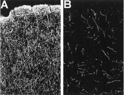
The serotonergic neurons of the mammalian brain comprise one of the most expansive chemical systems known. The cell bodies are largely confined to the midline (raphe) region of the brain stem in two general clusters: a superior group that consists of the dorsal raphe nucleus (B-7 and B-6), median raphe nucleus (B-8 and B-5), caudal linear nucleus (rostral B-8), and supralemniscal nucleus (B-9), and an inferior group that consists of nucleus raphe obscurus (B-2), nucleus raphe pallidus (B-1), nucleus raphe magnus (B-3), ventral lateral medulla (B-1/B-3), and the area postrema. The axons from these cells project throughout the neuroaxis from the spinal cord to the olfactory bulb and from the cerebral cortex to the hypothalamus. The development of this giant system begins very early in gestation and is influenced by a variety of growth regulatory factors, including the astroglial protein S-100 beta. Evidence will be presented that the serotonergic system plays a major role in the maturation of the brain by interacting with the 5-HT1A receptors which are most dense during these early developmental periods. The 5-HT1A receptor is located on both neurons and astrocytes, and in the latter cells may serve to stimulate release of S-100 beta. The developmental role of 5-HT appears to become dormant as the brain matures, and during aging and Alzheimer's disease, 5-HT receptors are significantly depressed. However, specific damage to 5-HT fibers in the adult brain by 5,7-dihydroxytryptamine produces a sharp fall in the levels of 5-HT which seems to reactivate the developmental signals in the brain. Not only are the serotonergic fibers encouraged to sprout and expand their territory, but the stimulation of the astrocytic growth factor by a 5-HT1A agonist is reinstated. The ability to recall developmental processes in the adult brain by interrupting the 5-HT fibers may provide important tools for understanding and treating the aged brain.

Publication Types:

* Review
* Review, Tutorial


PMID: 1752858 [PubMed - indexed for MEDLINE]
 
^ Wow, complete recovery. From the 3rd abstract:

"With respect to neuronal regeneration, following an initial 90% loss of 5-HT uptake sites after treatment with MDMA, the recovery of these sites occurred over a protracted period of time; a marked 25% reduction was seen at 6 months and the concentration of 5-HT uptake sites returned to control levels at 12 months following treatment with MDMA."

And that's from: "... (5-20 mg/kg twice daily for 4 consecutive days) ..."!
 
Be careful, the 3rd abstract is from an article quite some time before the first two.
 
any evidence on regrowth of 5HT2A sub populations?

i have heard, anecdotal, that l-deprenyl has been studied in this regard.
 
Not quite full recovery. A restoration of total pre-neurotoxicity levels of serotonin/SERT, but the distribution is abnormal, with elevated levels close to the brain stem (while more remote regions of the brain remain depleted.)

This is actually part of the evidence that significant axon loss is NOT occuring in humans under common patterns of MDMA use; abstinent users tend to have normal levels of SERT in ALL brain regions, not just by an overall average, suggesting a neuroadaptive change rather than structural injury.
 
TheDEA.org said:
Not quite full recovery. A restoration of total pre-neurotoxicity levels of serotonin/SERT, but the distribution is abnormal, with elevated levels close to the brain stem (while more remote regions of the brain remain depleted.)

This is actually part of the evidence that significant axon loss is NOT occuring in humans under common patterns of MDMA use; abstinent users tend to have normal levels of SERT in ALL brain regions, not just by an overall average, suggesting a neuroadaptive change rather than structural injury.

So.. excuse my limited knowledge but I have some questions:

1) Have there been any studies on humans (or monkeys) that show that the axons themselves degrade?
2) As far as I have read, there have been no studies in animals that show actual serotogenic NEURONS actually dying off?
3) Are there any studies that show an effective treatment to induce faster recovery from these brain changes in humans? (exercise, supplements, medicines, etc).

Thanks in advance.
 
1) Have there been any studies on humans (or monkeys) that show that the axons themselves degrade?

Yes, unambiguously. Here are some pictures (using silver staining I believe of biopsied brain slices):

friedmonkey.jpg


Normal vs. MDMA neurotox'ed monkey.

2) As far as I have read, there have been no studies in animals that show actual serotogenic NEURONS actually dying off?

I'm not aware of proof of cell body death in lab animals. The death of brain cells would be expected to increase levels of a protein called GFAP, but most animal MDMA neurotoxicity experiments haven't found increases in GFAP. MDMA neurotoxicity in lab animals typically involves dangerously high body temps, which can themselves kill brain cells, so it's not outside the bounds of possibility that overdoses could lead to some degree of brain cell death even without MDMA directly 'poisoning' the cells. MDMA can kill brain cells directly if you dump enough of it on a cell culture in a petri dish, but the very high concentrations used probably aren't relevant to recreational users.

3) Are there any studies that show an effective treatment to induce faster recovery from these brain changes in humans? (exercise, supplements, medicines, etc).

There's some discussion of drugs that may promote axon regrowth in animals HERE. At any rate, there is certainly no proven method, only a little theory.

Most (all?) people who think they've suffered neurotoxic damage are actually the victims of changes in brain function, particularly changes in the number of receptors in the brain. You brain tries to 'correct' for MDMA exposure (or any other psychoactive drug use) by making itself less sensitive to the neurotransmitters than MDMA releases; taken to extremes, the process can really screw people up. In most cases it doesn't seem to be permanent, but often takes months to largely recovery from.

If your mental health has been harmed by MDMA (or any other) drug use, the proven way to treat it is sobriety and time. :) (And I do mean *sobriety*. Cut out the alcohol binges, stop smoking pot, even try to quit caffeine.)
 
^^ Do you think this might have something to do with HPPD?

How would one go about returning the occpital 5HT2A receptors to normal?
http://www.ncbi.nlm.nih.gov/entrez/...d&dopt=Abstract&list_uids=11850153&query_hl=3
In recent MDMA users, post-synaptic 5-HT(2A) receptor densities were significantly lower in all cortical areas studied, while 5-HT(2A) receptor densities were significantly higher in the occipital cortex of ex-MDMA users. The combined results of this study suggest a compensatory upregulation of post-synaptic 5-HT(2A) receptors in the occipital cortex of ex-MDMA users due to low synaptic 5-HT levels.
 
*shrug* as far as I was awear HPPD was far more common in hallucinogen use than in MDMA use, so HPPD can't really be due to altered occpital lobe input, or at least can't depend on it.

Still, I can certainly see where you are comming from.
 
You're right in that it's more common in hallucinogen use.

It's odd, I've been doing a fair bit of hunting around the net. There's a forum dedicated to "visual snow". They get many of the effects of HPPD (e.g. visual static, afterimages, etc) but none of the "hallucinogen" effects (e.g. fractal patterns)... the odd thing is that their symptoms often commence without cause. A significant percentage of them had theirs triggered by SSRI administration.

There's a significant number of people on this board who report static vision, after images etc after long-term MDMA use, but again none of the typical hallucinogen symptoms.
 
^ I think your on to something V.

After my abuse I got an EEG done on my brain, and my occupital lobe had a VERY high brainwave feedback.

Also this study;
"Cowan and his colleagues examined brain activation during visual stimulation, using functional magnetic resonance imaging (fMRI), in subjects who had previously used Ecstasy (but not in the two weeks prior to imaging) and in subjects who had not previously used Ecstasy.

They found increased brain activation in three brain areas associated with visual processing in Ecstasy users with the highest lifetime exposure to the drug. The findings were consistent with the investigators' predictions based on results from animal models: that Ecstasy use is associated with a loss of serotonin signaling, which leads to hyper-excitability (increased activation) in the brain.

The hyper-excitability suggests a loss in brain efficiency, Cowan said, "meaning that it takes more brain area to process information or perform a task."

The investigators found that this shift in brain excitability did not return to normal in subjects who had not used Ecstasy in more than a year.

"We think this shift in cortical excitability may be chronic, long-lasting, and even permanent, which is a real worry," Cowan said, noting that the Ecstasy users in the study are young (18 to 35 years old). "The question is what will happen to their brains as they age over the next 60 years."

Cowan said that the pattern of hyper-excitability is similar to that observed in fMRI studies of individuals at risk for, or with early, Alzheimer's disease.

"I'm not saying that these people are at increased risk for dementia, but that there's a loss of brain efficiency in both recreational Ecstasy use and early Alzheimer's."

The findings suggest that brain hyper-excitability (increased activation in fMRI scans) may be a useful biomarker for Ecstasy-induced neurotoxicity, which the investigators will continue to study."
 
Top